Skip to main content Accessibility help
×
Hostname: page-component-78c5997874-s2hrs Total loading time: 0 Render date: 2024-11-19T22:05:39.482Z Has data issue: false hasContentIssue false

4 - Neonatal congenital heart disease

Published online by Cambridge University Press:  05 March 2012

Jeffrey C. Hellinger
Affiliation:
University of Pennsylvania
Mary M. K. Seshia
Affiliation:
University of Manitoba
Haresh Kirpalani
Affiliation:
Children's Hospital of Philadelphia
Monica Epelman
Affiliation:
Children's Hospital of Philadelphia
John Richard Mernagh
Affiliation:
McMaster University, Ontario
Get access

Summary

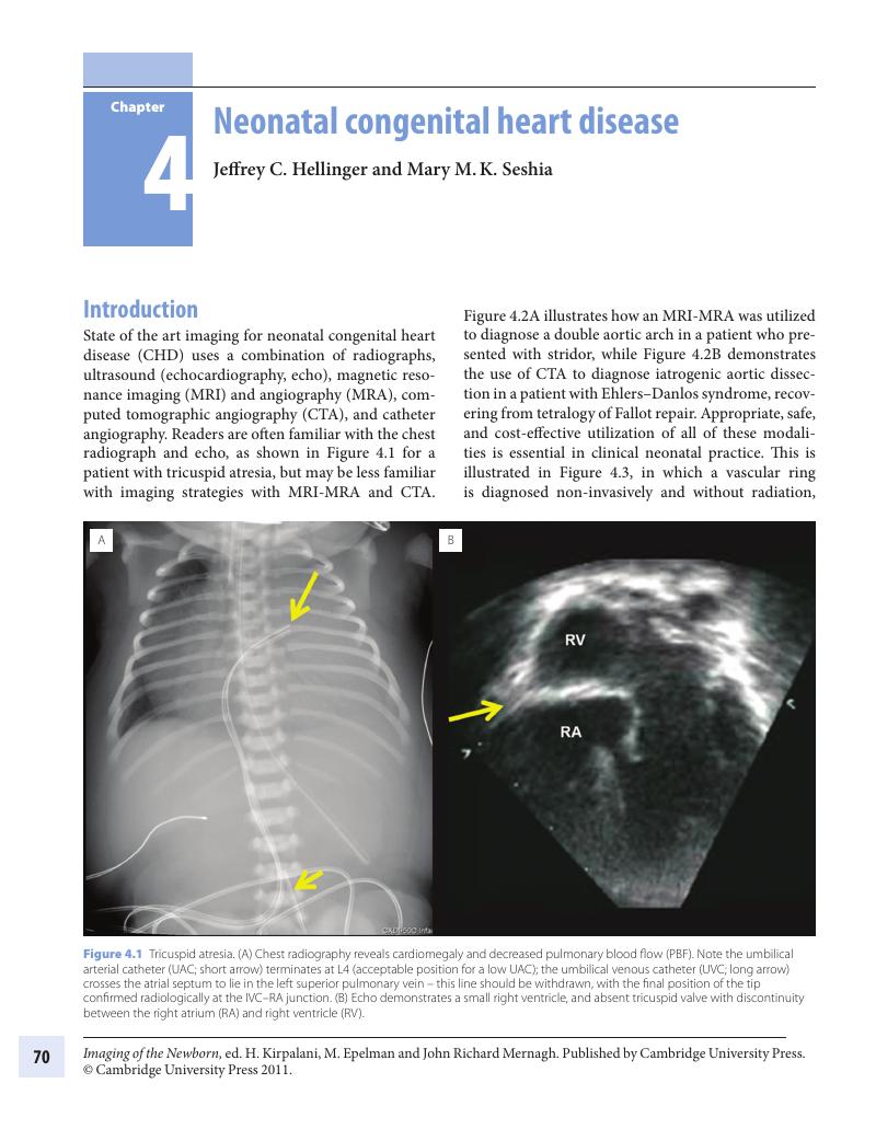
Image of the first page of this content. For PDF version, please use the ‘Save PDF’ preceeding this image.'
Type
Chapter
Information
Publisher: Cambridge University Press
Print publication year: 2011

Access options

Get access to the full version of this content by using one of the access options below. (Log in options will check for institutional or personal access. Content may require purchase if you do not have access.)

References

1. G., Nölker, K. J., Gutleben, H., Marschang, et al. Three dimensional left atrial and esophagus reconstruction using cardiac C-arm computed tomography with image integration into fluoroscopic views for ablation of atrial fibrillation: accuracy of a novel modality in comparison with multislice computed tomography. Heart Rhythm 2008; 5(12):1651–7.Google Scholar
2. J. C., Hellinger, A., Pena, M., Poon, F. P., Chan, M., Epelman. Pediatric computed tomographic angiography: imaging the cardiovascular system gently. Radiol Clin North Am 2010; 48(2):439–67.Google Scholar
3. E. M., Blass, L. B., Hoffmeyer. Sucrose as an analgesic for newborn infants. Pediatrics 1991; 87:215–18.Google Scholar
4. R., Van Praagh. The segmental approach clarified. Cardiovasc Intervent Radiol 1984; 7(6):320–5.Google Scholar
5. M. P., Blayney, D. R., Logan. First thoracic vertebral body as reference for endotracheal tube placement. Arch Dis Child 1994; 71:F32–5.Google Scholar
6. C., Lapierre, J., Déry, R., Guérin, et al. Segmental approach to imaging of congenital heart disease. RadioGraphics 2010; 30:397–411.Google Scholar
7. K., Murayama, H., Nagasaka, K., Tate, et al. Significant correlations between the flow volume of patent ductus venosus and early neonatal liver function: possible involvement of patent ductus venosus in postnatal liver function. Arch Dis Child Fetal Neonatal Ed 2006; 91:F175–9.Google Scholar
8. Z. J., Wang, G. P., Reddy, M. B., Gotway, et al. CT and MR imaging of pericardial disease. Radiographics 2003; 23:S167–80.Google Scholar
9. S. R., Pedra, J. F., Smallhorn, G., Ryan, et al. Fetal cardiomyopathies: pathogenic mechanisms, hemodynamic findings, and clinical outcome. Circulation 2002; 106(5):585–91.Google Scholar
10. J., Kohut, J., Krzystolik-Ładzińska, L., Szydłowski, et al. The diagnosis, clinical course, and follow-up of children with cardiac tumours – a single center experience. Kardiol Pol 2010; 68(3):304–9.Google Scholar
11. A. H., Friedman, M. A., Fogel, P., Stephens, et al. Identification, imaging, functional assessment, and management of congenital arterial abnormalities in children. Cardiol Young 2007; 17(Suppl. 2):56–67.Google Scholar
12. P. S., Rao. Diagnosis and management of cyanotic congenital heart disease: Part I. Indian J Pediatr 2009; 76(1):57–70.Google Scholar
13. P. S., Rao. Diagnosis and management of cyanotic congenital heart disease: Part II. Indian J Pediatr 2009; 76(3):297–308.Google Scholar
14. L., Kovacikova, K., Dakkak, P., Skrak, et al. Neonates with left-sided obstructive heart disease: clinical manifestation and management at primary care hospitals. Bratisl Lek Listy 2007; 108(7):316–19.Google Scholar
15. J. C., Hellinger, L. F., Cervantes, L. S., Medina (2009) Congenital disease of the aortic arch: coarctation and arch anomalies. In L. S., Medina, K. E., Applegate, C. C., Blackmore (Eds.) Evidence-Based Imaging in Pediatrics: Optimizing Imaging in Pediatric Patient Care, New York, Springer, pp. 359–80.Google Scholar
16. Z. J., Wang, G. P., Reddy, M. B., Gotway, et al. Cardiovascular shunts: MR imaging evaluation. Radiographics 2003; 23(Spec No):S181–94.Google Scholar
17. T. R., Johnson. Conotruncal cardiac defects: a clinical imaging perspective. Pediatr Cardiol 2010; 31(3):430–7.Google Scholar

Save book to Kindle

To save this book to your Kindle, first ensure [email protected] is added to your Approved Personal Document E-mail List under your Personal Document Settings on the Manage Your Content and Devices page of your Amazon account. Then enter the ‘name’ part of your Kindle email address below. Find out more about saving to your Kindle.

Note you can select to save to either the @free.kindle.com or @kindle.com variations. ‘@free.kindle.com’ emails are free but can only be saved to your device when it is connected to wi-fi. ‘@kindle.com’ emails can be delivered even when you are not connected to wi-fi, but note that service fees apply.

Find out more about the Kindle Personal Document Service.

Available formats
×

Save book to Dropbox

To save content items to your account, please confirm that you agree to abide by our usage policies. If this is the first time you use this feature, you will be asked to authorise Cambridge Core to connect with your account. Find out more about saving content to Dropbox.

Available formats
×

Save book to Google Drive

To save content items to your account, please confirm that you agree to abide by our usage policies. If this is the first time you use this feature, you will be asked to authorise Cambridge Core to connect with your account. Find out more about saving content to Google Drive.

Available formats
×