Introduction
Perkinsosis is the most severe disease in marine mollusks caused by protozoan endoparasites of the genus Perkinsus Machkin, 1950. The genus Perkinsus belongs to the phylum Perkinsozoa and comprises seven nominal species: Perkinsus marinus (Ray, Reference Ray1996), Perkinsus olseni (Lester and Davis, Reference Lester and Davis1981), Perkinsus qugwadi (Blackbourn et al., Reference Blackbourn, Bower and Meyer1998), Perkinsus chesapeaki (McLaughlin et al., Reference McLaughlin, Tall, Shaheen, Elsayed and Faisal2000), Perkinsus mediterraneus (Casas et al., Reference Casas, Grau, Reece, Apakupakul, Azevedo and Villalba2004), Perkinsus honshuensis (Dungan and Reece, Reference Dungan and Reece2006) and Perkinsus beihaiensis (Moss et al., Reference Moss, Xiao, Dungan and Reece2008). Since its discovery in 1950, Perkinsus spp. have been detected in mollusks around the world, such as in North America, South America, Europe, Asia, Oceania and Africa (Lester and Davis, Reference Lester and Davis1981; Ray, Reference Ray1996; Casas et al., Reference Casas, Grau, Reece, Apakupakul, Azevedo and Villalba2004; Moss et al., Reference Moss, Xiao, Dungan and Reece2008). Perkinsus marinus (syn. Dermocystidium marinum) is the causative agent of Dermo disease, which leads to seasonal mortality and a decline in the population of the eastern oyster, Crassostrea virginica, along the Gulf of Mexico coast of the United States (Ray, Reference Ray1996). Perkinsus olseni has a wide geographical distribution and a wide range of host, including Haliotis ruber, Tapes decussatus, Ruditapes philippinarum, Crassostrea hongkongensis, Pinctada martensii, Haliotis laevigata, as well as several other bivalves and gastropods (Goggin and Lester, Reference Goggin and Lester1995; Villalba et al., Reference Villalba, Reece, Camino Ordás, Casas and Figueras2004). Owing to the lethal and sublethal impact on commercially valuable mollusks, parasitic disease caused by P. olseni and P. marinus have been designated as internationally notifiable mollusk diseases by the World Organization for Animal Health (OIE, 2016).
Perkinsus beihaiensis was firstly described in the oysters Crassostrea ariakensis and C. hongkongensis of Southern China (Moss et al., Reference Moss, Xiao, Dungan and Reece2008). The reported hosts of P. beihaiensis was 15 species of mollusks include oysters, clams (Moss et al., Reference Moss, Xiao, Dungan and Reece2008; Sanil et al., Reference Sanil, Suja, Lijo and Vijayan2012; Ferreira et al., Reference Ferreira, Sabry, da Silva, Vasconcelos Gesteira, Romao, Paz, Feijo, Dantas Neto and Maggioni2015; Luz et al., Reference Luz, Carvalho, Oliveira and Boehs2017; Cui et al., Reference Cui, Ye, Wu and Wang2018; Pagenkopp Lohan et al., Reference Pagenkopp Lohan, Hill-Spanik, Torchin, Fleischer, Carnegie, Reece and Ruiz2018; Ye et al., Reference Ye, Wu, Lu, Yao, Wang, Shi, Yu and Zhao2022) and Mediterranean mussel (Mytilus galloprovincialis) (Itoh et al., Reference Itoh, Komatsua, Maeda, Hirase and Yoshinaga2019). The geographic distribution of P. beihaiensus extended to other countries such as India, Brazil, Panama and Japan (Sanil et al., Reference Sanil, Suja, Lijo and Vijayan2012; Ferreira et al., Reference Ferreira, Sabry, da Silva, Vasconcelos Gesteira, Romao, Paz, Feijo, Dantas Neto and Maggioni2015; Pagenkopp Lohan et al., Reference Pagenkopp Lohan, Hill-Spanik, Torchin, Fleischer, Carnegie, Reece and Ruiz2018; Itoh et al., Reference Itoh, Komatsua, Maeda, Hirase and Yoshinaga2019). Epidemiological investigation showed that the prevalence of P. beihaiensis was notably higher during the summer and autumn seasons compared to winter and spring (Yang, Reference Yang2022).
Over the past decades, the thick shelled mussel Mytilus coruscus has emerged as one of the most commercially important shellfish species in China due to its exceptional food quality, significant health benefits and ecological service value (Zhong et al., Reference Zhong, Qu and Chen2014; Liang et al., Reference Liang, Wang, Yu and Bi2015). However, in recent years, the large-scale and high-density cultivation of M. coruscus, coupled with changes in climate (such as global warming) and environment factors (such as seawater acidification), has led to various pathogenic challenges to this mussel species (Dong et al., Reference Dong, Chen, Lu, Wu and Qi2017). Therefore, it is necessary to investigate the presence of Perkinsus species in M. coruscus and determine their impacts on health of thick shelled mussels.
Materials and methods
Sample collection and process
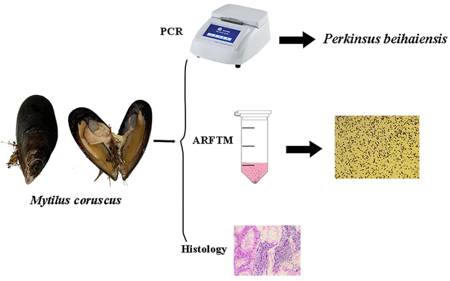
A total of 16 specimens (Supplementary Table S1) of M. coruscus (mean shell length ± s.d.: 9.6 ± 0.6 cm) were sampled in March 2023 from Gouqi island (E122°76′, N30°70′) in Zhoushan city, Zhejiang province, renowned as the hometown of M. coruscus. After shucking the shell, a portion of the mantle, gill and digestive gland were fixed in 4% PFA for histopathological examination. Another portion of the gill tissue was fixed in anhydrous ethanol for DNA extraction. The remaining soft body was weighed, shredded and subjected to ARFTM for quantifying the infection level of Perkinsus species.
Detection of Perkinsus species in ARFTM
Fifteen mL of ARFTM supplemented with antibiotics (chloramphenicol 200 μg mL−1, penicillin-streptomycin 500 U mL−1) and nystatin (4.6 μg mL−1) was added into each sample tube. After a week of incubation in the dark at room temperature (La Peyre et al., Reference La Peyre, Nickens, Volety, Tolley and La Peyre2003). The tissues were collected by centrifugation and then subjected to treatment with 2 M NaOH at 60°C until complete lysis (Choi et al., Reference Choi, Wilson, Lewis, Powell and Ray1989). Hypnospores or prezoosporangia were subsequently collected by centrifugation (4000 rpm, 20 min), followed by three washes with sterile seawater and fixed volume to 1 mL. Eighty μL of hypnospores suspension thorough mixed with 20 μL Lugol's iodine, then 10 μL of this mixture was transferred onto a hemocytometer to count the number of hypnospores with a bluish black spherical shape. Each sample underwent three counting repetitions.
The prevalence of infection by Perkinsus was expressed as the number of infected animals over the total number of sampled animals (Bush et al., Reference Bush, Lafferty, Lotz and Shostak1997), while the infection intensity was calculated as the number of Perkinsus cells per gram of tissue weight (Park and Choi, Reference Park and Choi2001).
Histological observation
The fixed tissues were dehydrated with aqueous ethanol through an ascending series of concentrations, cleared with xylene, embedded in paraffin and 6 μm thick sections were made using a Microtome (Leica, Germany). The sections were stained with hematoxylin & eosin, then mounted in Canada balsam for observation under a light microscope compound with brightfield optics (Leica, Germany).
DNA extraction, PCRs, sequencing and phylogenetic analysis
Genomic DNA was extracted from excised gill using a TIAN amp Genomic DNA Kit (TIANGEN, Beijing, China), following the manufacturer's protocols. The Perkinsus genus-specific internal transcribed spacer (ITS) ribosomal RNA primers (PerkITS-85: 5′-CCGCTTTGTTTGGATCCC-3′; PerkITS-750: 5′-ACATCAGGCCTTCTAATGA TG-3′) were applied to amplify the target sequence (Casas et al., Reference Casas, Villalba and Reece2002). The final volume of the polymerase chain reaction (PCR) was 20 μL containing 10 μL of 2xEs Taq Master Mix (Hangzhou, China), 10 pmol of each PCR primer, and 1 μL of the extracted DNA samples. The thermal cycler programme was as follows: 5 min at 94°C as the initial step; then 35 cycles of 30 s at 94°C, 45 s at 56°C, 45 s at 72°C. The final step was 7 min at 72°C. All PCR products were loaded on a 1.5% agarose gel. After purification, the positive PCR products were sequenced using the both PCR primers on an ABI 3730XL (Applied Biosystems, USA). The sequences were assembled manually with the software SeqMan (DNASTAR, USA). The homology of the generated sequences was analysed using the Basic Local Alignment Search Tool (BLAST) program available on the NCBI.
The maximum likelihood (ML) method was employed to construct phylogenetic trees using ITS sequences in IQ-tree (Nguyen et al., Reference Nguyen, Schmidt, von Haeseler and Minh2014). Based on the Bayesian information criterion (BIC), HKY + F + G4 was chosen as the optimal nucleotide substitution model for ITS in PhyloSuite (Zhang et al., Reference Zhang, Gao, Jakovlic, Zou, Zhang, Li and Wang2020). ML trees were obtained with 1000 bootstraps of the data.
Results
Identification of Perkinsus species in M. coruscus
According to the ARFTM assay, four positive samples exhibited enlarged blue–black hypnospores characteristic of Perkinsus sp. like organisms, and the prevalence of infection by Perkinsus sp. like organisms in M. coruscus was 25% (4/16). The diameter of hypnospores or prezoosporangia was 8–27 (15.6 ± 4.0, n = 111) μm (Fig. 1). The infection intensity ranged between 10.2 and 3.34 × 10⁵ cells per gram tissue. However, there were only two positive samples in the PCR assay, and the prevalence of infection was 12.5% (2/16). The PCR primers targeting the ITS region of Perkinsus spp. amplified 687 bp of products. Sequence alignment found that the two positive samples had the same ITS sequences, and the BLAST results revealed that the sequence had the highest homology with P. beihaiensis (Taizhou, China, accession number: MT908890) at 100. The nucleotide sequence obtained in the current study has been deposited in NCBI GenBank with accession numbers OR192847 and OR192848.

Figure 1. The hypnospores or prezoosporangia of Perkinsus beihaiensis in Mytilus coruscus. Scale bars = 100 μm.
Phylogenetic analysis
The ML phylogenetic tree (Fig. 2) indicated that the specimens of Perkinsus from Gouqi island cluster with P. beihaiensis from China, Panama and Brazil. Internal transcribed spacer region nucleotide sequences of P. beihaiensis, P. chesapeaki, P. olseni formed a monophyletic clade sister to a clade containing P. honshuensis and P. mediterraneus.

Figure 2. Phylogenetic relationships of Perkinsus species based on ITS sequences constructed by ML method. ‘*’ represented the sequences obtained in this study.
Histological observations
Perkinsus beihaiensis was observed in the visceral mass, gills and mantle (Fig. 3). The mature trophozoites, characterized by a large vacuolation and eccentric nucleus, were found in the three tissues, especially in visceral masses (Fig. 3A, B, D), and the schizonts were mainly observed in visceral mass (Fig. 3C). The infection of P. beihaiensis induced the hemocyte infiltration in the visceral mass (Fig. 3C, D). Although no apparent lesions were observed in the tissues, destruction of digestive tubules was evident (Fig. 3C, D).

Figure 3. Perkinsus beihaiensis infection in the gill (A), mantle (B) and visceral mass (C) of M. coruscus. (↑) and (▵) represents the trophozoite and schizont of the P. beihaiensis. (▴) represent the hemocyte of M. coruscus. Scale bars = 10 μm.
Discussion
Perkinsus beihaiensis, a cosmopolitan parasite species, has a wide geographical distribution, including Asia (China, Japan and India), South America (Brizal and Panama) (Moss et al., Reference Moss, Xiao, Dungan and Reece2008; Sanil et al., Reference Sanil, Suja, Lijo and Vijayan2012; Pagenkopp Lohan et al., Reference Pagenkopp Lohan, Hill-Spanik, Torchin, Fleischer, Carnegie, Reece and Ruiz2018; Itoh et al., Reference Itoh, Komatsua, Maeda, Hirase and Yoshinaga2019; Ye et al., Reference Ye, Wu, Lu, Yao, Wang, Shi, Yu and Zhao2022). In China, the main epidemic area of P. beihaiensis was in SCS (Moss et al., Reference Moss, Xiao, Dungan and Reece2008; Cui et al., Reference Cui, Ye, Wu and Wang2018; Wu et al., Reference Wu, Ye, Cui, Yao, Lu, Luo, Deng and Wang2018), and recently been reported in ECS and YBS (Ye et al., Reference Ye, Wu, Lu, Yao, Wang, Shi, Yu and Zhao2022). Although in previous study, P. beihaiensis has been detected in ESC, including Taizhou (Zhejiang province), Ningde and Putian (Fujian province) (Ye et al., Reference Ye, Wu, Lu, Yao, Wang, Shi, Yu and Zhao2022). However, the sample of collection location in the presently study was in Shengsi county (Zhoushan, Zhejiang province), the main production area of M. coruscus, that has not been investigated for Perkinsus species detection in ESC.
Perkinsus beihaiensis was firstly detected in a new host M. coruscus through ARFTM, histological observation and PCR in this study, expanding the host range of the parasite. The host specificity of P. beihaiensis is low (Ye et al., Reference Ye, Wu, Lu, Yao, Wang, Shi, Yu and Zhao2022). It has so far been reported in 15 bivalve species (Pagenkopp Lohan et al., Reference Pagenkopp Lohan, Hill-Spanik, Torchin, Fleischer, Carnegie, Reece and Ruiz2018; Itoh et al., Reference Itoh, Komatsua, Maeda, Hirase and Yoshinaga2019), and clams and oysters are the known susceptible hosts of the parasite. Perkinsus beihaiensis was the dominant Perkinsus species in the Mediterranean mussel (Itoh et al., Reference Itoh, Komatsua, Maeda, Hirase and Yoshinaga2019). Consistently, P. beihaiensis was the only Perkinsus species detected from M. coruscus, a congeneric species of M. galloprovincialis, in the present study, indicating that mussel seems to be highly susceptible to this parasite.
Mytilus coruscus is mainly distributed in the Hokkaido of Japan, Jeju Island of Korea, the Yellow Sea, Bohai, the East China Sea (ECS) and Taiwan (Dong et al., Reference Dong, Chen, Lu, Wu and Qi2017). With the breakthrough of artificial seedling cultivation technology, the breeding scale of M. coruscus has increased rapidly. It has become one of the most important native economic species in China recently with outstanding economic value. The cultivation of this mussel is raft farming charactered with a high degree of intensification and breeding density, which may provide conditions for the widespread of P. beihaiensis in M. coruscus.
Histological tissue section in oysters moderate infected with P. beihaiensis induced lesions and hemocyte defensive responses occurring in epithelia and connective tissues (Moss et al., Reference Moss, Xiao, Dungan and Reece2008). Contrastingly, the pathological effects caused by P. beihaiensis in M. coruscus appeared to be milder. Infection with this Perkinsus species could lead to inflammatory reaction of hemocyte in visceral mass and the destruction of digestive tubules was evident indicating that this parasite had negative physiological effects on M. coruscus. Therefore, close attention should be paid to the P. beihaiensis in the mussel to prevent harmful effects caused by this parasite to the local aquaculture industry.
Supplementary material
The supplementary material for this article can be found at https://doi.org/10.1017/S0031182024000702.
Data availability statement
Representative sequences obtained in this study were deposited in GenBank with the accession numbers OR192847–OR192848.
Authors’ contributions
All authors designed and conducted laboratory work and all of them were involved in the manuscript and approved the final version. Jia Y. Zhai and Peng Z. Qi These authors contributed equally to this article as co-first authors.
Financial support
This work was supported by the Natural Science Foundation of China (Nos. 41976111, 42176099, 42020104009 and 42076119), the Natural Science Foundation for Distinguished Young Scholars of Zhejiang province (Grant number: LR22D060002).
Competing interests
None.
Ethical standards
Not applicable.


