Article contents
Comparative proteomic analysis of Leishmania parasites isolated from visceral and cutaneous leishmaniasis patients
Published online by Cambridge University Press: 11 November 2021
Abstract

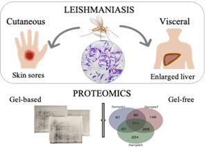
Leishmaniasis is an infectious disease in which different clinical manifestations are classified into three primary forms: visceral, cutaneous and mucocutaneous. These disease forms are associated with parasite species of the protozoan genus Leishmania. For instance, Leishmania infantum and Leishmania tropica are typically linked with visceral (VL) and cutaneous (CL) leishmaniasis, respectively; however, these two species can also cause other form to a lesser extent. What is more alarming is this characteristic, which threatens current medical diagnosis and treatment, is started to be acquired by other species. Our purpose was to address this issue; therefore, gel-based and gel-free proteomic analyses were carried out on the species L. infantum to determine the proteins differentiating between the parasites caused VL and CL. In addition, L. tropica parasites representing the typical cases for CL were included. According to our results, electrophoresis gels of parasites caused to VL were distinguishable regarding the repetitive down-regulation on some specific locations. In addition, a distinct spot of an antioxidant enzyme, superoxide dismutase, was shown up only on the gels of CL samples regardless of the species. In the gel-free approach, 37 proteins that were verified with a second database search using a different search engine, were recognized from the comparison between VL and CL samples. Among them, 31 proteins for the CL group and six proteins for the VL group were determined differentially abundant. Two proteins from the gel-based analysis, pyruvate kinase and succinyl-coA:3-ketoacid-coenzyme A transferase analysis were encountered in the protein list of the CL group.
- Type
- Research Article
- Information
- Copyright
- Copyright © The Author(s), 2021. Published by Cambridge University Press
References
- 1
- Cited by


