Hostname: page-component-586b7cd67f-g8jcs Total loading time: 0 Render date: 2024-11-30T07:35:42.953Z Has data issue: false hasContentIssue false

Monolithic quartz platform for cellular contact guidance

Published online by Cambridge University Press:  16 March 2020
Michael C. Robitaille
Affiliation:
Naval Research Laboratory, 4555 Overlook Ave SW, Washington, DC20375-5320, USA
Joseph A. Christodoulides
Affiliation:
Naval Research Laboratory, 4555 Overlook Ave SW, Washington, DC20375-5320, USA
Jinny L. Liu
Affiliation:
Naval Research Laboratory, 4555 Overlook Ave SW, Washington, DC20375-5320, USA
Wonmo Kang
Affiliation:
School for Engineering of Matter, Transport and Energy, Arizona State University, Tempe, AZ85287, USA
Jeff M. Byers
Affiliation:
Naval Research Laboratory, 4555 Overlook Ave SW, Washington, DC20375-5320, USA
Katarina Doctor
Affiliation:
Naval Research Laboratory, 4555 Overlook Ave SW, Washington, DC20375-5320, USA
Dmitry Kozak
Affiliation:
Naval Research Laboratory, 4555 Overlook Ave SW, Washington, DC20375-5320, USA
Marc P. Raphael*
Affiliation:
Naval Research Laboratory, 4555 Overlook Ave SW, Washington, DC20375-5320, USA
*
Address all correspondence to Marc P. Raphael at [email protected]
Get access

Abstract

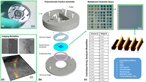
Contact guidance is vital to many physiological processes, yet is still poorly understood. This is partly due to the variability of experimental platforms, making comparisons difficult. To combat this, a multiplexed approach was used to fabricate topographical cues on single quartz coverslips for high-throughput screening. Furthermore, this method offers control of surface roughness and protein adsorption characterization, two critical aspects to the in vitro environment often overlooked in contact guidance platforms. The quartz surface can be regenerated, is compatible with versatile microscopy modes, and can scale up for manufacturing offering a novel platform that could serve as a potential standard assay.

Type
Research Letters
Copyright
Copyright © Materials Research Society 2020

Access options

Get access to the full version of this content by using one of the access options below. (Log in options will check for institutional or personal access. Content may require purchase if you do not have access.)

References

1.Harrison, R.G.: The reaction of embryonic cells to solid structures. J. Exp. Zool. 17, 521 (1914).CrossRefGoogle Scholar
2.Newgreen, D.: Physical influences on neural crest cell migration in avian embryos: contact guidance and spatial restriction. Dev. Biol. 131, 136 (1989).CrossRefGoogle ScholarPubMed
3.Li, J., Chen, J., and Kirsner, R.: Pathophysiology of acute wound healing. Clin. Dermatol. 25, 9 (2007).CrossRefGoogle ScholarPubMed
4.Provenzano, P.P., Eliceiri, K.W., Campbell, J.M., Inman, D.R., White, J.G., and Keely, P.J.: Collagen reorganization at the tumor-stromal interface facilitates local invasion. BMC Med. 4, 38 (2006).CrossRefGoogle ScholarPubMed
5.Lynch, K.J., Skalli, O., and Sabri, F.: Investigation of surface topography and stiffness on adhesion and neurites extension of PC12 cells on crosslinked silica aerogel substrates. PLoS ONE 12, e0185978 (2017).CrossRefGoogle ScholarPubMed
6.Clark, P., Connolly, P., Curtis, A., Dow, J., and Wilkinson, C.: Topographical control of cell behaviour: II. Multiple grooved substrata. Development 108, 635 (1990).Google ScholarPubMed
7.Rajnicek, A., Britland, S., and McCaig, C.: Contact guidance of CNS neurites on grooved quartz: influence of groove dimensions, neuronal age and cell type. J. Cell Sci. 110, 2905 (1997).Google ScholarPubMed
8.Teixeira, A.I., McKie, G.A., Foley, J.D., Bertics, P.J., Nealey, P.F., and Murphy, C.J.: The effect of environmental factors on the response of human corneal epithelial cells to nanoscale substrate topography. Biomaterials 27, 3945 (2006).CrossRefGoogle ScholarPubMed
9.Lord, M.S., Foss, M., and Besenbacher, F.: Influence of nanoscale surface topography on protein adsorption and cellular response. Nano Today 5, 66 (2010).CrossRefGoogle Scholar
10.Ross, A.M., Jiang, Z., Bastmeyer, M., and Lahann, J.: Physical aspects of cell culture substrates: topography, roughness, and elasticity. Small 8, 336 (2012).CrossRefGoogle ScholarPubMed
11.Christodoulides, J.A., Christophersen, M., Liu, J.L., Delehanty, J.B., Raghu, D., Robitaille, M., Byers, J.M., and Raphael, M.P.: Nanostructured substrates for multi-cue investigations of single cells. MRS Commun. 8, 49 (2018).CrossRefGoogle Scholar
12.Biggs, M.J.P., Richards, R.G., and Dalby, M.J.: Nanotopographical modification: a regulator of cellular function through focal adhesions. Nanomedicine 6, 619 (2010).CrossRefGoogle ScholarPubMed
13.Martinez, E., Engel, E., Planell, J., and Samitier, J.: Effects of artificial micro- and nano-structured surfaces on cell behaviour. Ann. Anat. 191, 126 (2009).CrossRefGoogle ScholarPubMed
14.Andersson, A.-S., Bäckhed, F., von Euler, A., Richter-Dahlfors, A., Sutherland, D., and Kasemo, B.: Nanoscale features influence epithelial cell morphology and cytokine production. Biomaterials 24, 3427 (2003).CrossRefGoogle ScholarPubMed
15.Charest, J.L., Bryant, L.E., Garcia, A.J., and King, W.P.: Hot embossing for micropatterned cell substrates. Biomaterials 25, 4767 (2004).CrossRefGoogle ScholarPubMed
16.Loesberg, W., Te Riet, J., Van Delft, F., Schön, P., Figdor, C., Speller, S., Van Loon, J., Walboomers, X., and Jansen, J.: The threshold at which substrate nanogroove dimensions may influence fibroblast alignment and adhesion. Biomaterials 28, 3944 (2007).CrossRefGoogle ScholarPubMed
17.Heydarkhan-Hagvall, S., Choi, C.-H., Dunn, J., Heydarkhan, S., Schenke-Layland, K., MacLellan, W.R., and Beygui, R.E.: Influence of systematically varied nano-scale topography on cell morphology and adhesion. Cell Commun. Adhes. 14, 181 (2007).CrossRefGoogle ScholarPubMed
18.Miller, C., Shanks, H., Witt, A., Rutkowski, G., and Mallapragada, S.: Oriented Schwann cell growth on micropatterned biodegradable polymer substrates. Biomaterials 22, 1263 (2001).CrossRefGoogle ScholarPubMed
19.Recknor, J.B., Recknor, J.C., Sakaguchi, D.S., and Mallapragada, S.K.: Oriented astroglial cell growth on micropatterned polystyrene substrates. Biomaterials 25, 2753 (2004).CrossRefGoogle ScholarPubMed
20.Voinova, M.V., Rodahl, M., Jonson, M., and Kasemo, B.: Viscoelastic acoustic response of layered polymer films at fluid-solid interfaces: continuum mechanics approach. Phys. Scr. 59, 391 (1999).CrossRefGoogle Scholar
21.Carpenter, A.E., Jones, T.R., Lamprecht, M.R., Clarke, C., Kang, I.H., Friman, O., Guertin, D.A., Chang, J.H., Lindquist, R.A., and Moffat, J.: CellProfiler: image analysis software for identifying and quantifying cell phenotypes. Genome Biol. 7, R100 (2006).CrossRefGoogle ScholarPubMed
22.Dimilla, P.A., Albelda, S.M., and Quinn, J.A.: Adsorption and elution of extracellular matrix proteins on non-tissue culture polystyrene Petri dishes. J. Colloid Interface Sci. 153, 212 (1992).CrossRefGoogle Scholar
23.Curtis, A.: The mechanism of adhesion of cells to glass: a study by interference reflection microscopy. J. Cell Biol. 20, 199 (1964).CrossRefGoogle ScholarPubMed
24.Klein, K., Rommel, C.E., Hirschfeld-Warneken, V.C., and Spatz, J.P.: Cell membrane topology analysis by RICM enables marker-free adhesion strength quantification. Biointerphases 8, 28 (2013).CrossRefGoogle ScholarPubMed
25.DiMilla, P.A., Stone, J.A., Quinn, J.A., Albelda, S.M., and Lauffenburger, D.A.: Maximal migration of human smooth muscle cells on fibronectin and type IV collagen occurs at an intermediate attachment strength. J. Cell Biol. 122, 729 (1993).CrossRefGoogle ScholarPubMed
26.Maheshwari, G., Brown, G., Lauffenburger, D.A., Wells, A., and Griffith, L.G.: Cell adhesion and motility depend on nanoscale RGD clustering. J. Cell Sci. 113, 1677 (2000).Google ScholarPubMed
27.Missirlis, D., Haraszti, T., Scheele, C.v.C., Wiegand, T., Diaz, C., Neubauer, S., Rechenmacher, F., Kessler, H., and Spatz, J.P.: Substrate engagement of integrins α5β1 and αvβ3 is necessary, but not sufficient, for high directional persistence in migration on fibronectin. Sci. Rep. 6, 23258 (2016).CrossRefGoogle Scholar
28.Teixeira, A.I., Abrams, G.A., Bertics, P.J., Murphy, C.J., and Nealey, P.F.: Epithelial contact guidance on well-defined micro- and nanostructured substrates. J. Cell Sci. 116, 1881 (2003).CrossRefGoogle ScholarPubMed
29.Azatov, M., Sun, X., Suberi, A., Fourkas, J.T., and Upadhyaya, A.: Topography on a subcellular scale modulates cellular adhesions and actin stress fiber dynamics in tumor associated fibroblasts. Phys. Biol. 14, 065003 (2017).CrossRefGoogle ScholarPubMed
30.Ray, A., Lee, O., Win, Z., Edwards, R.M., Alford, P.W., Kim, D.-H., and Provenzano, P.P.: Anisotropic forces from spatially constrained focal adhesions mediate contact guidance directed cell migration. Nat. Commun. 8, 14923 (2017).CrossRefGoogle ScholarPubMed
31.Petrie, R.J., Doyle, A.D., and Yamada, K.M.: Random versus directionally persistent cell migration. Nat. Rev. Mol. Cell Biol. 10, 538 (2009).CrossRefGoogle ScholarPubMed
32.Kim, D.-H. and Wirtz, D.: Focal adhesion size uniquely predicts cell migration. FASEB J. 27, 1351 (2013).CrossRefGoogle ScholarPubMed
Supplementary material: File

Robitaille et al. supplementary material

Robitaille et al. supplementary material

Download Robitaille et al. supplementary material(File)
File 1.7 MB