Hostname: page-component-586b7cd67f-tf8b9 Total loading time: 0 Render date: 2024-11-30T10:34:21.905Z Has data issue: false hasContentIssue false

Cell viability study of green synthesized CdTe/CdSe quantum dots against osteosarcoma cell line for improved therapeutic action

Published online by Cambridge University Press:  29 January 2019
Ncapayi Vuyelwa
Affiliation:
Department of Applied Chemistry, University of Johannesburg, Doornfontein Campus, P.O. Box 17011, Doornfontein, 2028, Johannesburg, South Africa Centre for Nanomaterials Science Research, University of Johannesburg, Johannesburg, 2028, South Africa
Oluwatobi S. Oluwafemi*
Affiliation:
Department of Applied Chemistry, University of Johannesburg, Doornfontein Campus, P.O. Box 17011, Doornfontein, 2028, Johannesburg, South Africa Centre for Nanomaterials Science Research, University of Johannesburg, Johannesburg, 2028, South Africa
*
Address all correspondence to Oluwatobi S. Oluwafemi at [email protected]
Get access

Abstract

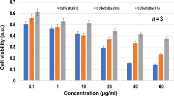
LM-8 is a murine osteosarcoma cell line associated with bone tumor in young adult and children. It contains the nuclear factor-κB which make this cell high resistance to irradiation thus limiting its treatment only to chemotherapy and surgery which has become a source of concerned for cancer therapy. In addressing this problem, we herein report the synthesis of CdTe/CdSe core/shell quantum dots (QDs) via a simple, reproducible method and its cytotoxic action against the LM-8 cell line. The cell viability study shows that the as-synthesized QDs are toxic to the LM-8 cell line in a concentration- and time-dependent manner.

Type
Research Letters
Copyright
Copyright © Materials Research Society 2019 

Access options

Get access to the full version of this content by using one of the access options below. (Log in options will check for institutional or personal access. Content may require purchase if you do not have access.)

References

1.Clift, M.J.D. and Stone, V.: Quantum dots: an insight and perspective of their biological interaction and how this relates to their relevance for clinical use. Theranostics 2, 668680 (2012).Google Scholar
2.Wang, Y., Wang, R., Liu, S., Yang, K., Zhou, L., and Li, H.: Synthesis and characterization of CdTe quantum dots by one step method. Bull. Chem. Soc. Ethiop. 27, 387393 (2013).Google Scholar
3.Lim, Y.T., Kim, S., Nakayama, A., Stott, N.E., Bawendi, M.G., and Frangioni, J.V.: Selection of quantum dot wavelengths for biomedical assays and imaging. Mol. Imaging 2, 50564 (2003).Google Scholar
4.Chandan, H.R., Jessica, D., Schiffman, R., and Geetha Balakrishna, R.: Quantum dots as fluorescent probes: Synthesis, surface chemistry, energy transfer mechanisms, and applications. Sens. Actuators, B 258, 11911214 (2018).Google Scholar
5.Chandan, H.R., Jessica, D., Schiffman, R., Geetha Balakrishna, R., Chen, N., He, Y., Su, Y., Li, X., Huang, Q., Wang, H., Zhang, X., Tai, R., and Fan, C.: The cytotoxicity of cadmium-based quantum dots. Biomaterials 33, 12381244 (2012).Google Scholar
6.Oh, E., Liu, R., Nel, A., Gemill, K.B., Bilal, M., Cohen, Y., and Medintz, I.L.: Meta-analysis of cellular toxicity for cadmium-containing quantum dots. Nat. Nanotechnol. 11, 479486 (2016).Google Scholar
7.Oluwatobi, S.O., Daramola, A.O., and Ncapayi, V.: A facile green synthesis of type II water soluble CdTe/CdS core shell nanoparticles. Mater. Lett. 133, 913 (2014).Google Scholar
8.Chen, R., Han, B., Yang, L., Yang, Y., Xu, Y., and Mai, Y.: Controllable synthesis and characterization of CdS quantum dots by a microemulsion-mediated hydrothermal method. J. Lumin. 172, 197200 (2016).Google Scholar
9.Jin, S., Hu, Y., Gu, Z., Liu, L., and Wu, H.C.: Application of quantum dots in biological imaging. J. Nanomater. 2011, 113 (2011).Google Scholar
10.Mohan, S. and Oluwafemi, O.S.: Simple synthesis of orange fluorescent CdSe-polycaprolactone nanofiber via a completely non-phosphine based route. Mater. Lett. 174, 157161 (2016).Google Scholar
11.Mussa Farkhani, S. and Valizadeh, A.: Review: three synthesis methods of CdX (X = Se, S or Te) quantum dots. IET Nanobiotechnol. 8, 5976 (2014).Google Scholar
12.Hardman, R.A.: A toxicologic review of quantum dots: toxicity depends on physicochemical and environmental factors. Environ. Health Perspect. 114, 165172 (2011).Google Scholar
13.Yong, K., Law, W., Roy, I., Jing, Z., Huang, H., Swihart, M.T., and Prasad, P.N.: Aqueous phase synthesis of CdTe quantum dots for biophotonics. J. Biophotonics 4, 920 (2011).Google Scholar
14.Ncapayi, V., Oluwafemi, S.O., Songca, S.P., and Kodama, T.: Optical and cytotoxicity properties of water-soluble type II CdTe/CdSe nanoparticles synthesised via a green method. Mater. Res. Soc. Symp. Proc. 1748, 1117 (2015).Google Scholar
15.Ncapayi, V., Parani, S., Songca, S.P., Kodama, T., and Oluwafemi, O.S.: Green synthesis of MPA-capped CdTe/CdSe quantum dots at different pH and its effect on the cell viability of fibroblast histiocytoma cells. Mater. Lett. 209, 299302 (2017).Google Scholar
16.Ncapayi, V., Parani, S., Songca, S.P., Kodama, T., and Oluwafemi, O.S.: Simple green synthesis of amino acid functionalized CdTe/CdSe/ZnSe core-multi shell with improved cell viability for cellular imaging. Mater. Lett. 189, 168171 (2017).Google Scholar
17.Hosono, K., Nishida, Y., Knudson, W., Knudson, C.B., Naruse, T., Suzuki, Y., and Ishiguro, N.: Hyaluronan oligosaccharides inhibit tumorigenicity of osteosarcoma cell lines MG-63 and LM-8 in vitro and in vivo via perturbation of hyaluronan-rich pericellular matrix of the cells. Am. J. Pathol. 171, 274286 (2007).Google Scholar
18.Asai, T., Ueda, T., Itoh, K., Yoshioka, K., Aoki, Y., Mori, S., and Yoshikawa, H.: Establishment and characterization of a murine osteosarcoma cell line (LM8) with high metastatic potential to the lung. Int. J. Cancer 76, 418422 (1998).Google Scholar
19.Sugiyasu, K., Nanno, K., Tamai, N., Hashimoto, N., Kishida, Y., Yoshikawa, H. & Myoui, A.: Radio-sensitization of the murine osteosarcoma cell line LM8 with parthenolide, a natural inhibitor of NF-κB. Oncol. Lett. 2, 407412 (2011).Google Scholar
20.Yu, W.W., Qu, L., Guo, W., and Peng, X.: Experimental determination of the extinction coefficient of CdTe, CdSe, and CdS nanocrystals. Chem. Mater. 15, 28542860 (2003).Google Scholar