Crossref Citations
This article has been cited by the following publications. This list is generated based on data provided by
Crossref.
Kaniukov, Egor Yu.
Yakimchuk, Dzmitry V.
Bundyukova, Victoria D.
Shumskaya, Alena E.
Amirov, Abdulkarim A.
and
Demyanov, Sergey E.
2018.
Peculiarities of Charge Transfer in SiO2(Ni)/Si Nanosystems.
Advances in Condensed Matter Physics,
Vol. 2018,
Issue. ,
p.
1.
Yakimchuk, Dzmitry
Kaniukov, Egor
Bundyukova, Victoria
Shumskaya, Alena
Kutuzau, Maksim
Demaynov, Sergey
and
Sivakov, Vladimir
2018.
The Problem of Optimal Plasmonic Nanostructures Choice for SERS Applications.
p.
1.
Bandarenka, Hanna V.
Girel, Kseniya V.
Zavatski, Sergey A.
Panarin, Andrei
and
Terekhov, Sergei N.
2018.
Progress in the Development of SERS-Active Substrates Based on Metal-Coated Porous Silicon.
Materials,
Vol. 11,
Issue. 5,
p.
852.
Yakimchuk, Dzmitry V.
Kaniukov, Egor Yu
Lepeshov, Sergey
Bundyukova, Victoria D.
Demyanov, Sergey E.
Arzumanyanm, Grigory M.
Doroshkevich, Nelya V.
Mamatkulov, Kahramon Z.
Bochmann, Arne
Presselt, Martin
Stranik, Ondrej
Khubezhov, Soslan A.
Krasnok, Aleksander E.
Alù, Andrea
and
Sivakov, Vladimir A.
2019.
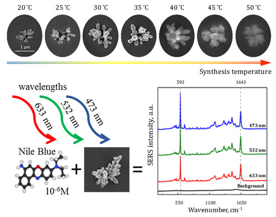
Self-organized spatially separated silver 3D dendrites as efficient plasmonic nanostructures for surface-enhanced Raman spectroscopy applications.
Journal of Applied Physics,
Vol. 126,
Issue. 23,
Bundyukova, Victoria
Kaniukov, Egor
Shumskaya, Alena
Smirnov, Andrey
Kravchenko, Maksim
Yakimchuk, Dzmitry
Bobrikov, I.
Chudoba, V.
Derenovskaya, O.
Friesen, A.
and
Verkheev, A.
2019.
Ellipsometry as an express method for determining the pore parameters of ion-track SiO2templates on a silicon substrate.
EPJ Web of Conferences,
Vol. 201,
Issue. ,
p.
01001.
Bundyukova, V. D.
Yakimchuk, D. V.
Shlimas, D. I.
and
Khubezhov, S. A.
2019.
Deposition of Gold Nanostructures into Porous SiO2∕Si Templates from the Electrolyte Based on Au(I) Sulfite Complex.
International Journal of Nanoscience,
Vol. 18,
Issue. 03n04,
p.
1940065.
Yakimchuk, D.
Bundyukova, V.
Borgekov, D.
Zdorovets, M.
Kozlovskiy, A.
Khubezhov, S.A.
Magkoev, T.T.
Bliev, A.P.
Belonogov, E.
Demyanov, S.
and
Kaniukov, E.
2019.
A simple way to control the filling degree of the SiO2/Si template pores with nickel.
Materials Today: Proceedings,
Vol. 7,
Issue. ,
p.
860.
Kalkabay, Gulnar
Kozlovskiy, Artem
Zdorovets, Maxim
Borgekov, Daryn
Kaniukov, Egor
and
Shumskaya, Alena
2019.
Influence of temperature and electrodeposition potential on structure and magnetic properties of nickel nanotubes.
Journal of Magnetism and Magnetic Materials,
Vol. 489,
Issue. ,
p.
165436.
Myndrul, Valerii
and
Iatsunskyi, Igor
2019.
Nanosilicon-Based Composites for (Bio)sensing Applications: Current Status, Advantages, and Perspectives.
Materials,
Vol. 12,
Issue. 18,
p.
2880.
Zavatski, Sergey
Khinevich, Nadia
Girel, Kseniya
Redko, Sergey
Kovalchuk, Nikolai
Komissarov, Ivan
Lukashevich, Vladimir
Semak, Igor
Mamatkulov, Kahramon
Vorobyeva, Maria
Arzumanyan, Grigory
and
Bandarenka, Hanna
2019.
Surface Enhanced Raman Spectroscopy of Lactoferrin Adsorbed on Silvered Porous Silicon Covered with Graphene.
Biosensors,
Vol. 9,
Issue. 1,
p.
34.
Kaniukov, Egor
Yakimchuk, Dzmitry
Bundyukova, Victoria
Petrov, Alexander
Belonogov, Evgenii
and
Demyanov, Sergey
2019.
Nanophotonics, Nanooptics, Nanobiotechnology, and Their Applications.
Vol. 222,
Issue. ,
p.
3.
Yakimchuk, D V
Khubezhov, S A
Bundyukova, V D
Kozlovskiy, A L
Zdorovets, M V
Shlimas, D I
Tishkevich, D I
and
Kaniukov, E Yu
2019.
Copper nanostructures into pores of SiO2/Si template: galvanic displacement, chemical and structural characterization.
Materials Research Express,
Vol. 6,
Issue. 10,
p.
105058.
El-Desoky, Hanaa
Abdel-Galeil, Mohamed
and
Khalifa, Aya
2019.
Mesoporous SiO2 (SBA-15) modified graphite electrode as highly sensitive sensor for ultra trace level determination of Dapoxetine hydrochloride drug in human plasma.
Journal of Electroanalytical Chemistry,
Vol. 846,
Issue. ,
p.
113157.
Yakimchuk, Dzmitry
Kaniukov, Egor
Bundyukova, Victoria
Demyanov, Sergey
and
Sivakov, Vladimir
2019.
Fundamental and Applied Nano-Electromagnetics II.
p.
75.
Bundyukova, Victoria D.
Yakimchuk, Dzmitry V.
Kozlovskiy, Artem
Shlimas, Dmitriy I.
Tishkevich, Daria I.
and
Kaniukov, Egor Yu.
2019.
Synthesis of gold nanostructures using wet chemical deposition in SiO2/Si template.
Lithuanian Journal of Physics,
Vol. 59,
Issue. 3,
Khinevich, Nadia
Zavatski, Sergey
Kholyavo, Victor
and
Bandarenka, Hanna
2019.
Bimetallic nanostructures on porous silicon with controllable surface plasmon resonance.
The European Physical Journal Plus,
Vol. 134,
Issue. 2,
Anisovich, M. V.
Shumskaya, A. E.
Bundyukova, V. D.
Yakimchuk, D. V.
and
Kaniukov, E. Yu.
2019.
Study of Safety of SiO2(Ag)–Si System by Cytoflourometric Method of Analysis of Reactive Oxygen Species and Cell Death in Culture.
International Journal of Nanoscience,
Vol. 18,
Issue. 03n04,
p.
1940080.
Yakimchuk, Dzmitry
Bundyukova, Victoria
Smirnov, Andrey
and
Kaniukov, Egor
2019.
Express Method of Estimation of Etched Ion Track Parameters in Silicon Dioxide Template.
physica status solidi (b),
Vol. 256,
Issue. 5,
Kaniukov, E.Yu
Shumskaya, A.E.
Kutuzau, M.D.
Bundyukova, V.D.
Yakimchuk, D.V.
Borgekov, D.B.
Ibragimova, M.A.
Korolkov, I.V.
Giniyatova, ShG.
Kozlovskiy, A.L.
and
Zdorovets, M.V.
2019.
Degradation mechanism and way of surface protection of nickel nanostructures.
Materials Chemistry and Physics,
Vol. 223,
Issue. ,
p.
88.
Kaniukov, Egor
Bundyukova, Victoria
Kutuzau, Maksim
and
Yakimchuk, Dzmitry
2019.
Fundamental and Applied Nano-Electromagnetics II.
p.
41.
