Hostname: page-component-cd9895bd7-dzt6s Total loading time: 0 Render date: 2024-12-23T11:41:12.617Z Has data issue: false hasContentIssue false

Post-process composition and biological responses of laser sintered PMMA and β-TCP composites

Published online by Cambridge University Press:  21 May 2018

Rajkumar Velu
Affiliation:
Additive Manufacturing Research Centre, Auckland University of Technology, Auckland 1010, New Zealand
Banu Pradheepa Kamarajan
Affiliation:
Department of Biotechnology, PSG College of Technology, Coimbatore 641004, India
Muthusamy Ananthasubramanian
Affiliation:
Department of Biotechnology, PSG College of Technology, Coimbatore 641004, India
Truc Ngo
Affiliation:
Shiley-Marcos School of Engineering, University of San Diego, San Diego, California 92110, USA
Sarat Singamneni*
Affiliation:
Additive Manufacturing Research Centre, Auckland University of Technology, Auckland 1010, New Zealand
*
a)Address all correspondence to this author. e-mail: [email protected]
Get access

Abstract

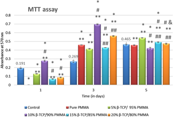
PMMA/β-TCP composites were evaluated to be suitable for laser sintering earlier, but the possible after effects are not known yet. Effects of sintering on the biological nature and the influences of critical compositions and process parameters have not been investigated so far. The current research attempts this, first identifying experimentally the most suitable laser process conditions for the specific grades of PMMA and β-TCP and then subjecting single layer sintered samples to FTIR analysis and in vitro studies involving MTT and ALP assays, alizarin red S tests, and real-time PCR analyses. While the laser interactions are not detrimental, the biological responses are generally positive proving the selective laser sintering of PMMA/β-TCP composites to be a potential approach for specific medical applications.

Type
Article
Copyright
Copyright © Materials Research Society 2018 

Access options

Get access to the full version of this content by using one of the access options below. (Log in options will check for institutional or personal access. Content may require purchase if you do not have access.)

References

REFERENCES

Logeart-Avramoglou, D., Anagnostou, F., Bizios, R., and Petite, H.: Engineering bone: Challenges and obstacles. J. Cell Mol. Med. 9, 972984 (2005).CrossRefGoogle ScholarPubMed
Ben-Nissan, B.: Natural bioceramics: From coral to bone and beyond. Curr. Opin. Solid State Mater. Sci. 7, 283288 (2003).CrossRefGoogle Scholar
Lane, J.M., Tomin, E., and Bostrom, M.P.G.: Biosynthetic bone grafting. Clin. Orthop. Relat. Res. 367, S107S117 (1999).CrossRefGoogle Scholar
Pelker, R.R. and Friedlaender, G.E.: Biomechanical aspects of bone autografts and allografts. Orthop. Clin. N. Am. 18, 235239 (1987).Google ScholarPubMed
Mankin, H.J., Gebhardt, M.C., Jennings, L.C., Springfield, D.S., and Tomford, W.W.: Long term results of allograft replacement in the management of bone tumors. Clin. Orthop. Relat. Res. 324, 8697 (1996).CrossRefGoogle Scholar
Strong, D.M., Friedlaender, G.E., Tomford, W.W., Springfield, D.S., Shives, T.C., Bur-chardt, H., Enneking, W.F., and Mankin, H.J.: Immunologic responses in human recipients of osseous and osteochondral allografts. Clin. Orthop. Relat. Res. 326, 107114 (1996).CrossRefGoogle Scholar
Simonds, R.J., Holmberg, S.D., Hurwitz, R.L., Coleman, T.R., Bottenfield, S., Conley, L.J., Kohlenberg, S.H., Castro, K.G., Dahan, B.A., Schable, C.A., Rayfield, M.A., and Rogers, M.F.: Transmission of human-immunodeficiency-virus type-1 from a seronegative organ and tissue donor. N. Engl. J. Med. 326, 726732 (1992).CrossRefGoogle ScholarPubMed
Jensen, S., Aarboe, M., Pinholt, E., Hjorting-Hansen, E., Melsen, F., and Ruyter, I.: Tissue reaction and material characteristics of four bone substitues. Int. J. Oral Maxillofac. Implants 11, 5566 (1996).Google Scholar
Moore, W.R., Graves, S.E., and Bain, G.I.: Synthetic bone graft substitutes. Aust. N. Z. J. Surg. 71, 354361 (2001).CrossRefGoogle ScholarPubMed
Albee, F.H. and Morrison, H.F.: Studies in bone growth: Triple CaP as a stimulus to osteogenesis. Ann. Surg. 71, 3239 (1920).CrossRefGoogle Scholar
Goodridge, R.D., Dalgarno, K.W., and Wood, D.J.: Indirect selective laser sintering of an appetite-mullite glass-ceramic for potential use in bone replacement applications. Proc. Inst. Mech. Eng., Part H 220, 5768 (2006).CrossRefGoogle ScholarPubMed
Asti, A. and Gioglio, L.: Natural and synthetic biodegradable polymers: Different scaffolds for cell expansion and tissue formation. Int. J. Artif. Organs 37, 187205 (2014).Google ScholarPubMed
Vroman, I. and Tighzert, L.: Biodegradable polymers. Materials 2, 307344 (2009).CrossRefGoogle Scholar
Taş, A.C., Korkusuz, F., Timuçin, M., and Akkaş, N.: An investigation of the chemical synthesis and high-temperature sintering behaviour of calcium hydroxyapatite (HA) and tricalcium phosphate (TCP) bioceramics. J. Mater. Sci.: Mater. Med. 8, 9196 (1997).Google Scholar
Gauthier, O., Goyenvalle, E., Bouler, J.M., Guicheux, J., Pilet, P., Weiss, P., and Daculsi, G.: Macroporous biphasic calcium phosphate ceramics versus injectable bone substitute: A comparative study 3 and 8 weeks after implantation in rabbit bone. J. Mater. Sci. Mater. Med. 12, 385390 (2001).CrossRefGoogle ScholarPubMed
Baxter, R.M., Macdonald, D.W., Kurtz, S.M., and Steinbeck, M.J.: Severe impingement of lumbar disc replacements increases the functional biological activity of polyethylene wear debris. J. Bone Jt. Surg. Am. Vol. 95, e751e759 (2013).CrossRefGoogle ScholarPubMed
Kim, H., Kim, H.M., Jang, J.E., Kim, C.M., Kim, E.Y., Lee, D., and Khang, G.: Osteogenic differentiation of bone marrow stem cell in poly(lactic-co-glycolic acid) scaffold loaded various ratio of hydroxyapatite. Int. J. Stem Cells 6, 6774 (2013).CrossRefGoogle ScholarPubMed
Seunarine, K., Gadegaard, N., Tormen, M., Meredith, D.O., Riehle, M.O., and Wilkinson, C.D.W.: 3D polymer scaffolds for tissue engineering. Nanomedicine 1, 281296 (2006).CrossRefGoogle ScholarPubMed
Agrawal, C.M. and Ray, R.B.: Biodegradable polymeric scaffolds for musculoskeletal tissue engineering. J. Biomed. Mater. Res. 55, 141150 (2001).3.0.CO;2-J>CrossRefGoogle ScholarPubMed
Mooney, D.J. and Langer, R.S.: Engineering biomaterials for tissue engineering. In The Biomedical Engineering Handbook, Bronzino, J.D., ed. (CRC Press, Boca Raton, 1995); pp. 109/1109/8.Google Scholar
Bonfield, W., Wang, M., and Tanner, K.E.: Interfaces in analogue biomaterials. Acta Mater. 46, 25092518 (1998).CrossRefGoogle Scholar
Thomson, R.C., Shung, A.K., Yaszemski, M.J., and Mikos, A.G.: Polymer scaffold processing. In Principles of Tissue Engineering, Lanza, R.P., Langer, R., and Vacanti, J., eds. (Academic Press, San Diego, 2000); pp. 251262.CrossRefGoogle Scholar
Widmer, M.S. and Mikos, A.G.: Fabrication of biodegradable polymer scaffolds for tissue engineering. In Frontiers in Tissue Engineering, Patrick, C.W., Mikos, A.G., and Mcintyre, L.V., eds. (Elsevier Sciences, New York, 1998); pp. 107120.CrossRefGoogle Scholar
Niklason, L.E. and Langer, R.: Prospects for organ and tissue replacement. JAMA, J. Am. Med. Assoc. 285, 573576 (2001).CrossRefGoogle ScholarPubMed
Leong, K.F., Cheah, C.M., and Chua, C.K.: Solid freeform fabrication of three- dimensional scaffolds for engineering replacement tissues and organs. Biomaterials 24, 23632378 (2003).CrossRefGoogle ScholarPubMed
Chua, C.K. and Leong, K.F.: Rapid Prototyping: Principles and Applications in Manu-facturing, 2nd ed. (World Scientific, Singapore, 2003).CrossRefGoogle Scholar
Naing, M.W., Chua, C.K., and Leong, K.F.: Computer aided tissue engineering scaffold fabrication. In Virtual Prototyping & Bio Manufacturing in Medical Applications (Springer, Boston, MA, 2008); pp. 6785.Google Scholar
Hao, L., Savalani, M.M., Zhang, Y., Tanner, K.E., and Harris, R.A.: Selective laser sintering of hydroxyapatite reinforced polyethylene composites for bioactive implants and tissue scaffold development. Proc. Inst. Mech. Eng., Part H 220, 521531 (2006).CrossRefGoogle ScholarPubMed
Savalani, M.M., Hao, L., and Harris, R.A.: Evaluation of CO2 and Nd:YAG lasers for the selective laser sintering of HAPEX®. Proc. IME B J. Eng. Manufact. 220, 171182 (2006).CrossRefGoogle Scholar
Jarcho, M., Kay, J.F., and Gumaer, K.l.: Tissue cellular and subcellular events at a bone ceramic hydroxyapatite interface. J. Bioeng. 1, 79 (1977).Google Scholar
Kalita, S.J., Bose, S., Hosick, H.L., and Bandyopadhyay, A.: Development of controlled porosity polymer–ceramic composite scaffolds via fused deposition modeling. Mater. Sci. Eng., C 23, 611620 (2003).CrossRefGoogle Scholar
Duan, B., Wang, M., Zhou, W.Y., Cheung, W.L., Li, Z.Y., and Lu, W.W.: Three-dimensional nanocomposite scaffolds fabricated via selective laser sintering for bone tissue engineering. Acta Biomater. 6, 44954505 (2010).CrossRefGoogle ScholarPubMed
Lewis, G.: Properties of antibiotic-loaded acrylic bone cements for use in cemented arthroplasties. J. Biomed. Mater. Res., Part B 89, 558574 (2009).CrossRefGoogle ScholarPubMed
Puska, M., Kokkari, A., Närhi, T., and Vallittu, P.: Mechanical properties of oligomer modified acrylic bone cement. Biomaterials 24, 417425 (2003).CrossRefGoogle ScholarPubMed
Descamps, M., Richart, O., Hardouin, P., Hornez, J.C., and Leriche, A.: Synthesis of macroporous β-tricalcium phosphate with controlled porous architectural. Ceram. Int. 34, 11311137 (2008).CrossRefGoogle Scholar
Velu, R. and Singamneni, S.: Selective laser sintering of polymer biocomposites based on polymethyl methacrylate. J. Mater. Res., 29, 18831892 (2014).CrossRefGoogle Scholar
Lohfeld, S., Cahill, S., Barron, V., McHugh, P., Dürselen, L., Kreja, L., Bausewein, C., and Ignatius, A.: Fabrication, mechanical and in vivo performance of polycaprolactone/tricalciumphosphate composite scaffolds. Acta Biomater. 8, 34463456 (2012).CrossRefGoogle Scholar
Velu, R. and Singamneni, S.: Evaluation of the influences of process parameters while selective laser sintering PMMA powders. Proc. Inst. Mech. Eng., Part C 229, 603613 (2014).CrossRefGoogle Scholar
Rudin, A. and Choi, P.: The Elements of Polymer Science & Engineering (Academic Press, Chicago, 2012).Google Scholar
Rantuch, P., Kačíková, D., and Nagypál, B.: Investigation of activation energy of polypropylene composite thermos oxidation by model-free methods. Eur. J. Environ. Saf. Sci. 2, 1218 (2014).Google Scholar
Jang, D., Nguyen, T., Sarkar, S.K., and Lee, B.: Microwave sintering and in vitro study of defect-free stable porous multi-layered Hap-ZrO2 artificial bone scaffold. Sci. Technol. Adv. Mater. 13, 035009 (9pp) (2012).CrossRefGoogle Scholar
Gregory, C.A., Gunn, W.G., Peister, A., and Prockop, D.J.: An Alizarin red-based assay of mineralization by adherent cells in culture: Comparison with cetylpyridinium chloride extraction. Anal. Biochem. 329, 7784 (2004).CrossRefGoogle ScholarPubMed
Ma, R., Tang, S., Tan, H., Lin, W., Wang, Y., Wei, J., Zhao, L., Zhao, L., and Preparation, T.T.: Characterization, and in vitro osteoblast functions of a nano-hydroxyapatite/polyetheretherketone biocomposite as orthopedic implant material. Int. J. Nanomed. 9, 39493961 (2014).Google ScholarPubMed
Tsai, S., Liou, H., Lin, C., Kuo, K., Hung, Y., Weng, R., and Hsu, F.: MG63 osteoblast-like cells exhibit different behavior when grown on electrospun collagen matrix versus electrospun gelatin matrix. PLoS One 7, e31200 (2012).CrossRefGoogle ScholarPubMed
Schmittgen, T.D. and Livak, K.J.: Analyzing real-time PCR data by the comparative CT method. Nat. Protoc. 3, 11011108 (2008).CrossRefGoogle Scholar
Yokozeki, H., Hayashi, T., Nakagawa, T., Kurosawa, H., Shibuya, K., and Ioku, K.: Influence of surface microstructure on the reaction of the active ceramics in vivo. J. Mater. Sci. Mater. Med. 9, 381384 (1998).CrossRefGoogle ScholarPubMed
Hao, L., Lawrence, J., and Chian, K.S.: On the effects of CO2 laser irradiation on the surface properties of a magnesia partially stabilised zirconia (MgO-PSZ) bioceramic and the subsequent improvements in human osteoblast cell adhesion. J. Biomater. Appl. 19, 81105 (2004).CrossRefGoogle ScholarPubMed
Tsang, V.L. and Bhatia, S.N.: Three-dimensional tissue fabrication. Adv. Drug Deliv. Rev. 56, 16351647 (2004).CrossRefGoogle ScholarPubMed
Duan, G., Zhang, C., Li, A., Yang, X., Lu, L., and Wang, X.: Preparation and characterization of mesoporous zirconia made by using a poly(methyl methacrylate) template. Nanoscale Res. Lett. 3, 118 (2008).CrossRefGoogle ScholarPubMed
Di Silvio, L., Dalby, M.J., and Bonfield, W.: In vitro response of osteoblasts to hydroxyapatite-reinforced polyethylene composites. J. Mater. Sci. Mater. Med. 9, 845848 (1998).CrossRefGoogle ScholarPubMed
Muhammad, K.B., Abas, W.A., Kim, K.H., Pingguan-Murphy, B., Zain, N.M., and Akram, H.: In vitro comparative study of white and dark polycaprolactone trifumarate in situ cross-linkable scaffolds seeded with rat bone marrow stromal cells. Clinics 67, 629637 (2012).CrossRefGoogle ScholarPubMed
Kim, S.S., Park, M.S., Jeon, O., Choi, C.Y., and Kim, B.S.: Poly(lactide-co-glycolide)/hydroxyapatite composite scaffolds for bone tissue engineering. Biomaterials 27, 399409 (2006).CrossRefGoogle ScholarPubMed
Sun, L., Gan, B., Fan, Y., Zhuang, F., and Hu, Q.: The proliferation and gene expression in MC3T3-E1 under simulated microgravity. In Complex Medical Engineering, 2007. CME 2007. IEEE/ICME International Conference (IEEE, Beijing, China, 2007); pp. 18031806.Google Scholar
Sulaiman, S.B., Keong, T.K., Cheng, C.H., Saim, A.B., and Idrus, R.B.H.: Tricalcium phosphate/hydroxyapatite (TCP–HA) bone scaffold as potential candidate for the formation of tissue engineered bone. Indian J. Med. Res. 137, 10931101 (2013).Google ScholarPubMed
Gumusderelioglu, M., Tuncay, E.O., Kaynak, G., Demirtas, T.T., Aydin, S.T., and Hakki, S.S.: Encapsulated boron as an osteconductive agent for bone scaffolds. J. Trace Elem. Med. Biol. 31, 120128 (2015).CrossRefGoogle Scholar
Yamano, S., Lin, T.Y., Dai, J., Fabella, K., and Moursi, A.M.: Bioactive collagen membrane as a carrier for sustained release of PDGF. J. Tissue Sci. Eng. 2, 110 (2011).CrossRefGoogle Scholar
Xu, L., Lv, K., Zhang, W., Zhang, X., Jiang, X., and Zhang, F.: The healing of critical-size calvarial bone defects in rat with rhPDGF-BB, BMSCs, and β-TCP scaffolds. J. Mater. Sci. Mater. Med. 23, 10731084 (2012).CrossRefGoogle ScholarPubMed
Balamurugan, A., Kannan, S., Selvaraj, V., and Rajeswari, S.: Development and spectral characterization of poly(methyl methacrylate)/hydroxyapatite composite for biomedical applications. Trends Biomater. Artif. Organs 18, 4145 (2004).Google Scholar