Hostname: page-component-cd9895bd7-mkpzs Total loading time: 0 Render date: 2024-12-23T12:05:29.244Z Has data issue: false hasContentIssue false

Improved performance of the functionalized nitinol as a prospective bone implant material

Published online by Cambridge University Press:  19 July 2018

Sarmita Sinha
Affiliation:
Department of Mechanical Engineering, Jadavpur University, Kolkata-700032, India
Howa Begam
Affiliation:
School of Bioscience and Engineering, Jadavpur University, Kolkata-700032, India
Vinod Kumar
Affiliation:
Department of Veterinary Surgery & Radiology, West Bengal University of Animal and Fishery Sciences, Kolkata-700037, India
Samit Kumar Nandi*
Affiliation:
Department of Veterinary Surgery & Radiology, West Bengal University of Animal and Fishery Sciences, Kolkata-700037, India
Jerzy Kubacki*
Affiliation:
A. Chełkowski Institute of Physics, University of Silesia, Katowice 40-007, Poland
Abhijit Chanda*
Affiliation:
Department of Mechanical Engineering, Jadavpur University, Kolkata-700032, India
*
a)Address all correspondence to these authors. e-mail: [email protected]
Get access

Abstract

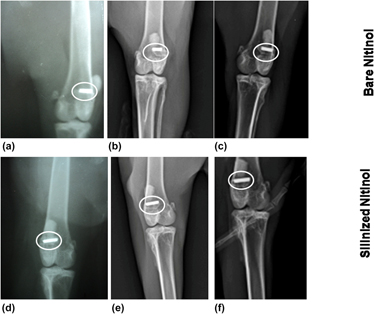
Nitinol, being a shape memory and super elastic alloy, is used in medical industry. Surface modification of nitinol helps to reduce the nickel ion leaching in physiological environment. The purpose of this study is to modify the nitinol surface by the silanization technique and to conduct a comparative investigation with the bare nitinol in the aspect of leaching of nickel ion, hemocompatibility, and in vivo animal response. X-ray photoelectron spectroscopy and energy dispersive X-ray spectroscopy studies confirmed the addition of organofunctional alkoxysilane molecules through the silanization process. The histological study showed the presence of adequate number of osteoblasts in silanized nitinol. The fluorochrome labeling study depicted more new bone formation (8 and 21% higher) in silanized nitinol specimens than bare one at one and three months postoperatively. Radiology and SEM study also proved the better performance of silanized samples. The cumulative in vivo results indicate its suitability as the potential bioimplant in various orthopedic surgical uses.

Type
Article
Copyright
Copyright © Materials Research Society 2018 

Access options

Get access to the full version of this content by using one of the access options below. (Log in options will check for institutional or personal access. Content may require purchase if you do not have access.)

References

REFERENCES

Duerig, T.W., Pelton, A., and Stokel, D.: An overview of nitinol medical applications. Mater. Sci. Eng., A 273, 149 (1999).CrossRefGoogle Scholar
Chrzanowski, W., Walke, W., Armitage, D.A., and Knowles, J.C.: Study on bioactivity of NiTinol after surface treatment. Arch. Mater. Sci. 6, 6 (2008).Google Scholar
Kobayashi, S., Ohgoe, Y., Ozeki, K., Sato, K., Sumiya, T., Hirakuri, K.K., and Aoki, H.: Diamond-like carbon coatings on orthodontic archwires. Diamond Relat. Mater. 14, 1094 (2005).CrossRefGoogle Scholar
Kujala, S.: Biocompatibility and Biomechanical Aspects of Nitinol Shape Memory Metal Implants (Oulu University Press, Oulu Finland, 2003).Google Scholar
Kwok, D.T.K., Schulz, M., Hu, T., Chu, C., and Chu, P.: Surface treatments of nearly equiatomic NiTi alloy (nitinol) for surgical implants. In Biomedical Engineering, Trends in Materials Science, Laskovski, A., (Ed.) Vol. 269 (InTech, Intechoopen, London, United Kingdom, 2011); p. 282.Google Scholar
Shabalovskaya, S.A.: Physicochemical and biological aspects of nitinol as a biomaterial. Int. Mater. Rev. 46, 233 (2001).CrossRefGoogle Scholar
Morgan, N.B.: Medical shape memory alloy applications at the market and its products. Materi. Sci. Eng., A 378, 16 (2004).CrossRefGoogle Scholar
Shabalovskaya, S.A.: Surface, corrosion and biocompatibility aspects of nitinol as an implant material. Bio-Med. Mater. Eng. 12, 69 (2002).Google ScholarPubMed
Bernard, S.A., Balla, V.K., Davies, N.M., Bose, S., and Bandyopadhyay, A.: Bone cell-materials interactions and Ni ion release of anodized equiatomic NiTi alloy. Acta Biomater. 7, 1902 (2011).CrossRefGoogle ScholarPubMed
Barrett, R.D., Bishara, S.E., and Quinn, J.K.: Biodegradation of orthodontic appliances. Part I. Biodegradation of nickel and chromium in vitro. Am. J. Orthod. Dentofac. Orthop. 103, 8 (1993).CrossRefGoogle ScholarPubMed
Katić, J., Metikoš-Huković, M., Babić, R., and Marciuš, M.: Sol–gel derived biphasic calcium phosphate ceramics on nitinol for medical applications. Int. J. Electrochem. Sci. 8, 1394 (2013).Google Scholar
Rondelli, G.: Corrosion resistance tests on NiTi shape memory alloy. Biomaterials 17, 2003 (1996).CrossRefGoogle ScholarPubMed
Trepanier, C., Venugopalan, R., and Pelton, A.R.: Corrosion resistance and biocompatibility of passivated NiTi. In Shape Memory Implants, Yahia, L., (Ed.) Vol. 35 (Springer, Berlin, Heidelberg, New York, 2000).Google Scholar
Gill, P., Musaramthota, V., Munroe, N., Datye, A., Dua, R., Haider, W., McGoron, A., and Rokicki, R.: Surface modification of Ni–Ti alloys for stent application after magnetoelectropolishing. Mater. Sci. Eng., C 50, 37 (2015).CrossRefGoogle ScholarPubMed
Gu, Y.W., Tay, B.Y., Lim, C.S., and Yong, M.S.: Biomimetic deposition of apatite coating on surface-modified NiTi alloy. Biomaterials 26, 6916 (2005).CrossRefGoogle ScholarPubMed
Kim, J.H., Shin, J.H., Shin, D.H., Moon, M.W., Park, K., Kim, T.H., Shin, K.M., Won, Y.H., Han, D.K., and Lee, K.R.: Comparison of diamond-like carbon-coated nitinol stents with or without polyethylene glycol grafting and uncoated nitinol stents in a canine iliac artery model. Br. J. Radiol. 84, 210 (2011).CrossRefGoogle ScholarPubMed
Kong, X., Grabitz, R.G., Van Oeveren, W., Klee, D., Van Kooten, T.G., Freudenthal, F., Qing, M., Von Bernuth, G., and Seghaye, M.C.: Effect of biologically active coating on biocompatibility of nitinol devices designed for the closure of intra-atrial communications. Biomaterials 23, 1775 (2002).CrossRefGoogle ScholarPubMed
Lahann, J., Klee, D., Pluester, W., and Hoecker, H.: Bioactive immobilization of r-hirudin on CVD-coated metallic implant devices. Biomaterials 22, 817 (2001).CrossRefGoogle ScholarPubMed
Muhonen, V., Kujala, S., Vuotikka, A., Äaritalo, V., Peltola, T., Areva, S., Närhi, T., and Tuukkanen, J.: Biocompatibility of sol–gel-derived titania-silica coated intramedullary NiTi nails. Acta Biomater. 5, 785 (2009).CrossRefGoogle ScholarPubMed
Pérez, L.M., Arruebo, M., Irusta, S., Gracia-Villa, L., Santamaría, J., and Puértolas, J.A.: Mechanochemical characterisation of silica-based coatings on nitinol substrates. Microporous Mesoporous Mater. 98, 292 (2007).CrossRefGoogle Scholar
Shen, Y., Wang, G., Chen, L., Li, H., Yu, P., Bai, M., Zhang, Q., Lee, J., and Yu, Q.: Investigation of surface endothelialization on biomedical nitinol (NiTi) alloy: Effects of surface micropatterning combined with plasma nanocoatings. Acta Biomater. 5, 3593 (2009).CrossRefGoogle ScholarPubMed
Shibli, S.M.A., Beenakumari, K.S., and Suma, N.D.: Nano nickel oxide/nickel incorporated nickel composite coating for sensing and estimation of acetylcholine. Biosens. Bioelectron. 22, 633 (2006).CrossRefGoogle ScholarPubMed
Tang, C.J., Wang, G.X., Shen, Y., Wan, L.J., Xiao, L., Zhang, Q., Yu, Q.S., Liu, L.S., and Wen, G.B.: A study on surface endothelialization of plasma coated intravascular stents. Surf. Coat. Technol. 204, 1487 (2010).CrossRefGoogle Scholar
Tolomeo, D., Slater, T., and Wu, P.: Predictive modelling of radial strength for superelastic stents. In SMST-2000: Proceedings of the International Conference on Shape Memory and Superelastic Technologies, Russell, S.M. and Pelton, A.R., eds. (International Organization on SMST, Pacific Grove, California, 2000); p. 517.Google Scholar
Yeung, K.W.K., Poon, R.W.Y., Liu, X.M., Chu, P.K., Chung, C.Y., Liu, X.Y., Chan, S., Lu, W.W., Chan, D., and Luk, K.D.K.: Nitrogen plasma-implanted nickel titanium alloys for orthopedic use. Surf. Coat. Technol. 201, 5607 (2007).CrossRefGoogle Scholar
Bakhshi, R., Darbyshire, A., Evans, J.E., You, Z., Lu, J., and Seifalian, A.M.: Polymeric coating of surface modified nitinol stent with POSS-nanocomposite polymer. Colloids Surf., B 86, 93 (2011).CrossRefGoogle ScholarPubMed
Dubruel, P., Vanderleyden, E., Bergada, M., De Paepe, I., Chen, H., Kuypers, S., Luyten, J., Schrooten, J., Van Hoorebeke, L., and Schacht, E.: Comparative study of silanization reactions for the biofunctionalisation of Ti-surfaces. Surf. Sci. 600, 2562 (2006).CrossRefGoogle Scholar
Bakhsheshi-Rad, H.R., Hamzah, E., Daroonparvar, M., Yajid, M.A.M., Kasiri-Asgarani, M., Abdul-Kadir, M.R., and Medraj, M.: In vitro degradation behavior of Mg alloy coated by fluorine doped hydroxyapatite and calcium deficient hydroxyapatite. Trans. Nonferrous Met. Soc. China 24, 2516 (2014).CrossRefGoogle Scholar
Yeh, H.Y. and Lin, J.C.: Bioactivity and platelet adhesion study of a human thrombomodulin-immobilized nitinol surface. J. Biomater. Sci., Polym. Ed. 20, 807 (2009).CrossRefGoogle ScholarPubMed
Yu, H., Yan, J., Ma, H., Zeng, X., Liu, Y., and Zhao, X.: Creating poly(ethylene glycol) film on the surface of NiTi alloy by gamma irradiation. Radiat. Phys. Chem. 112, 199 (2015).CrossRefGoogle Scholar
Haider, W., Munroe, N., Tek, V., Gill, P.K.S., Tang, Y., and McGoron, A.J.: Cytotoxicity of metal ions released from nitinol alloys on endothelial cells. J. Mater. Eng. Perform. 20, 816 (2011).CrossRefGoogle ScholarPubMed
Kao, C.T., Ding, S.J., He, H., Chou, M.Y., and Huang, T.H.: Cytotoxicity of orthodontic wire corroded in fluoride solution in vitro. Angle Orthod. 77, 349 (2007).CrossRefGoogle ScholarPubMed
McMahon, R.E., Ma, J., Verkhoturov, S.V., Munoz-Pinto, D., Karaman, I., Rubitschek, F., Maier, H.J., and Hahn, M.S.: A comparative study of the cytotoxicity and corrosion resistance of nickel–titanium and titanium–niobium shape memory alloys. Acta Biomater. 8, 2863 (2012).CrossRefGoogle ScholarPubMed
Pulletikurthi, C., Gill, P.M., Pandya, S., Persaud, D., Haider, W., Iyer, K., and McGoron, A.: Cytotoxicity of Ni from surface-treated porous nitinol (PNT) on osteoblast cells. J. Mater. Eng. Perform. 20, 824 (2011).CrossRefGoogle ScholarPubMed
Oyane, A., Kim, H.M., Furuya, T., Kokubo, T., Miyazaki, T., and Nakamura, T.: Preparation and assessment of revised simulated body fluids. J. Biomed. Mater. Res., Part A 65, 188 (2003).CrossRefGoogle ScholarPubMed
Green, A.A.: The preparation of acetate and phosphate buffer solutions of known pH and ionic strength. J. Am. Chem. Soc. 55, 2331 (1933).CrossRefGoogle Scholar
Nandi, S.K., Ghosh, S.K., Kundu, B., De, D.K., and Basu, D.: Evaluation of new porous β-tri-calcium phosphate ceramic as bone substitute in goat model. Small Rumin. Res. 75, 144 (2008).CrossRefGoogle Scholar
Roy, R.K., Choi, H.W., Yi, J.W., Moon, M.W., Lee, K.R., Han, D.K., Shin, J.H., Kamijo, A., and Hasebe, T.: Hemocompatibility of surface-modified, silicon-incorporated, diamond-like carbon films. Acta Biomater. 5, 249 (2009).CrossRefGoogle ScholarPubMed
Shabalovskaya, S.A., Siegismund, D., Heurich, E., and Rettenmayr, M.: Evaluation of wettability and surface energy of native nitinol surfaces in relation to hemocompatibility. Mater. Sci. Eng., C 33, 127 (2013).CrossRefGoogle ScholarPubMed
Sinha, S., Pramanik, P.C., Begam, H., and Chanda, A.: Study on the effect of strain rate and temperature on mechanical properties of nitinol. Appl. Mech. Mater. 592, 1185 (2014).CrossRefGoogle Scholar
Beck, G.R., Ha, S.W., Camalier, C.E., Yamaguchi, M., Li, Y., Lee, J.K., and Weitzmann, M.N.: Bioactive silica-based nanoparticles stimulate bone-forming osteoblasts, suppress bone-resorbing osteoclasts, and enhance bone mineral density in vivo. Nanomed.: Nanotechnol. Biol. Med. 8, 793 (2012).CrossRefGoogle ScholarPubMed
Carlisle, E.M.: In vivo requirement for silicon in articular cartilage and connective tissue formation in the chick. J. Nutr. 106, 478 (1976).CrossRefGoogle ScholarPubMed
Carlisle, E.M.: Silicon: A requirement in bone formation independent of vitamin D1. Calcif. Tissue Int. 33, 27 (1981).CrossRefGoogle ScholarPubMed
Carlisle, E.M.: Silicon as an essential trace element in animal nutrition. Silicon Biochem. 703, 123 (2008).Google Scholar
Jugdaohsingh, R.: Silicon and bone health. J. Nutr. Health Aging 11, 99 (2007).Google ScholarPubMed
Mladenović, Ž., Johansson, A., Willman, B., Shahabi, K., Björn, E., and Ransjö, M.: Soluble silica inhibits osteoclast formation and bone resorption in vitro. Acta Biomater. 10, 406 (2014).CrossRefGoogle ScholarPubMed
Price, C.T., Koval, K.J., and Langford, J.R.: Silicon: A review of its potential role in prevention and treatment of postmenopausal osteoporosis. Int. J. Endocrinol. 2013, 1 (2013).CrossRefGoogle ScholarPubMed
Reffitt, D.M., Ogston, N., Jugdaohsingh, R., Cheung, H.F.J., Evans, B.A.J., Thompson, R.P.H., Powell, J.J., and Hampson, G.N.: Orthosilicic acid stimulates collagen type 1 synthesis and osteoblastic differentiation in human osteoblast-like cells in vitro. Bone 32, 127 (2003).CrossRefGoogle ScholarPubMed
Schwarz, K.: A bound form of silicon in glycosaminoglycans and polyuronides. Proc. Natl. Acad. Sci. U. S. A. 70, 1608 (1973).CrossRefGoogle ScholarPubMed