Skip to main content Accessibility help
×
Hostname: page-component-745bb68f8f-mzp66 Total loading time: 0 Render date: 2025-01-18T20:19:47.245Z Has data issue: false hasContentIssue false

1 - Cytology of the uterine cervix and corpus

Published online by Cambridge University Press:  05 July 2013
Robert A. Soslow
Affiliation:
Memorial Sloan-Kettering Cancer Center, New York
Teri A. Longacre
Affiliation:
Stanford University School of Medicine, California
M. Fujiwara
Affiliation:
Fellow, Department of Pathology
C. S. Kong
Affiliation:
Associate Professor, Department of Pathology
Get access

Summary

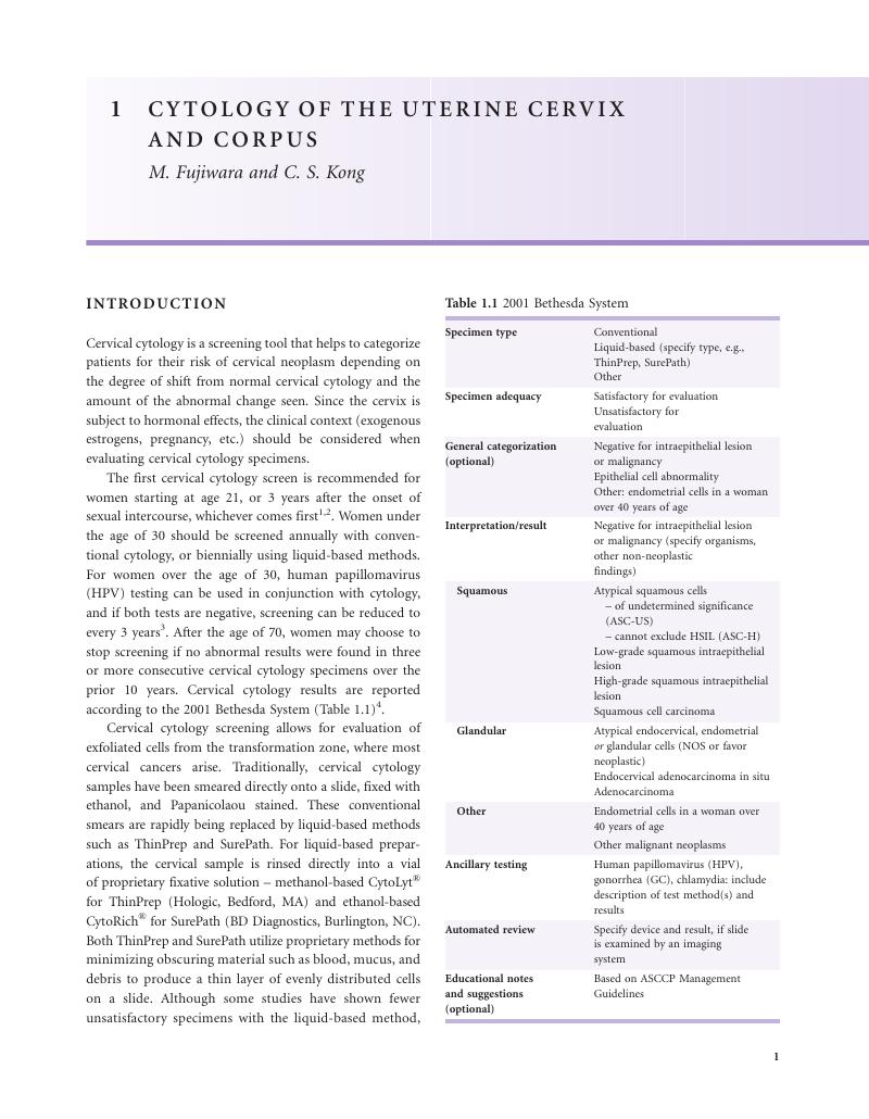
Image of the first page of this content. For PDF version, please use the ‘Save PDF’ preceeding this image.'
Type
Chapter
Information
Uterine Pathology , pp. 1 - 18
Publisher: Cambridge University Press
Print publication year: 2012

Access options

Get access to the full version of this content by using one of the access options below. (Log in options will check for institutional or personal access. Content may require purchase if you do not have access.)

References

Saslow, D, Runowicz, CD, Solomon, D, et al. American Cancer Society guideline for the early detection of cervical neoplasia and cancer. J Low Genit Tract Dis 2003;7:67–86.CrossRefGoogle ScholarPubMed
Smith, RA, Cokkinides, V, Brooks, D, et al. Cancer screening in the United States, 2011: a review of current American Cancer Society guidelines and issues in cancer screening. CA Cancer J Clin 2011;61:8–30.CrossRefGoogle ScholarPubMed
Wright, TC, Massad, LS, Dunton, CJ, et al. 2006 Consensus guidelines for the management of women with abnormal cervical cancer screening tests. Am J Obstet Gynecol 2007;197:346–55.CrossRefGoogle ScholarPubMed
Solomon, D, Davey, D, Kurman, R, et al. The 2001 Bethesda System: terminology for reporting results of cervical cytology. Jama 2002;287:2114–19.CrossRefGoogle ScholarPubMed
Davey, E, d'Assuncao, J, Irwig, L, et al. Accuracy of reading liquid based cytology slides using the ThinPrep Imager compared with conventional cytology: prospective study. BMJ 2007;335:31.CrossRefGoogle ScholarPubMed
Siebers, AG, Klinkhamer, PJ, Grefte, JM, et al. Comparison of liquid-based cytology with conventional cytology for detection of cervical cancer precursors: a randomized controlled trial. JAMA 2009;302:1757–64.CrossRefGoogle ScholarPubMed
,ACOG Committee on Practice Bulletins—Gynecology. ACOG Practice Bulletin no. 109: Cervical cytology screening. Obstet Gynecol 2009;114:1409–20.CrossRefGoogle Scholar
DeMay, RM. Cytopathology of false negatives preceding cervical carcinoma. Am J Obstet Gynecol 1996;175:1110–13.CrossRefGoogle ScholarPubMed
Cuzick, J, Mayrand, MH, Ronco, G, Snijders, P, Wardle, J. Chapter 10: New dimensions in cervical cancer screening. Vaccine 2006;24(Suppl 3):S90–7.CrossRefGoogle ScholarPubMed
Cox, JT, Schiffman, M, Solomon, D. Prospective follow-up suggests similar risk of subsequent cervical intraepithelial neoplasia grade 2 or 3 among women with cervical intraepithelial neoplasia grade 1 or negative colposcopy and directed biopsy. Am J Obstet Gynecol 2003;188:1406–12.CrossRefGoogle ScholarPubMed
Sherman, ME, Castle, PE, Solomon, D. Cervical cytology of atypical squamous cells-cannot exclude high-grade squamous intraepithelial lesion (ASC-H): characteristics and histologic outcomes. Cancer 2006;108:298–305.CrossRefGoogle ScholarPubMed
Evans, MF, Adamson, CS, Papillo, JL, et al. Distribution of human papillomavirus types in ThinPrep Papanicolaou tests classified according to the Bethesda 2001 terminology and correlations with patient age and biopsy outcomes. Cancer 2006;106:1054–64.CrossRefGoogle ScholarPubMed
Levine, L, Lucci, JA, Dinh, TV. Atypical glandular cells: new Bethesda Terminology and Management Guidelines. Obstet Gynecol Surv 2003;58:399–406.CrossRefGoogle ScholarPubMed
Westin, MC, Derchain, SF, Rabelo-Santos, SH, et al. Atypical glandular cells and adenocarcinoma in situ according to the Bethesda 2001 classification: cytohistological correlation and clinical implications. Eur J Obstet Gynecol Reprod Biol 2008;139:79–85.CrossRefGoogle ScholarPubMed
Mathers, ME, Johnson, SJ, Wadehra, V. How predictive is a cervical smear suggesting glandular neoplasia?Cytopathology 2002;13:83–91.CrossRefGoogle ScholarPubMed
Chhieng, DC, Gallaspy, S, Yang, H, Roberson, J, Eltoum, I. Women with atypical glandular cells: a long-term follow-up study in a high-risk population. Am J Clin Pathol 2004;122:575–9.CrossRefGoogle Scholar
Zhao, C, Florea, A, Onisko, A, Austin, RM. Histologic follow-up results in 662 patients with Pap test findings of atypical glandular cells: results from a large academic womens hospital laboratory employing sensitive screening methods. Gynecol Oncol 2009;114:383–9.CrossRefGoogle ScholarPubMed
,ASCCP. HPV Genotyping Clinical Update; 2011. (Accessed Oct 14 2011, at http://www.asccp.org/ConsensusGuidelines/HPVGenotypingClinicalUpdate/tabid/5963/Default.aspx.)
Ho, GY, Bierman, R, Beardsley, L, Chang, CJ, Burk, RD. Natural history of cervicovaginal papillomavirus infection in young women. N Engl J Med 1998;338:423–8.CrossRefGoogle ScholarPubMed
Solomon, D, Schiffman, M, Tarone, R. Comparison of three management strategies for patients with atypical squamous cells of undetermined significance: baseline results from a randomized trial. J Natl Cancer Inst 2001;93:293–9.CrossRefGoogle ScholarPubMed
Cremoux, P, Coste, J, Sastre-Garau, X, et al. Efficiency of the hybrid capture 2 HPV DNA test in cervical cancer screening. A study by the French Society of Clinical Cytology. Am J Clin Pathol 2003;120:492–9.CrossRefGoogle ScholarPubMed
Einstein, MH, Martens, MG, Garcia, FA, et al. Clinical validation of the Cervista HPV HR and 16/18 genotyping tests for use in women with ASC-US cytology. Gynecol Oncol 2010;118:116–22.CrossRefGoogle ScholarPubMed
Day, SP, Hudson, A, Mast, A, et al. Analytical performance of the Investigational Use Only Cervista HPV HR test as determined by a multi-center study. J Clin Virol 2009;45(Suppl 1):S63–72.CrossRefGoogle ScholarPubMed
Belinson, JL, Wu, R, Belinson, SE, et al. A population-based clinical trial comparing endocervical high-risk HPV testing using hybrid capture 2 and Cervista from the SHENCCAST II study. Am J Clin Pathol 2011;135:790–5.CrossRefGoogle ScholarPubMed
Galan-Sanchez, F, Rodriguez-Iglesias, MA. Use of Cervista HPV HR assay for detection of human papillomavirus in samples with hybrid capture borderline negative results. Apmis 2010;118:681–4.CrossRefGoogle ScholarPubMed
Einstein, MH, Garcia, FA, Mitchell, AL, Day, SP. Age-stratified performance of the Cervista HPV 16/18 genotyping test in women with ASC-US cytology. Cancer Epidemiol Biomarkers Prev 2011; 20:1185–9.CrossRefGoogle ScholarPubMed
Bartholomew, DA, Luff, RD, Quigley, NB, Curtis, M, Olson, MC. Analytical performance of Cervista HPV 16/18 genotyping test for cervical cytology samples. J Clin Virol 2011;51:38–43.CrossRefGoogle ScholarPubMed
Kinney, W, Stoler, MH, Castle, PE. Special commentary: patient safety and the next generation of HPV DNA tests. Am J Clin Pathol 2010;134:193–9.CrossRefGoogle ScholarPubMed
Davis-Devine, S, Day, SJ, Freund, GG. Test performance comparison of inform HPV and hybrid capture 2 high-risk HPV DNA tests using the SurePath liquid-based Pap test as the collection method. Am J Clin Pathol 2005;124:24–30.CrossRefGoogle ScholarPubMed
Hesselink, AT, Brule, AJ, Brink, AA, et al. Comparison of hybrid capture 2 with in situ hybridization for the detection of high-risk human papillomavirus in liquid-based cervical samples. Cancer 2004;102:11–18.CrossRefGoogle ScholarPubMed
Kurtycz, DF, Smith, M, He, R, Miyazaki, K, Shalkham, J. Comparison of methods trial for high-risk HPV. Diagn Cytopathol 2010;38:104–8.Google ScholarPubMed
Siddiqui, MT, Hornaman, K, Cohen, C, Nassar, A. ProEx C immunocytochemistry and high-risk human papillomavirus DNA testing in Papanicolaou tests with atypical squamous cell (ASC-US) cytology: correlation study with histologic biopsy. Arch Pathol Lab Med 2008;132:1648–52.Google ScholarPubMed
Sandri, MT, Lentati, P, Benini, E, et al. Comparison of the Digene HC2 assay and the Roche AMPLICOR human papillomavirus (HPV) test for detection of high-risk HPV genotypes in cervical samples. J Clin Microbiol 2006;44:2141–6.CrossRefGoogle ScholarPubMed
Bentz, JS. Liquid-based cytology for cervical cancer screening. Expert Rev Mol Diagn 2005;5:857–71.CrossRefGoogle ScholarPubMed
Passamonti, B, Bulletti, S, Camilli, M, et al. Evaluation of the FocalPoint GS system performance in an Italian population-based screening of cervical abnormalities. Acta Cytol 2007;51:865–71.CrossRefGoogle Scholar
Chivukula, M, Saad, RS, Elishaev, E, et al. Introduction of the Thin Prep Imaging System (TIS): experience in a high volume academic practice. Cytojournal 2007;4:6.Google Scholar
Zhang, FF, Banks, HW, Langford, SM, Davey, DD. Accuracy of ThinPrep Imaging System in detecting low-grade squamous intraepithelial lesions. Arch Pathol Lab Med 2007;131:773–6.Google ScholarPubMed
Jayamohan, Y, Karabakhtsian, RG, Banks, HW, Davey, DD. Accuracy of Thinprep Imaging System in detecting atypical glandular cells. Diagn Cytopathol 2009;37:479–82.CrossRefGoogle ScholarPubMed
Papillo, JL, St John, TL, Leiman, G. Effectiveness of the ThinPrep Imaging System: clinical experience in a low risk screening population. Diagn Cytopathol 2008;36:155–60.CrossRefGoogle Scholar
Kardos, TF. The FocalPoint System: FocalPoint slide profiler and FocalPoint GS. Cancer 2004;102:334–9.CrossRefGoogle ScholarPubMed
Chute, DJ, Lim, H, Kong, CS. BD FocalPoint slide profiler performance with atypical glandular cells on SurePath Papanicolaou smears. Cancer Cytopathol 2010;118:68–74.CrossRefGoogle ScholarPubMed
Wilbur, DC, Black-Schaffer, WS, Luff, RD, et al. The Becton Dickinson FocalPoint GS Imaging System: clinical trials demonstrate significantly improved sensitivity for the detection of important cervical lesions. Am J Clin Pathol 2009;132:767–75.CrossRefGoogle ScholarPubMed
Halford, JA. Cytological features of chronic follicular cervicitis in liquid-based specimens: a potential diagnostic pitfall. Cytopathology 2002;13:364–70.CrossRefGoogle ScholarPubMed
Al-Nourhji, O, Beckmann, MJ, Markwell, SJ, Massad, LS. Pathology correlates of a Papanicolaou diagnosis of low-grade squamous intraepithelial lesion, cannot exclude high-grade squamous intraepithelial lesion. Cancer 2008;114:469–73.CrossRefGoogle ScholarPubMed
Difurio, MJ, Mailhiot, T, Sundborg, MJ, Nauschuetz, KK. Comparison of the clinical significance of the Papanicolaou test interpretations LSIL cannot rule out HSIL and ASC-H. Diagn Cytopathol 2010;38:313–17.Google ScholarPubMed
Owens, CL, Moats, DR, Burroughs, FH, Gustafson, KS. “Low-grade squamous intraepithelial lesion, cannot exclude high-grade squamous intraepithelial lesion” is a distinct cytologic category: histologic outcomes and HPV prevalence. Am J Clin Pathol 2007;128:398–403.CrossRefGoogle ScholarPubMed
Elsheikh, TM, Kirkpatrick, JL, Wu, HH. The significance of “low-grade squamous intraepithelial lesion, cannot exclude high-grade squamous intraepithelial lesion” as a distinct squamous abnormality category in Papanicolaou tests. Cancer 2006;108:277–81.CrossRefGoogle ScholarPubMed
Ducatman, BS, Wang, HH, Jonasson, JG, Hogan, CL, Antonioli, DA. Tubal metaplasia: a cytologic study with comparison to other neoplastic and non-neoplastic conditions of the endocervix. Diagn Cytopathol 1993;9:98–103; discussion –5.CrossRefGoogle ScholarPubMed
Novotny, DB, Maygarden, SJ, Johnson, , Frable, WJ. Tubal metaplasia. A frequent potential pitfall in the cytologic diagnosis of endocervical glandular dysplasia on cervical smears. Acta Cytol 1992;36:1–10.Google ScholarPubMed
Szyfelbein, WM, Baker, PM, Bell, DA. Superficial endometriosis of the cervix: a source of abnormal glandular cells on cervicovaginal smears. Diagn Cytopathol 2004;30:88–91.CrossRefGoogle ScholarPubMed
Sauder, K, Wilbur, DC, Duska, L, Tambouret, RH. An approach to post-radical trachelectomy vaginal-isthmus cytology. Diagn Cytopathol 2009;37:437–42.CrossRefGoogle ScholarPubMed

Save book to Kindle

To save this book to your Kindle, first ensure [email protected] is added to your Approved Personal Document E-mail List under your Personal Document Settings on the Manage Your Content and Devices page of your Amazon account. Then enter the ‘name’ part of your Kindle email address below. Find out more about saving to your Kindle.

Note you can select to save to either the @free.kindle.com or @kindle.com variations. ‘@free.kindle.com’ emails are free but can only be saved to your device when it is connected to wi-fi. ‘@kindle.com’ emails can be delivered even when you are not connected to wi-fi, but note that service fees apply.

Find out more about the Kindle Personal Document Service.

Available formats
×

Save book to Dropbox

To save content items to your account, please confirm that you agree to abide by our usage policies. If this is the first time you use this feature, you will be asked to authorise Cambridge Core to connect with your account. Find out more about saving content to Dropbox.

Available formats
×

Save book to Google Drive

To save content items to your account, please confirm that you agree to abide by our usage policies. If this is the first time you use this feature, you will be asked to authorise Cambridge Core to connect with your account. Find out more about saving content to Google Drive.

Available formats
×