Skip to main content Accessibility help
×
Hostname: page-component-586b7cd67f-vdxz6 Total loading time: 0 Render date: 2024-11-22T17:26:52.088Z Has data issue: false hasContentIssue false

Chapter 4 - Congenital Heart Disease (I)

Published online by Cambridge University Press:  19 August 2019

Michael T. Ashworth
Affiliation:
Great Ormond Street Hospital for Children, London
Get access

Summary

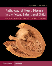
This chapter, the first of two devoted to congenital heart disease, deals with the commoner forms including ventricular septal defect, atrioventricular septal defect, tetralogy of Fallot and hypoplastic left heart. The pathological features are described in detail and profusely illustrated, including images of the histopathology, where relevant.

Type
Chapter
Information
Pathology of Heart Disease in the Fetus, Infant and Child
Autopsy, Surgical and Molecular Pathology
, pp. 75 - 117
Publisher: Cambridge University Press
Print publication year: 2019

Access options

Get access to the full version of this content by using one of the access options below. (Log in options will check for institutional or personal access. Content may require purchase if you do not have access.)

References

Diller, GP, Kempny, A, Alonso-Gonzalez, R et al. Survival prospects and circumstances of death in contemporary adult congenital heart disease patients under follow-up at a large tertiary centre. Circulation 2015; 132: 21182125.CrossRefGoogle Scholar
Jordan, SC, Scott, O. Incidence, aetiology and recurrence of congenital heart disease. In Jordan, SC, Scott, O (eds) Heart Disease in Paediatrics, 3rd edn. Oxford: Butterworth Heniemann; 1989, pp. 39.Google Scholar
Kempny, A, Dimopoulos, K, Uebing, A et al. Outcome of cardiac surgery in patients with congenital heart disease in England between 1997 and 2015. PLoS ONE 2017; 12: e0178963.Google Scholar
Arey, JB. Malformations of the ventricular septum. In Arey, JB (ed.) Cardiovascular Pathology in Infants and Children. Philadelphia: WB Saunders Company; 1984, pp. 77111.Google Scholar
McCarthy, KP, Ho, SY, Anderson, RH. Categorisation of ventricular septal defects: review of the perimembranous morphology. Images Paediatr Cardiol 2000; 2: 2440.Google ScholarPubMed
Al-Marsafawy, HMF, Ho, SY, Redington, AN, Anderson, RH. The relationship of the outlet septum to the aortic outflow tract in hearts with interruption of the aortic arch. J Thorac Cardiovasc Surg 1995; 109: 12251236.CrossRefGoogle Scholar
McCarthy, KP, Ho, SY, Anderson, RH. Ventricular septal defects: morphology of the doubly committed juxtaarterial and muscular variants. Images Paediatr Cardiol 2000; 2: 523.Google Scholar
Alpert, BS, Cook, DH, Varghese, PJ, Rowe, RD. Spontaneous closure of small ventricular septal defects: ten year follow up. Pediatrics 1979; 63: 204206.Google Scholar
McKay, R, Smith, A. Ventricular septal defect (interventricular communication). In McKay, R, Smith, A (eds) A Practical Atlas of Congenital Heart Disease. London: Springer; 2004: pp. 105107.Google Scholar
Yang, J, Yang, L, Yu, S et al. Transcatheter versus surgical closure of perimembranous ventricular septal defects in children: a randomized controlled trial. J Am Coll Cardiol 2014; 63: 11591168.CrossRefGoogle ScholarPubMed
Becker, AE, Anderson, RH. Atrioventricular septal defects. What’s in a name? J Thorac Cardiovasc Surg 1982; 83: 461469.Google Scholar
Rastelli, G, Kirklin, JW, Titus, JL. Anatomic observations on complete form of persistent common atrioventricular canal with special reference to atrioventricular valves. Mayo Clin Proc 1966; 41: 296.Google ScholarPubMed
Anderson, RH, Ho, SY, Falcao, S. The diagnostic features of atrioventricular septal defect with common atrioventricular orifice. Cardiol Young 1998; 8: 3349.Google Scholar
Karl, TR. Atrioventricular septal defect with Tetralogy of Fallot or double-outlet right ventricle: surgical considerations. Semin Thorac Cardiovasc Surg 1997: 9: 2634.Google ScholarPubMed
Thiene, G, Wenink, AC, Frescura, C et al. The surgical anatomy of the conduction tissues in atrioventricular defects. J Thorac Cardiovasc Surg 1981; 82: 928937.Google Scholar
Weintraub, RG, Brawn, WJ, Venables, AW, Mee, RB. Two patch repair of complete atrioventricular septal defect in the first year of life: results and sequential assessment of atrioventricular valve function. J Thorac Cardiovasc Surg 1990: 99: 320326.CrossRefGoogle ScholarPubMed
Mourato, FA, Villachan, LR, Mattos, Sda S. Prevalence and profile of congenital heart disease and pulmonary hypertension in Down syndrome in a pediatric cardiology service. Rev Paul Pediatr 2014; 32: 159163.Google Scholar
McKay, R, Battistessa, SA, Wilkinson, JL, Wright, JP. A communication from the left ventricle to the right atrium: a defect in the central fibrous body. Int J Cardiol 1989; 23: 117123.Google Scholar
Hagen, PT, Scholz, DG, Edwards, WD. Incidence and size of patent foramen ovale during the first decades of life; an autopsy study of 965 normal hearts. Mayo Clin Proc 1984; 59: 14891494.CrossRefGoogle ScholarPubMed
Fisher, DC, Fisher, EA, Budd, JH et al. The incidence of patent foramen ovale in 1,000 consecutive patients. A contrast transesophageal echocardiography study. Chest 1995; 107: 15041509.Google Scholar
Geva, T, Martins, JD, Wald, RM. Atrial septal defects. Lancet 2014; 383: 19211932.Google Scholar
Freeman, SB, Bean, LH, Allen, EG et al. Ethnicity, sex, and the incidence of congenital heart defects: a report from the National Down Syndrome Project. Genet Med 2008; 10: 173180.Google Scholar
Lee, ME, Sade, RM. Coronary sinus septal defect: surgical considerations. J Thorac Cardiovasc Surg 1979; 78: 563569.CrossRefGoogle ScholarPubMed
Al, Zaghal AM, Li, J, Anderson, RH et al. Anatomic criteria for the diagnosis of sinus venosus defects. Heart 1997; 78: 298304.Google Scholar
Attenhofer, Jost CH, Connolly, HM, Danielson, GK et al. Sinus venosus atrial septal defect: long-term postoperative outcome for 115 patients. Circulation 2005; 112: 19531958.Google Scholar
Bush, D, Abman, SH, Galambos, C. Prominent intrapulmonary bronchopulmonary anastomoses and abnormal lung development in infants and children with Down syndrome. J Pediatr 2017; 180: 156162.Google Scholar
Hanslik, A, Pospisil, U, Salzer-Muhar, U, Greber-Platzer, S, Male, C. Predictors of spontaneous closure of isolated secundum atrial septal defect in children: a longitudinal study. Pediatrics 2006; 118: 15601565.Google Scholar
Ellesøe, SG, Johansen, MM, Bjerre, JV et al. Familial atrial septal defect and sudden cardiac death: identification of a novel NKX2–5 mutation and a review of the literature. Congenit Heart Dis 2016; 11: 283290.Google Scholar
Silver, MM, Freedom, RM, Silver, MD, Olley, PM. The morphology of the human newborn ductus arteriosus: a reappraisal of its structure and closure with special reference to prostaglandin E1. Hum Pathol 1981; 12: 11231136.Google Scholar
Rudolph, AM. The ductus arteriosus and persistent patency of the ductus arteriosus. In Rudolph, AM (ed.) Congenital Diseases of the Heart: Clinical-Physiological Considerations, 2nd edn. Armonk: Futura Publishing Co; 2001, pp. 155196.Google Scholar
Ishida, H, Inamura, N, Kawazu, Y, Kayatani, F. Clinical features of the complete closure of the ductus arteriosus prenatally. Congenit Heart Dis 2011; 6: 5156.CrossRefGoogle ScholarPubMed
Ishida, H, Kawazu, Y, Kayatani, F, Inamura, N. Prognostic factors of premature closure of the ductus arteriosus in utero: a systematic literature review. Cardiol Young 2017; 27: 634638.CrossRefGoogle ScholarPubMed
Schiessl, B, Schneider, KT, Zimmerman, A, et al. Prenatal constriction of the fetal ductus arteriosus – related to maternal pain medication. Z Geburtshilfe Neonatol 2005; 209: 6568.Google Scholar
Tynan, M. The ductus arteriosus and its closure. N Eng J Med 1993; 329: 15701572.CrossRefGoogle ScholarPubMed
Bancalari, E. Changes in the pathogenesis and prevention of chronic lung disease of prematurity. Am J Perinatol 2001; 18: 19.Google Scholar
Forsey, JT, Elmasry, OA, Martin, RP. Patent arterial duct. Orphanet J Rare Dis 2009; 4: 17.Google Scholar
Lam, JY, Lopushinsky, SR, Ma, IWY, Dicke, F, Brindle, ME. Treatment options for pediatric patent ductus arteriosus: systematic review and meta-analysis. Chest 2015; 148: 784793.Google Scholar
Dyamenahalli, U, Smallhorn, JF, Geva, T et al. Isolated ductus arteriosus aneurysm in the fetus and infant: a multi-institutional experience. J Am Coll Cardiol 2000; 36: 262269.CrossRefGoogle ScholarPubMed
Lund, JT, Hansen, D, Brocks, V, Jensen, MB, Jacobsen, JR. Aneurysm of the ductus arteriosus in the neonate: three case reports with a review of the literature. Pediatr Cardiol 1992; 13: 222226.Google Scholar
Sheridan, RM, Michelfelder, EC, Choe, KA et al. Ductus arteriosus aneurysm with massive thrombosis of pulmonary artery and fetal hydrops. Pediatr Dev Pathol 2012; 15: 7985.Google Scholar
Walker, JC, Dikkers, R, Halmos, GB, Berger, RM, du Marchie, Sarvaas GJ. Ductus arteriosus aneurysm and vocal cord paralysis. Circulation 2015; 131: 17131714.Google Scholar
Pellegrino, A, Deverall, PB, Anderson, RH et al. Aortic coarctation in the first three months of life. Anatomopathological study with respect to treatment. J Thorac Cardiovasc Surg 1985; 89: 121127.Google Scholar
Rosenberg, HS. Coarctation as a deformation. In Jaffe, R (ed.) Forefront of Pediatric Pathology. New York: Hemisphere Publishing Corporation; 1990, pp. 103115.Google Scholar
Becker, AE, Becker, MJ, Edwards, JE. Anomalies associated with coarctation of the aorta. Particular reference to infancy. Circulation 1970; 41: 10671075.CrossRefGoogle ScholarPubMed
Eckhauser, A, South, ST, Meyers, L, Bleyl, SB, Botto, LD. Turner syndrome in girls presenting with coarctation of the aorta. J Pediatr 2015; 167: 10621066.Google Scholar
Iwaki, R, Matsuhisa, H, Hoshino, M, Oshima, Y. Three-dimensional evaluation of ductal tissue in coarctation of the aorta using X-ray phase-contrast tomography. J Thorac Cardiovasc Surg 2016; 152: 14541456.CrossRefGoogle ScholarPubMed
Elzenga, NJ, Gittenberger de, Groot AC. Localised coarctation of the aorta. An age dependent spectrum. Br Heart J 1983; 49: 317323.Google Scholar
Russell, GA, Berry, PJ, Watterson, K, Dhasmana, JP, Wisheart, JD. Patterns of ductal tissue in coarctation of the aorta in the first three months of life. J Thorac Cardiovasc Surg 1991; 102: 368369.Google Scholar
Wiegand, G, Schlensak, C, Hofbeck, M. Pitfalls in echocardiography: coarctation of the aorta presenting as dilated cardiomyopathy. Ultraschall Med 2016; 37: 482486.Google ScholarPubMed
Hornberger, LK, Sanders, SP, Sahn, DJ et al. In utero pulmonary artery and aortic growth and potential for progression of pulmonary outflow tract obstruction in tetralogy of Fallot. J Am Coll Cardiol 1995; 25: 739745.Google Scholar
Daubeney, PE, Delaney, DJ, Anderson, RH et al. Pulmonary atresia with intact ventricular septum: range of morphology in a population based study. J Am Coll Cardiol 2002; 39: 16701679.Google Scholar
Gittenberger-de, Groot AC, Eralp, I, Lie-Venema, H, Bartelings, MM, Poelmann, RE. Development of the coronary vasculature and its implications for coronary abnormalities in general and specifically in pulmonary atresia without ventricular septal defect. Acta Pediatr Suppl 2004; 93: 1319.Google Scholar
O’Connor, WN, Stahr, BJ, Cottrill, CM, Todd, EP, Noonan, JA. Ventriculocoronary connections in hypoplastic right heart syndrome: autopsy serial section study of six cases. J Am Coll Cardiol 1988; 11: 10611072.Google Scholar
Tworetzky, W, McElhinney, DB, Marx, GR et al. In utero valvuloplasty for pulmonary atresia with hypoplastic right ventricle: techniques and outcomes. Pediatrics 2009; 124: 510518.Google Scholar
Stamm, C, Anderson, RH, Ho, YS. Clinical anatomy of the normal pulmonary root compared with that in isolated pulmonary valvular stenosis. J Am Coll Cardiol 1998; 31: 14201425.Google Scholar
Sreeram, N, Kitchener, D, Smith, A. Spectrum of valvular abnormalities in Noonan’s syndrome – a pathologic study. Cardiol Young 1994; 4: 6266.Google Scholar
Anderson, RH, Allwork, SP, Ho, SY, Lenox, CC, Zuberbuhler, JR. Surgical anatomy of Tetralogy of Fallot. J Thorac Cardiovasc Surg 1981: 81: 887896.Google Scholar
Emmanouilides, GC, Thanopoulos, B, Siassi, B, Fishbein, M. Agenesis of ductus arteriosus associated with the syndrome of tetralogy of Fallot and absent pulmonary valve. Am J Cardiol 1976; 37: 403409.CrossRefGoogle Scholar
Zhao, HX, Miller, DC, Reitz, BA, Shumway, NE. Surgical repair of tetralogy of Fallot. Long term follow up with particular emphasis on late death and reoperation. J Thorac Cardiovasc Surg 1985; 89: 204220.Google Scholar
Wagenvoort, CA, Nauta, J, Van der, Schaar PJ. Vascular changes in pulmonic stenosis and tetralogy of Fallot studied in lung biopsies. Circulation 1967; 36: 924932.Google Scholar
Loomba, RS, Buelow, MW, Woods, RK. Complete repair of tetralogy of Fallot in the neonatal versus non-neonatal period: a meta-analysis. Pediatr Cardiol 2017; 38: 893901.Google Scholar
Liao, PK, Edwards, WD, Julsrud, PR et al. Pulmonary blood supply in patients with pulmonary atresia and ventricular septal defect. J Am Coll Cardiol 1985; 6: 13431350.Google Scholar
Thiene, G, Frescura, C, Bini, RM, Valente, M, Gallucci, V. Histology of pulmonary arterial supply in pulmonary atresia with ventricular septal defect. Circulation 1979; 60: 10661074.Google Scholar
Davies, B, Mussa, S, Davies, P et al. Unifocalization of major aortopulmonary collateral arteries in pulmonary atresia with ventricular septal defect is essential to achieve excellent outcomes irrespective of native pulmonary artery morphology. J Thorac Cardiovasc Surg 2009; 138: 12691275.Google Scholar
Tenisch, E, Raboisson, MJ, Rypens, F, et al. Significance of lung anomalies in fetuses affected by tetralogy of Fallot with absent pulmonary valve syndrome. Cardiol Young 2017; 27: 17401747.Google Scholar
Edwards, JE. Pathology of left ventricular outflow tract obstruction. Circulation 1965; 31: 586599.Google Scholar
McKay, R, Smith, A, Leung, MP et al. Morphology of the ventriculoaortic junction in critical aortic stenosis. Implications for hemodynamic function and clinical management. J Thorac Cardiovasc Surg 1992; 104: 434442.Google Scholar
Durán, AC, Daliento, L, Frescura, C et al. Unicommissural aortic valve in neonates and its association with other congenital heart disease. Cardiol Young 1995; 5: 132139.Google Scholar
Bartram, U, Bartelings, MM, Kramer, HH et al. Congenital polyvalvular disease: a review. Pediatr Cardiol 2001; 22: 93101.Google Scholar
Kyndt, F, Gueffet, JP, Probst, V et al. Mutations in the gene encoding filamin A as a cause for familial cardiac valvular dystrophy. Circulation 2007; 115: 4049.CrossRefGoogle ScholarPubMed
McKellar, SH, Tester, DJ, Yagubyan, M et al. Novel NOTCH1 mutations in patients with bicuspid aortic valve disease and thoracic aortic aneurysms. J Thorac Cardiovasc Surg 2007; 134: 290296.CrossRefGoogle ScholarPubMed
Warren, AE, Boyd, ML, O’Connell, C, Dodds, L. Dilatation of the ascending aorta in paediatric patients with bicuspid aortic valve: frequency, rate of progression and risk factors. Heart 2006; 92: 14961500.Google Scholar
Treibel, TA, López, B, González, A et al. Reappraising myocardial fibrosis in severe aortic stenosis: an invasive and non-invasive study in 133 patients. Eur Heart J 2018; 39: 699709.Google Scholar
Lupinetti, FM. Left ventricular outflow tract obstruction. Semin Thorac Cardiovasc Surg Pediatr Card Surg Annu 2004; 7: 102106.Google Scholar
Peterson, TA, Todd, DB, Edwards, JE. Supravalvular aortic stenosis. J Thorac Cardiovasc Surg 1965; 50: 734741.Google Scholar
Morris, CA, Mervis, CB. Williams syndrome and related disorders. Annu Rev Genomics Hum Genet 2000; 1: 461484.CrossRefGoogle ScholarPubMed
Li, DY, Toland, AE, Boak, BB, et al. Elastin point mutations cause an obstructive vascular disease, supravalvular aortic stenosis. Hum Mol Genet 1997; 6: 10211028.Google Scholar
Van Son, JAM, Edwards, WD, Danielson, GK. Pathology of coronary arteries, myocardium and great arteries in supravalvular aortic stenosis. Report of five cases with implications for surgical treatment. J Thorac Cardiovasc Surg 1994; 108: 2128.Google Scholar
Salmon, AP. Hypoplastic left heart syndrome: outcome and management. Arch Dis Child 2001; 85: 450451.Google Scholar
Sedmera, D, Cook, AC, Shirali, G, McQuinn, TC. Current issues and perspectives in hypoplasia of the left heart. Cardiol Young 2005; 15: 5672.Google Scholar
O’Connor, WN, Cash, JB, Cottrill, CM. Ventriculocoronary connections in hypoplastic left hearts: an autopsy microscopic study. Circulation 1992; 66: 10781086.Google Scholar
Sauer, U, Gittenberger de Groot, AC, Geishauser, M et al. Coronary arteries in the hypoplastic left heart syndrome. Histopathologic and histometrical study and implications for surgery. Circulation 1989; 80: I168-I 176Google Scholar
Elzenga, NJ, Gittenberger de Groot, AC. Coarctation and related aortic arch anomalies in hypoplastic left heart syndrome. Int J Cardiol 1985; 8: 379393.Google Scholar
Dewan, S, Krishnamurth, A, Kole, D et al. Model of human fetal growth in hypoplastic left heart syndrome: reduced ventricular growth due to decreased ventricular filling and altered shape. Front Pediatr 2017; 5: 25.CrossRefGoogle ScholarPubMed
Cole, CR, Eghtesady, P. The myocardial and coronary histopathology and pathogenesis of hypoplastic left heart syndrome. Cardiol Young 2016; 26: 1929.Google Scholar
Ghanayem, NS, Allen, KR, Tabbutt, S et al.; Pediatric Heart Network Investigators. Interstage mortality after the Norwood procedure: results of the multicenter Single Ventricle Reconstruction trial. J Thorac Cardiovasc Surg 2012; 144: 896906.Google Scholar
Wilder, TJ, McCrindle, BW, Hickey, EJ et al.; Congenital Heart Surgeons’ Society. Is a hybrid strategy a lower-risk alternative to stage-1 Norwood operation? J Thoracic Cardiovasc Surg 2017; 153: 163172.Google Scholar
Allwork, SP, Bentall, HH, Becker, AE et al. Congenitally corrected transposition of the great arteries: morphologic study of 32 cases. Am J Cardiol 1976; 38: 910923.Google Scholar
Yacoub, MH, Radley, Smith R. Anatomy of the coronary arteries in transposition of the great arteries and methods of their transfer in anatomical correction. Thorax 1978; 33: 418424.CrossRefGoogle ScholarPubMed
Anderson, RH, Henry, GW, Becker, AE. Morphologic aspects of complete transposition. Cardiol Young 1991; 1: 41.Google Scholar
Konstantinov, IE. Taussig-Bing anomaly: from original description to the current era. Tex Heart Inst J 2009; 36: 580585.Google Scholar
Fraser, CD Jr. The neonatal arterial switch operation: technical pearls. Semin Thorac Cardiovasc Surg Pediatr Card Surg Annu 2017; 20: 3842.Google Scholar
Pasquali, SK, Hasselblad, V, Li, JS, Kong, DF, Sanders, SP. Coronary artery pattern and outcome of arterial switch operation for transposition of the great arteries: a meta-analysis. Circulation 2002; 106: 25752580.Google Scholar
Lacour-Gayet, F. Complexity stratification of the arterial switch operation: a second learning curve. Cardiol Young 2012; 22 : 739744.Google Scholar
Legendre, A, Losay, J, Touchot-Koné, A et al. Coronary events after arterial switch operation for transposition of the great arteries. Circulation 2003; 108 (Suppl 1): II186190.Google Scholar
Dodge-Khatami, A, Mavroudis, C, Mavroudis, CD, Jacobs, JP. Past, present, and future of the arterial switch operation: historical review. Cardiol Young 2012; 22: 724731.CrossRefGoogle ScholarPubMed
Roofthooft, MT, Bergman, KA, Waterbolk, TW et al. Persistent pulmonary hypertension of the newborn with transposition of the great arteries. Ann Thorac Surg 2007; 83: 14461450.Google Scholar
Warnes, CA. Transposition of the great arteries. Circulation 2006; 114: 26992709.Google Scholar
Kammeraad, JA, van Deurzen, CH, Sreeram, N et al. Predictors of sudden cardiac death after Mustard or Senning repair for transposition of the great arteries. J Am Coll Cardiol 2004; 44: 10951102.Google Scholar
Ebenroth, ES, Hurwitz, RA, Cordes, TM. Late onset of pulmonary hypertension after successful Mustard surgery for d-transposition of the great arteries. Am J Cardiol 2000; 85: 127130.Google Scholar
Kutty, S, Danford, DA, Diller, GP, Tutarel, O. Contemporary management and outcomes in congenitally corrected transposition of the great arteries. Heart 2018; 104: 11481155.CrossRefGoogle ScholarPubMed
Collett, RW, Edwards, JE. Persistent truncus arteriosus: a classification according to anatomic types. Surg Clin N Amer 1949; 29: 12451270.Google Scholar
Crupi, G, McCartney, FJ, Anderson, RH. Persistent truncus arteriosus. A study of 66 autopsy cases with special reference to definition and morphogenesis. Am J Cardiol 1977; 40: 569578.Google Scholar
de la Cruz, MV, Cayre, R, Angelini, P, Noriega-Ramos, N, Sadowinski, S. Coronary arteries in truncus arteriosus. Am J Cardiol 1990; 66: 14821486.Google Scholar

Save book to Kindle

To save this book to your Kindle, first ensure [email protected] is added to your Approved Personal Document E-mail List under your Personal Document Settings on the Manage Your Content and Devices page of your Amazon account. Then enter the ‘name’ part of your Kindle email address below. Find out more about saving to your Kindle.

Note you can select to save to either the @free.kindle.com or @kindle.com variations. ‘@free.kindle.com’ emails are free but can only be saved to your device when it is connected to wi-fi. ‘@kindle.com’ emails can be delivered even when you are not connected to wi-fi, but note that service fees apply.

Find out more about the Kindle Personal Document Service.

Available formats
×

Save book to Dropbox

To save content items to your account, please confirm that you agree to abide by our usage policies. If this is the first time you use this feature, you will be asked to authorise Cambridge Core to connect with your account. Find out more about saving content to Dropbox.

Available formats
×

Save book to Google Drive

To save content items to your account, please confirm that you agree to abide by our usage policies. If this is the first time you use this feature, you will be asked to authorise Cambridge Core to connect with your account. Find out more about saving content to Google Drive.

Available formats
×