Skip to main content Accessibility help
×
Hostname: page-component-586b7cd67f-2plfb Total loading time: 0 Render date: 2024-11-29T10:43:41.799Z Has data issue: false hasContentIssue false

8 - Plasma Cell Myeloma and Related Disorders

Published online by Cambridge University Press:  01 February 2018
Anna Porwit
Affiliation:
Lunds Universitet, Sweden
Marie Christine Béné
Affiliation:
Université de Nantes, France
Get access

Summary

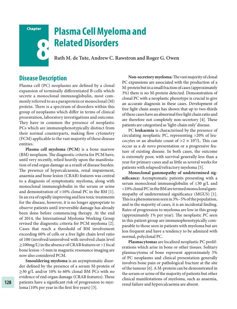
Image of the first page of this content. For PDF version, please use the ‘Save PDF’ preceeding this image.'
Type
Chapter
Information
Publisher: Cambridge University Press
Print publication year: 2018

Access options

Get access to the full version of this content by using one of the access options below. (Log in options will check for institutional or personal access. Content may require purchase if you do not have access.)

References

Swerdlow, S.H., Campo, E., Harris, N.L., et al. WHO classification of tumours of haematopoietic and lymphoid tissues. (4th ed. Lyon, Internat. Agency for Research on Cancer; 2008).Google Scholar
Rajkumar, S.V., Updated diagnostic criteria and staging system for multiple myeloma. Am Soc Clin Oncol Educ Book, 36 (2016), e41823.CrossRefGoogle Scholar
Rajkumar, S.V., Landgren, O. and Mateos, M-V.. Smoldering multiple myeloma. Blood, 125 (2015), 3069–75.CrossRefGoogle ScholarPubMed
Drayson, M., Tang, L.X., Drew, R., Mead, G.P., et al. Serum free light-chain measurements for identifying and monitoring patients with nonsecretory multiple myeloma. Blood, 97 (2001), 2900–2.CrossRefGoogle ScholarPubMed
Simeon, V., Todoerti, K., Rocca, F., et al. Molecular classification and pharmacogenetics of primary plasma cell leukemia: an initial approach toward precision medicine. Int J Mol Sci, 16 (2015), 17514–34.CrossRefGoogle ScholarPubMed
Soutar, R., Lucraft, H., Jackson, G., et al. Guidelines on the diagnosis and management of solitary plasmacytoma of bone and solitary extramedullary plasmacytoma. Br J Haematol, 124 (2004), 717–26.CrossRefGoogle ScholarPubMed
Gertz, M.A., Immunoglobulin light chain amyloidosis: 2011 update on diagnosis, risk-stratification, and management. Am J Hematol, 86 (2011), 180–6.Google ScholarPubMed
Dispenzieri, A., POEMS syndrome: 2014 update on diagnosis, risk-stratification, and management. Am J Hematol, 89 (2014), 214–23.CrossRefGoogle ScholarPubMed
Rawstron, A.C., de Tute, R.M., Haughton, J. and Owen, R.G., Measuring disease levels in myeloma using flow cytometry in combination with other laboratory techniques: lessons from the past 20 years at the Leeds Haematological Malignancy Diagnostic Service: MRD DETECTION BY FLOW CYTOMETRY AND OTHER LABORATORY TECHNIQUES. Cytometry B Clin Cytom, 90 (2016), 5460.CrossRefGoogle ScholarPubMed
Rawstron, A.C., Orfao, A., Beksac, M., et al. Report of the European Myeloma Network on multiparametric flow cytometry in multiple myeloma and related disorders. Haematologica, 93 (2008), 431–8.CrossRefGoogle ScholarPubMed
Stetler-Stevenson, M., Paiva, B., Stoolman, L., et al. Consensus guidelines for myeloma minimal residual disease sample staining and data acquisition. Cytometry B Clin Cytom, 90 (2016), 2630.CrossRefGoogle ScholarPubMed
Arroz, M., Came, N., Lin, P., et al. Consensus guidelines on plasma cell myeloma minimal residual disease analysis and reporting. Cytometry B Clin Cytom, 90 (2016), 31–9.CrossRefGoogle ScholarPubMed
Paiva, B., Almeida, J., Pérez-Andrés, M., et al. Utility of flow cytometry immunophenotyping in multiple myeloma and other clonal plasma cell-related disorders. Cytometry B Clin Cytom, 78 (2010), 239–52.Google ScholarPubMed
van Dongen, J.J.M., Lhermitte, L.L., Böttcher, S., et al. EuroFlow antibody panels for standardized n-dimensional flow cytometric immunophenotyping of normal, reactive and malignant leukocytes. Leukemia, 26 (2012), 1908–75.CrossRefGoogle ScholarPubMed
Liu, D., Lin, P., Hu, Y., et al. Immunophenotypic heterogeneity of normal plasma cells: comparison with minimal residual plasma cell myeloma. J Clin Pathol, 65 (2012), 823–9.CrossRefGoogle ScholarPubMed
Paiva, B., Gutiérrez, N-C., Chen, X., et al. Clinical significance of CD81 expression by clonal plasma cells in high-risk smoldering and symptomatic multiple myeloma patients. Leukemia, 26 (2012), 1862–9.CrossRefGoogle ScholarPubMed
Tembhare, P.R., Yuan, C.M., Venzon, D., et al. Flow cytometric differentiation of abnormal and normal plasma cells in the bone marrow in patients with multiple myeloma and its precursor diseases. Leuk Res, 38 (2014), 371–6.CrossRefGoogle ScholarPubMed
Raja, K.R.M., Kovarova, L. and Hajek, R.. Review of phenotypic markers used in flow cytometric analysis of MGUS and MM, and applicability of flow cytometry in other plasma cell disorders. Br J Haematol, 149 (2010), 334–51.CrossRefGoogle ScholarPubMed
Robillard, N., Jego, G., Pellat-Deceunynck, C., et al. CD28, a marker associated with tumoral expansion in multiple myeloma. Clin Cancer Res, 4 (1998), 1521–6.Google ScholarPubMed
Paiva, B., Vidriales, M-B., Mateo, G., et al. The persistence of immunophenotypically normal residual bone marrow plasma cells at diagnosis identifies a good prognostic subgroup of symptomatic multiple myeloma patients. Blood, 114 (2009), 4369–72.CrossRefGoogle ScholarPubMed
Paiva, B., Pérez-Andrés, M., Vídriales, M-B., et al. Competition between clonal plasma cells and normal cells for potentially overlapping bone marrow niches is associated with a progressively altered cellular distribution in MGUS vs myeloma. Leukemia, 25 (2011), 697706.CrossRefGoogle ScholarPubMed
Paiva, B., Paino, T., Sayagues, J-M., et al. Detailed characterization of multiple myeloma circulating tumor cells shows unique phenotypic, cytogenetic, functional, and circadian distribution profile. Blood, 122 (2013), 3591–8.CrossRefGoogle ScholarPubMed
Kumar, S., Rajkumar, S.V., Kyle, R.A., et al. Prognostic value of circulating plasma cells in monoclonal gammopathy of undetermined significance. J Clin Oncol, 23 (2005), 5668–74.CrossRefGoogle Scholar
Gonsalves, W.I., Rajkumar, S.V., Dispenzieri, A., et al. Quantification of circulating clonal plasma cells via multiparametric flow cytometry identifies patients with smoldering multiple myeloma at high risk of progression. Leukemia. (26 July 2016), doi: 10.1038/leu.2016.205.Google Scholar
Bianchi, G., Kyle, R.A., Larson, D.R., et al. High levels of peripheral blood circulating plasma cells as a specific risk factor for progression of smoldering multiple myeloma. Leukemia, 27 (2013), 680–5.CrossRefGoogle Scholar
Bataille, R., Pellat-Deceunynck, C., Robillard, N., et al. CD117 (c-kit) is aberrantly expressed in a subset of MGUS and multiple myeloma with unexpectedly good prognosis. Leuk Res, 32 (2008), 379–82.CrossRefGoogle Scholar
Schmidt-Hieber, M., Perez-Andres, M., Paiva, B., et al. CD117 expression in gammopathies is associated with an altered maturation of the myeloid and lymphoid hematopoietic cell compartments and favorable disease features. Haematologica, 96 (2011), 328–32.CrossRefGoogle ScholarPubMed
Mateo, G., Montalbán, M.A., Vidriales, M-B., et al. Prognostic value of immunophenotyping in multiple myeloma: a study by the PETHEMA/GEM cooperative study groups on patients uniformly treated with high-dose therapy. J Clin Oncol, 26 (2008), 2737–44.CrossRefGoogle ScholarPubMed
An, G., Xu, Y., Shi, L., et al. t(11;14) multiple myeloma: a subtype associated with distinct immunological features, immunophenotypic characteristics but divergent outcome. Leuk Res, 37 (2013), 1251–7.CrossRefGoogle ScholarPubMed
Mateo, G., Castellanos, M., Rasillo, A., et al. Genetic abnormalities and patterns of antigenic expression in multiple myeloma. Clin Cancer Res, 11 (2005), 3661–7.CrossRefGoogle ScholarPubMed
Moreau, P., Robillard, N., Avet-Loiseau, H., et al. Patients with CD45 negative multiple myeloma receiving high-dose therapy have a shorter survival than those with CD45 positive multiple myeloma. Haematologica, 89 (2004), 547–51.Google Scholar
Paiva, B., Chandia, M., Vidriales, M-B., et al. Multiparameter flow cytometry for staging of solitary bone plasmacytoma: new criteria for risk of progression to myeloma. Blood, 124 (2014), 1300–3.CrossRefGoogle Scholar
Hill, Q.A., Rawstron, A.C., de Tute, R.M. and Owen, R.G.. Outcome prediction in plasmacytoma of bone: a risk model utilizing bone marrow flow cytometry and light-chain analysis. Blood, 124 (2014), 1296–9.CrossRefGoogle Scholar
Shingles, J., de Tute, R.M., Rawstron, A.C. and Owen, R.G.. CD319 and CD38 expression patterns in Waldenstrom's macroglobulinaemia (WM), myeloma and MGUS: implications for antibody therapy. Br J Haematol, 173S1 (2016), 79, Abstract 190.Google Scholar
Flores-Montero, J., de Tute, R., Paiva, B., et al. Immunophenotype of normal vs. myeloma plasma cells: toward antibody panel specifications for MRD detection in multiple myeloma. Cytometry B Clin Cytom, 90 (2016), 6172.CrossRefGoogle ScholarPubMed
Domingo, E., Moreno, C., Sánchez-Ibarrola, A., et al. Enhanced sensitivity of flow cytometry for routine assessment of minimal residual disease. Haematologica, 95 (2010), 691–2.CrossRefGoogle ScholarPubMed
Paiva, B., Cedena, M-T., Puig, N., et al. Minimal residual disease monitoring and immune profiling in multiple myeloma in elderly patients. Blood, 127 (2016), 3165–74.CrossRefGoogle Scholar
San Miguel, J.F., Almeida, J., Mateo, G., et al. Immunophenotypic evaluation of the plasma cell compartment in multiple myeloma: a tool for comparing the efficacy of different treatment strategies and predicting outcome. Blood, 99 (2002):1853–6.CrossRefGoogle ScholarPubMed
Rawstron, A.C., Davies, F.E., DasGupta, R., et al. Flow cytometric disease monitoring in multiple myeloma: the relationship between normal and neoplastic plasma cells predicts outcome after transplantation. Blood, 100 (2002), 3095–100.CrossRefGoogle ScholarPubMed
Rawstron, A.C., Child, J.A., de Tute, R.M., et al. Minimal residual disease assessed by multiparameter flow cytometry in multiple myeloma: impact on outcome in the Medical Research Council Myeloma IX Study. J Clin Oncol, 31 (2013), 2540–7.CrossRefGoogle ScholarPubMed
Paiva, B., Vidriales, M-B., Cerveró, J., et al. Multiparameter flow cytometric remission is the most relevant prognostic factor for multiple myeloma patients who undergo autologous stem cell transplantation. Blood, 112 (2008), 4017–23.Google ScholarPubMed
Paiva, B., Martinez-Lopez, J., Vidriales, M-B., et al. Comparison of immunofixation, serum free light chain, and immunophenotyping for response evaluation and prognostication in multiple myeloma. J Clin Oncol, 29 (2011), 1627–33.CrossRefGoogle ScholarPubMed
Paiva, B., Gutiérrez, N.C., Rosiñol, L., et al. High-risk cytogenetics and persistent minimal residual disease by multiparameter flow cytometry predict unsustained complete response after autologous stem cell transplantation in multiple myeloma. Blood, 119 (2012), 687–91.CrossRefGoogle ScholarPubMed
Rawstron, A.C., Gregory, W.M., de Tute, R.M., et al. Minimal residual disease in myeloma by flow cytometry: independent prediction of survival benefit per log reduction. Blood, 125 (2015), 1932–5.CrossRefGoogle ScholarPubMed
Oldaker, T.A., Wallace, P.K. and Barnett, D.. Flow cytometry quality requirements for monitoring of minimal disease in plasma cell myeloma. Cytometry B Clin Cytom, 90 (2016), 40–6.CrossRefGoogle ScholarPubMed

Save book to Kindle

To save this book to your Kindle, first ensure [email protected] is added to your Approved Personal Document E-mail List under your Personal Document Settings on the Manage Your Content and Devices page of your Amazon account. Then enter the ‘name’ part of your Kindle email address below. Find out more about saving to your Kindle.

Note you can select to save to either the @free.kindle.com or @kindle.com variations. ‘@free.kindle.com’ emails are free but can only be saved to your device when it is connected to wi-fi. ‘@kindle.com’ emails can be delivered even when you are not connected to wi-fi, but note that service fees apply.

Find out more about the Kindle Personal Document Service.

Available formats
×

Save book to Dropbox

To save content items to your account, please confirm that you agree to abide by our usage policies. If this is the first time you use this feature, you will be asked to authorise Cambridge Core to connect with your account. Find out more about saving content to Dropbox.

Available formats
×

Save book to Google Drive

To save content items to your account, please confirm that you agree to abide by our usage policies. If this is the first time you use this feature, you will be asked to authorise Cambridge Core to connect with your account. Find out more about saving content to Google Drive.

Available formats
×