Skip to main content Accessibility help
×
Hostname: page-component-599cfd5f84-jhfc5 Total loading time: 0 Render date: 2025-01-07T07:48:06.229Z Has data issue: false hasContentIssue false

13 - Flow Cytometry in Myelodysplastic Syndromes

Published online by Cambridge University Press:  01 February 2018
Anna Porwit
Affiliation:
Lunds Universitet, Sweden
Marie Christine Béné
Affiliation:
Université de Nantes, France
Get access

Summary

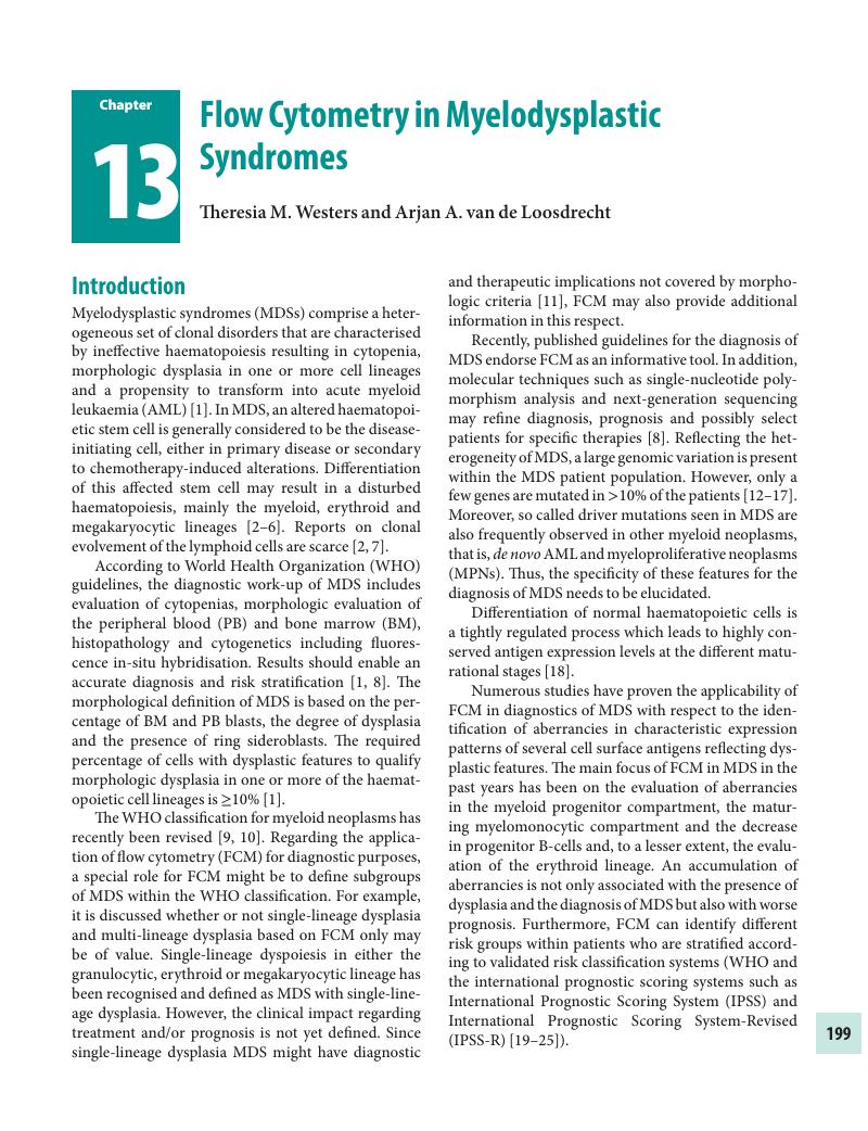
Image of the first page of this content. For PDF version, please use the ‘Save PDF’ preceeding this image.'
Type
Chapter
Information
Publisher: Cambridge University Press
Print publication year: 2018

Access options

Get access to the full version of this content by using one of the access options below. (Log in options will check for institutional or personal access. Content may require purchase if you do not have access.)

References

Brunning, R., Orazi, A., Germing, U., et al. Myelodysplastic syndromes/neoplasms. In, Swerdlow, et al., editor. WHO Classification of Tumours and Haematopoietic and Lymphoid Tissues. (Lyon, IARC, 2008):88103.Google Scholar
Nilsson, L., Astrand-Grundstrom, I., Arvidsson, I., et al. Isolation and characterization of hematopoietic progenitor/stem cells in 5q-deleted myelodysplastic syndromes, evidence for involvement at the hematopoietic stem cell level. Blood, 96 (2000), 2012–21.CrossRefGoogle ScholarPubMed
Nilsson, L., Eden, P., Olsson, E., et al. The molecular signature of MDS stem cells supports a stem-cell origin of 5q myelodysplastic syndromes. Blood, 110 (2007), 3005–14.CrossRefGoogle ScholarPubMed
Will, B., Zhou, L., Vogler, T.O., et al. Stem and progenitor cells in myelodysplastic syndromes show aberrant stage-specific expansion and harbor genetic and epigenetic alterations. Blood, 120 (2012), 2076–86.CrossRefGoogle ScholarPubMed
Pang, W.W., Pluvinage, J.V., Price, E.A., et al. Hematopoietic stem cell and progenitor cell mechanisms in myelodysplastic syndromes. Proc Natl Acad Sci U S A, 110 (2013), 3011–16.CrossRefGoogle ScholarPubMed
Woll, P.S., Kjallquist, U., Chowdhury, O., et al. Myelodysplastic syndromes are propagated by rare and distinct human cancer stem cells in vivo. Cancer Cell, 25 (2014), 794808.CrossRefGoogle Scholar
White, N.J., Nacheva, E., Asimakopoulos, F.A. et al. Deletion of chromosome 20q in myelodysplasia can occur in a multipotent precursor of both myeloid cells and B cells. Blood, 83 (1994), 2809–16.CrossRefGoogle Scholar
Malcovati, L., Hellstrom-Lindberg, E., Bowen, D., et al. Diagnosis and treatment of primary myelodysplastic syndromes in adults, recommendations from the European LeukemiaNet. Blood, 122 (2013), 2943–64.CrossRefGoogle ScholarPubMed
Arber, D.A. and Hasserjian, R.P.. Reclassifying myelodysplastic syndromes, what's where in the new WHO and why. Hematology Am Soc Hematol Educ Program, 2015 (2015), 294–8.Google ScholarPubMed
Arber, D.A., Orazi, A., Hasserjian, R.P., et al. The 2016 revision to the World Health Organization classification of myeloid neoplasms and acute leukemia. Blood, 127 (2016), 2391–405.CrossRefGoogle Scholar
Knipp, S., Strupp, C., Gattermann, N., et al. Presence of peripheral blasts in refractory anemia and refractory cytopenia with multilineage dysplasia predicts an unfavourable outcome. Leuk Res, 32 (2008), 33–7.CrossRefGoogle ScholarPubMed
Langemeijer, S.M., Kuiper, R.P., Berends, M., et al. Acquired mutations in TET2 are common in myelodysplastic syndromes. Nat Genet, 41 (2009), 838–42.CrossRefGoogle Scholar
Bejar, R., Stevenson, K., Abdel-Wahab, O., et al. Clinical effect of point mutations in myelodysplastic syndromes. N Engl J Med, 364 (2011), 2496–506.CrossRefGoogle ScholarPubMed
Papaemmanuil, E., Cazzola, M., Boultwood, J., et al. Somatic SF3B1 mutation in myelodysplasia with ring sideroblasts. N Engl J Med, 365 (2011), 1384–95.CrossRefGoogle ScholarPubMed
Malcovati, L., Papaemmanuil, E., Bowen, D.T., et al. Clinical significance of SF3B1 mutations in myelodysplastic syndromes and myelodysplastic/myeloproliferative neoplasms. Blood, 118 (2011), 6239–46.CrossRefGoogle ScholarPubMed
Papaemmanuil, E., Gerstung, M., Malcovati, L., et al. Clinical and biological implications of driver mutations in myelodysplastic syndromes. Blood, 122 (2013), 3616–27.CrossRefGoogle Scholar
Haferlach, T., Nagata, Y., Grossmann, V., et al. Landscape of genetic lesions in 944 patients with myelodysplastic syndromes. Leukemia, 28 (2014), 241–7.CrossRefGoogle ScholarPubMed
van Lochem, E.G., van der Velden, V.H.J., Wind, H.K., et al. Immunophenotypic differentiation patterns of normal hematopoiesis in human bone marrow, reference patterns for age-related changes and disease-induced shifts. Cytometry B Clin Cytom, 60 (2004), 113.CrossRefGoogle ScholarPubMed
Wells, D.A., Benesch, M., Loken, M.R., et al. Myeloid and monocytic dyspoiesis as determined by flow cytometric scoring in myelodysplastic syndrome correlates with the IPSS and with outcome after hematopoietic stem cell transplantation. Blood, 102 (2003), 394403.CrossRefGoogle ScholarPubMed
van de Loosdrecht, A.A., Westers, T.M., Westra, A.H., et al. Identification of distinct prognostic subgroups in low- and intermediate-1-risk myelodysplastic syndromes by flow cytometry. Blood, 111 (2008), 1067–77.CrossRefGoogle Scholar
Scott, B.L., Wells, D.A., Loken, M.R., et al. Validation of a flow cytometric scoring system as a prognostic indicator for posttransplantation outcome in patients with myelodysplastic syndrome. Blood, 112 (2008), 2681–6.CrossRefGoogle ScholarPubMed
Matarraz, S., Lopez, A., Barrena, S., et al. Bone marrow cells from myelodysplastic syndromes show altered immunophenotypic profiles that may contribute to the diagnosis and prognostic stratification of the disease, a pilot study on a series of 56 patients. Cytometry B Clin Cytom, 78 (2010), 154–68.Google Scholar
Chu, S.C., Wang, T.F., Li, C.C., et al. Flow cytometric scoring system as a diagnostic and prognostic tool in myelodysplastic syndromes. Leuk Res, 357 (2011), 868–73.Google Scholar
Alhan, C., Westers, T.M., Cremers, E.M.P., et al. High flow cytometric scores identify adverse prognostic subgroups within the revised international prognostic scoring system for myelodysplastic syndromes. Br J Haematol, 167 (2014), 100–9.CrossRefGoogle ScholarPubMed
Kern, W., Bacher, U., Haferlach, C., et al. Multiparameter flow cytometry provides independent prognostic information in patients with suspected myelodysplastic syndromes, a study on 804 patients. Cytometry B Clin Cytom, 88 (2015), 154–64.CrossRefGoogle Scholar
van de Loosdrecht, A.A., Alhan, C., Bene, M.C., et al. Standardization of flow cytometry in myelodysplastic syndromes, report from the first European LeukemiaNet working conference on flow cytometry in myelodysplastic syndromes. Haematologica, 94 (2009), 1124–34.CrossRefGoogle ScholarPubMed
Westers, T.M., Ireland, R., Kern, W., et al. Standardization of flow cytometry in myelodysplastic syndromes, a report from an international consortium and the European LeukemiaNet Working Group. Leukemia, 26 (2012), 1730–41.CrossRefGoogle Scholar
Kern, W., Bacher, U., Schnittger, S., et al. Multiparameter flow cytometry reveals myelodysplasia-related aberrant antigen expression in myelodysplastic/myeloproliferative neoplasms. Cytometry B Clin Cytom, 84 (2013), 194–7.Google Scholar
Westers, T.M., van der Velden, V.H.J., Alhan, C., et al. Implementation of flow cytometry in the diagnostic work-up of myelodysplastic syndromes in a multicenter approach, report from the Dutch Working Party on Flow Cytometry in MDS. Leuk Res, 36 (2012), 422–30.CrossRefGoogle Scholar
van Dongen, J.J., Lhermitte, L., Bottcher, S., et al. EuroFlow antibody panels for standardized n-dimensional flow cytometric immunophenotyping of normal, reactive and malignant leukocytes. Leukemia, 26 (2012), 1908–75.CrossRefGoogle ScholarPubMed
Mathis, S., Chapuis, N., Debord, C., et al. Flow cytometric detection of dyserythropoiesis, a sensitive and powerful diagnostic tool for myelodysplastic syndromes. Leukemia, 27 (2013), 1981–7.CrossRefGoogle ScholarPubMed
Porwit, A. and Rajab, A.. Flow cytometry immunophenotyping in integrated diagnostics of patients with newly diagnosed cytopenia, one tube 10-color 14-antibody screening panel and 3-tube extensive panel for detection of MDS-related features. Int J Lab Hematol, 37 Suppl 1 (2015), 133–43.CrossRefGoogle ScholarPubMed
Bardet, V., Wagner-Ballon, O., Guy, J., et al. Multicentric study underlining the interest of adding CD5, CD7 and CD56 expression assessment to the flow cytometric Ogata score in myelodysplastic syndromes and myelodysplastic/myeloproliferative neoplasms. Haematologica, 100 (2015), 472–8.CrossRefGoogle Scholar
Alhan, C., Westers, T.M., Cremers, E.M.P., et al. Application of flow cytometry for myelodysplastic syndromes, pitfalls and technical considerations. Cytometry B Clin Cytom, 90 (2016), 358–67.CrossRefGoogle ScholarPubMed
Jevremovic, D., Timm, M.M., Reichard, K.K., et al. Loss of blast heterogeneity in myelodysplastic syndrome and other chronic myeloid neoplasms. Am J Clin Pathol, 142 (2014), 292–8.CrossRefGoogle ScholarPubMed
Terwijn, M., Feller, N., van Rhenen, A., et al. Interleukin-2 receptor alpha-chain (CD25) expression on leukaemic blasts is predictive for outcome and level of residual disease in AML. Eur J Cancer, 45 (2009), 1692–9.CrossRefGoogle ScholarPubMed
Alhan, C., Westers, T.M., van der Helm, L.H., et al. Absence of aberrant myeloid progenitors by flow cytometry is associated with favorable response to azacitidine in higher risk myelodysplastic syndromes. Cytometry B Clin Cytom 86 (2014), 207–15.CrossRefGoogle ScholarPubMed
Monreal, M.B., Pardo, M.L., Pavlovsky, M.A., et al. Increased immature hematopoietic progenitor cells CD34+/CD38dim in myelodysplasia. Cytometry B Clin Cytom, 70 (2006), 6370.CrossRefGoogle ScholarPubMed
Goardon, N., Nikolousis, E., Sternberg, A., et al. Reduced CD38 expression on CD34+ cells as a diagnostic test in myelodysplastic syndromes. Haematologica, 94 (2009), 1160–3.CrossRefGoogle ScholarPubMed
Falco, P., Levis, A., Stacchini, A., et al. Prognostic relevance of cytometric quantitative assessment in patients with myelodysplastic syndromes. Eur J Haematol, 87 (2011), 409–18.CrossRefGoogle ScholarPubMed
Ogata, K., Kakumoto, K., Matsuda, A., et al. Differences in blast immunophenotypes among disease types in myelodysplastic syndromes, a multicenter validation study. Leuk Res, 36 (2012), 1229–36.CrossRefGoogle ScholarPubMed
Sutherland, D.R., Illingworth, A., Keeney, M., et al. High-sensitivity detection of PNH red blood cells, red cell precursors, and white blood cells. Curr Protoc Cytom, 72 (2015), 630.Google Scholar
Stachurski, D., Smith, B.R., Pozdnyakova, O., et al. Flow cytometric analysis of myelomonocytic cells by a pattern recognition approach is sensitive and specific in diagnosing myelodysplastic syndrome and related marrow diseases, emphasis on a global evaluation and recognition of diagnostic pitfalls. Leuk Res, 32 (2008), 215–24.CrossRefGoogle ScholarPubMed
Oelschlaegel, U., Westers, T.M., Mohr, B., et al. Myelodysplastic syndromes with a deletion 5q display a characteristic immunophenotypic profile suitable for diagnostics and response monitoring. Haematologica, 100 (2015), e936.CrossRefGoogle ScholarPubMed
Pradhan, A., Mijovic, A., Mills, K., et al. Differentially expressed genes in adult familial myelodysplastic syndromes. Leukemia, 18 (2004), 449–59.CrossRefGoogle ScholarPubMed
Briggs, R.C., Shults, K.E., Flye, L.A., et al. Dysregulated human myeloid nuclear differentiation antigen expression in myelodysplastic syndromes, evidence for a role in apoptosis. Cancer Res, 66 (2006), 4645–51.CrossRefGoogle ScholarPubMed
Clintock-Treep, S.A., Briggs, R.C., Shults, K.E., et al. Quantitative assessment of myeloid nuclear differentiation antigen distinguishes myelodysplastic syndrome from normal bone marrow. Am J Clin Pathol, 135 (2011), 380–5.Google Scholar
Bellos, F., Alpermann, T., Gouberman, E., et al. Evaluation of flow cytometric assessment of myeloid nuclear differentiation antigen expression as a diagnostic marker for myelodysplastic syndromes in a series of 269 patients. Cytometry B Clin Cytom, 82 (2012), 295304.CrossRefGoogle Scholar
Bellos, F. and Kern, W.. Flow cytometry in the diagnosis of myelodysplastic syndromes (MDS) and the value of myeloid nuclear differentiation antigen (MNDA). Cytometry B Clin Cytom (2015), DOI: 10.1002/cyto.b.21190.Google ScholarPubMed
Shen, Q., Ouyang, J., Tang, G., et al. Flow cytometry immunophenotypic findings in chronic myelomonocytic leukemia and its utility in monitoring treatment response. Eur J Haematol, 95 (2015), 168–76.CrossRefGoogle Scholar
Xu, Y., McKenna, R.W., Karandikar, N.J., et al. Flow cytometric analysis of monocytes as a tool for distinguishing chronic myelomonocytic leukemia from reactive monocytosis. Am J Clin Pathol, 124 (2005), 799806.CrossRefGoogle ScholarPubMed
Subira, D., Font, P., Villalon, L., et al. Immunophenotype in chronic myelomonocytic leukemia, is it closer to myelodysplastic syndromes or to myeloproliferative disorders? Transl Res, 151 (2008), 240–5.CrossRefGoogle ScholarPubMed
Kern, W., Bacher, U., Haferlach, C., et al. Acute monoblastic/monocytic leukemia and chronic myelomonocytic leukemia share common immunophenotypic features but differ in the extent of aberrantly expressed antigens and amount of granulocytic cells. Leuk Lymphoma, 52 (2011), 92100.CrossRefGoogle ScholarPubMed
Sconocchia, G., Fujiwara, H., Rezvani, K., et al. G-CSF-mobilized CD34+ cells cultured in interleukin-2 and stem cell factor generate a phenotypically novel monocyte. J Leukoc Biol, 76 (2004), 1214–19.CrossRefGoogle ScholarPubMed
Wood, B.L.. Myeloid malignancies, myelodysplastic syndromes, myeloproliferative disorders, and acute myeloid leukemia. Clin Lab Med, 27 (2007), 551–75.CrossRefGoogle ScholarPubMed
Selimoglu-Buet, D., Wagner-Ballon, O., Saada, V., et al. Characteristic repartition of monocyte subsets as a diagnostic signature of chronic myelomonocytic leukemia. Blood, 125 (2015), 3618–26.CrossRefGoogle ScholarPubMed
Stetler-Stevenson, M., Arthur, D.C., Jabbour, N., et al. Diagnostic utility of flow cytometric immunophenotyping in myelodysplastic syndrome. Blood, 98 (2001), 979–87.CrossRefGoogle ScholarPubMed
Malcovati, L., Della Porta, M.G., Lunghi, M., et al. Flow cytometry evaluation of erythroid and myeloid dysplasia in patients with myelodysplastic syndrome. Leukemia, 19 (2005), 776–83.CrossRefGoogle ScholarPubMed
Della Porta, M.G., Malcovati, L., Invernizzi, R., et al. Flow cytometry evaluation of erythroid dysplasia in patients with myelodysplastic syndrome. Leukemia, 20 (2006), 549–55.Google ScholarPubMed
Kern, W., Haferlach, C., Schnittger, S., et al. Clinical utility of multiparameter flow cytometry in the diagnosis of 1013 patients with suspected myelodysplastic syndrome, correlation to cytomorphology, cytogenetics, and clinical data. Cancer, 116 (2010), 4549–63.CrossRefGoogle ScholarPubMed
Lorand-Metze, I., Ribeiro, E., Lima, C.S., et al. Detection of hematopoietic maturation abnormalities by flow cytometry in myelodysplastic syndromes and its utility for the differential diagnosis with non-clonal disorders. Leuk Res, 31 (2007), 147–55.CrossRefGoogle ScholarPubMed
Xu, F., Wu, L., He, Q., et al. Immunophenotypic analysis of erythroid dysplasia and its diagnostic application in myelodysplastic syndromes. Intern Med J, 42 (2012), 401–11.CrossRefGoogle ScholarPubMed
Hu, J., Liu, J., Xue, F., et al. Isolation and functional characterization of human erythroblasts at distinct stages, implications for understanding of normal and disordered erythropoiesis in vivo. Blood, 121 (2013), 3246–53.CrossRefGoogle ScholarPubMed
Fajtova, M., Kovarikova, A., Svec, P., et al. Immunophenotypic profile of nucleated erythroid progenitors during maturation in regenerating bone marrow. Leuk Lymphoma, 54 (2013), 2523–30.CrossRefGoogle Scholar
Eidenschink Brodersen, L., Menssen, A.J., Wangen, J.R., et al. Assessment of erythroid dysplasia by ‘difference from normal’ in routine clinical flow cytometry work-up. Cytometry B Clin Cytom, 88 (2015), 125–35.CrossRefGoogle Scholar
Laranjeira, P., Rodrigues, R., Carvalheiro, T., et al. Expression of CD44 and CD35 during normal and myelodysplastic erythropoiesis. Leuk Res, 39 (2014), 361–70.Google ScholarPubMed
Westers, T.M., Cremers, E.M.P., Oelschlaegel, U., et al. Immunophenotypic analysis of erythroid dysplasia in myelodysplastic syndromes. A report from the IMDSFlow Working Group. Haematologica, 102 (2017) 308–19.CrossRefGoogle ScholarPubMed
Cremers, E.M.P., Westers, T.M., Alhan, C., et al. Implementation of erythroid analysis by flow cytometry in diagnostic models for myelodysplastic syndromes. Haematologica, 102 (2017), 320–6.CrossRefGoogle ScholarPubMed
Sandes, A.F., Yamamoto, M., Matarraz, S., et al. Altered immunophenotypic features of peripheral blood platelets in myelodysplastic syndromes. Haematologica, 97 (2012), 895902.CrossRefGoogle ScholarPubMed
Ogata, K., Della Porta, M.G., Malcovati, L., et al. Diagnostic utility of flow cytometry in low-grade myelodysplastic syndromes, a prospective validation study. Haematologica, 94 (2009), 1066–74.CrossRefGoogle ScholarPubMed
Della Porta, M.G., Picone, C., Pascutto, C., et al. Multicenter validation of a reproducible flow cytometric score for the diagnosis of low-grade myelodysplastic syndromes, results of a European LeukemiaNET study. Haematologica, 97 (2012), 1209–17.CrossRefGoogle ScholarPubMed
Kuranda, K., Vargaftig, J., de la Rochere, P., et al. Age-related changes in human hematopoietic stem/progenitor cells. Aging Cell, 10 (2011), 542–6.CrossRefGoogle Scholar
Parmentier, S., Schetelig, J., Lorenz, K., et al. Assessment of dysplastic hematopoiesis, lessons from healthy bone marrow donors. Haematologica, 97 (2012), 723–30.CrossRefGoogle ScholarPubMed
Della Porta, M.G., Travaglino, E., Boveri, E., et al. Minimal morphological criteria for defining bone marrow dysplasia, a basis for clinical implementation of WHO classification of myelodysplastic syndromes. Leukemia, 29 (2015), 6675.CrossRefGoogle ScholarPubMed
Primo, D., Sanchez, M.L., Espinosa, A.B., et al. Lineage involvement in chronic myeloid leukaemia, comparison between MBCR/ABL and mBCR/ABL cases. Br J Haematol, 132 (2006), 736–9.CrossRefGoogle Scholar
Aalbers, A.M., van den Heuvel-Eibrink, M.M., de Haas, V., et al. Applicability of a reproducible flow cytometry scoring system in the diagnosis of refractory cytopenia of childhood. Leukemia, 27 (2013), 1923–5.CrossRefGoogle ScholarPubMed
Aalbers, A.M., van den Heuvel-Eibrink, M.M., Baumann, I., et al. Bone marrow immunophenotyping by flow cytometry in refractory cytopenia of childhood. Haematologica, 100 (2015), 315–23.CrossRefGoogle ScholarPubMed
Cremers, E.M.P., Westers, T.M., Alhan, C., et al. Multiparameter flow cytometry is instrumental to distinguish myelodysplastic syndromes from non-neoplastic cytopenias. Eur J Cancer, 54 (2016), 4956.CrossRefGoogle Scholar
Xu, F., Guo, J., Wu, L.Y., et al. Diagnostic application and clinical significance of FCM progress scoring system based on immunophenotyping in CD34+ blasts in myelodysplastic syndromes. Cytometry B Clin Cytom, 84 (2013), 267–78.Google ScholarPubMed
Sandes, A.F., Kerbauy, D.M., Matarraz, S., et al. Combined flow cytometric assessment of CD45, HLA-DR, CD34, and CD117 expression is a useful approach for reliable quantification of blast cells in myelodysplastic syndromes. Cytometry B Clin Cytom, 84 (2013), 157–66.Google ScholarPubMed
Porwit, A., van de Loosdrecht, A.A., Bettelheim, P., et al. Revisiting guidelines for integration of flow cytometry results in the WHO classification of myelodysplastic syndromes-proposal from the International/European LeukemiaNet Working Group for Flow Cytometry in MDS. Leukemia, 28 (2014), 1793–8.CrossRefGoogle Scholar
Kern, W., Haferlach, C., Schnittger, S., et al. Serial assessment of suspected myelodysplastic syndromes, significance of flow cytometric findings validated by cytomorphology, cytogenetics, and molecular genetics. Haematologica, 98 (2013), 201–7.CrossRefGoogle ScholarPubMed
Garcia-Manero, G.. Myelodysplastic syndromes, 2014 update on diagnosis, risk-stratification, and management. Am J Hematol, 89 (2014), 97108.CrossRefGoogle ScholarPubMed
Cutler, J.A., Wells, D.A., van de Loosdrecht, A.A., et al. Phenotypic abnormalities strongly reflect genotype in patients with unexplained cytopenias. Cytometry B Clin Cytom, 80 (2011), 150–7.Google ScholarPubMed
Westers, T.M., Alhan, C., Chamuleau, M.E.D., et al. Aberrant immunophenotype of blasts in myelodysplastic syndromes is a clinically relevant biomarker in predicting response to growth factor treatment. Blood, 115 (2010), 1779–84.CrossRefGoogle Scholar
Ossenkoppele, G.J., van de Loosdrecht, A.A. and Schuurhuis, G.J.. Review of the relevance of aberrant antigen expression by flow cytometry in myeloid neoplasms. Br J Haematol, 153 (2011), 421–36.CrossRefGoogle ScholarPubMed

Save book to Kindle

To save this book to your Kindle, first ensure [email protected] is added to your Approved Personal Document E-mail List under your Personal Document Settings on the Manage Your Content and Devices page of your Amazon account. Then enter the ‘name’ part of your Kindle email address below. Find out more about saving to your Kindle.

Note you can select to save to either the @free.kindle.com or @kindle.com variations. ‘@free.kindle.com’ emails are free but can only be saved to your device when it is connected to wi-fi. ‘@kindle.com’ emails can be delivered even when you are not connected to wi-fi, but note that service fees apply.

Find out more about the Kindle Personal Document Service.

Available formats
×

Save book to Dropbox

To save content items to your account, please confirm that you agree to abide by our usage policies. If this is the first time you use this feature, you will be asked to authorise Cambridge Core to connect with your account. Find out more about saving content to Dropbox.

Available formats
×

Save book to Google Drive

To save content items to your account, please confirm that you agree to abide by our usage policies. If this is the first time you use this feature, you will be asked to authorise Cambridge Core to connect with your account. Find out more about saving content to Google Drive.

Available formats
×