Skip to main content Accessibility help
×
Hostname: page-component-745bb68f8f-hvd4g Total loading time: 0 Render date: 2025-01-23T02:27:15.264Z Has data issue: false hasContentIssue false

1 - Ultrasound principles

Published online by Cambridge University Press:  05 May 2016
László Csiba
Affiliation:
Department of Neurology, Debreceni Egyetem, Hungary
Claudio Baracchini
Affiliation:
Department of Neuroscience, Università degli Studi di Padova, Italy
Get access

Summary

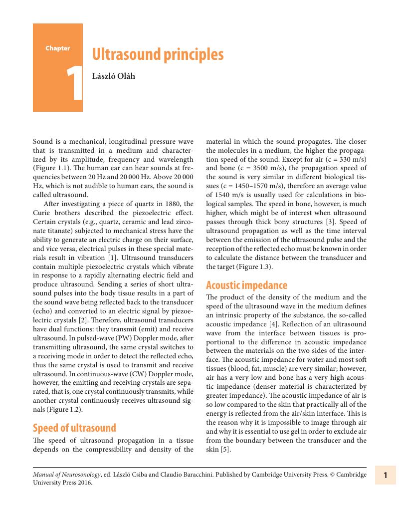
Image of the first page of this content. For PDF version, please use the ‘Save PDF’ preceeding this image.'
Type
Chapter
Information
Publisher: Cambridge University Press
Print publication year: 2016

Access options

Get access to the full version of this content by using one of the access options below. (Log in options will check for institutional or personal access. Content may require purchase if you do not have access.)

References

Otto, CM. Principles of echocardiographic image acquisition and Doppler analysis. In: Otto, CM, ed. Textbook of Clinical Echocardiography, 2nd ed. Philadelphia, PA: WB Saunders; 2000, 129.Google Scholar
Weyman, AE. Physical principles of ultrasound. In: Weyman, AE, ed. Principles and Practice of Echocardiography, 2nd ed. Philadelphia, PA: Lippincott Williams & Wilkins; 1994, 328.Google Scholar
Evans, DH. Physical and technical principles. In: Baumgartner, RW, ed. Handbook on Neurovascular Ultrasound. Basel: Karger; 2006, 118.Google Scholar
Schäberle, W. Fundamental principles. In: Ultrasonography in Vascular Diagnosis. Berlin: Springer-Verlag; 2005, 144.Google Scholar
Chan, V, Perlas, A. Basics of ultrasound imaging. In: Narouze, SN, ed. Atlas of Ultrasound-guided Procedures in Interventional Pain Management. New York: Springer Science+Business Media, LLC; 2011, 1319.Google Scholar
Kossoff, G. Basic physics and imaging characteristics of ultrasound. World J Surg. 2000;24:134142.CrossRefGoogle ScholarPubMed
Neumyer, MM, Alexandrov, AV. Cerebrovascular anatomy and principles of extracranial ultrasound examination. In: Alexandrov, AV, ed. Cerebrovascular Ultrasound in Stroke Prevention and Treatment. New York: Blackwell Publishing; 2004, 316.Google Scholar
Doppler, CA. Über das farbige licht der Doppelsterne und einiger anderer Gestirne des Himmels. Abhandlungen der königl. böhm. Gesellschaft der Wissenschaften. 1843;2:465482.Google Scholar
Williams, J. VASCPROG 560. Vascular Imaging Techniques. Module 5. Doppler Imaging. [Internet] 2013 [last accessed December 4, 2013]. Available from: www.robarts.ca/CIHR_VTP/Vascular_Imaging/Module5/Doppler%20Imaging.pdfGoogle Scholar
Støylen, A. Basic Ultrasound, Echocardiography and Doppler for Clinicians. [Internet] 2013 [updated 2013 November; last accessed December 7, 2013]. Available from: http://folk.ntnu.no/stoylen/strainrate/Ultrasound/Google Scholar
Alexandrov, AV, Neumyer, MM. Diagnostic criteria for cerebrovascular ultrasound. In: Alexandrov, AV, ed. Cerebrovascular Ultrasound in Stroke Prevention and Treatment. New York: Blackwell Publishing; 2004, 79129.Google Scholar
Ralls, PW. Tissue Harmonic Imaging – Updated. [Internet] 2004 [updated 2004; last accessed December 10, 2013]. Available from: www.sonocredits.com/sonocredits/article.asp?TestID=41Google Scholar
Deshpande, N, Needles, A, Willmann, JK. Molecular ultrasound imaging: current status and future directions. Clin Radiol. 2010;65:567581.CrossRefGoogle ScholarPubMed
McCulloch, M, Gresser, C, Moos, S, et al. Ultrasound contrast physics: a series on contrast echocardiography, article 3. J Am Soc Echocardiogr. 2000;13:959967.CrossRefGoogle ScholarPubMed
Klibanov, AL. Molecular imaging with targeted ultrasound contrast microbubbles. Ernst Schering Res Found Workshop. 2005;49:171191.Google Scholar
Ries, F, Honisch, C, Lambertz, M, Schlief, R. A transpulmonary contrast medium enhances the transcranial Doppler signal in humans. Stroke. 1993;24:19031909.Google Scholar
Wikipedia. Cavitation. [Internet] 2014 [last accessed January 9, 2014]. Available from: http://en.wikipedia.org/wiki/CavitationGoogle Scholar
Dijkmans, PA, Juffermans, LJM, Musters, RJP, et al. Microbubbles and ultrasound: from diagnosis to therapy. Eur J Echocardiography. 2004;5:245256.CrossRefGoogle ScholarPubMed
Blomley, MJK, Cooke, JC, Unger, EC, Monaghan, MJ, Cosgrove, DO. Microbubble contrast agents: a new era in ultrasound. BMJ. 2001;322:12221225.Google Scholar
Qin, S, Caskey, CF, Ferrara, KW. Ultrasound contrast microbubbles in imaging and therapy: physical principles and engineering. Phys Med Biol. 2009;54:R27R57.Google Scholar
Wei, K. Future applications of contrast ultrasound. J Cardiovasc Ultrasound. 2011;19:107114.Google Scholar
Schmitz, G. Ultrasonic imaging of molecular targets. Basic Res Cardiol. 2008;103:174181.CrossRefGoogle ScholarPubMed
Correas, JM, Hélénon, O, Pourcelot, L, Moreau, JF. Ultrasound contrast agents. Acta Radiologica. 1997;38(Suppl 412):101112.Google Scholar
Kang, ST, Yeh, CK. Ultrasound microbubble contrast agents for diagnostic and therapeutic applications: current status and future design. Chang Gung Med J. 2012;35:125139.Google ScholarPubMed
Sirsi, SR, Borden, MA. Advances in ultrasound mediated gene therapy using microbubble contrast agents. Theranostics. 2012;2:12081222.CrossRefGoogle ScholarPubMed
Zhou, Y, Yang, K, Cui, J, Ye, JY, Deng, CX. Controlled permeation of cell membrane by single bubble acoustic cavitation. J Control Release. 2011;157:103111.CrossRefGoogle ScholarPubMed
Lanza, GM, Wickline, SA. Targeted ultrasonic contrast agents for molecular imaging and therapy. Curr Probl Cardiol. 2003;28:625653.Google Scholar
Wei, K, Jayaweera, AR, Firoozan, S, et al. Quantification of myocardial blood flow with ultrasound induced destruction of microbubbles administered as a constant venous infusion. Circulation. 1998;97:473483.CrossRefGoogle ScholarPubMed
Meairs, S. Advances in neurosonology – brain perfusion, sonothrombolysis and CNS drug delivery. Perspect Med. 2012;1:510.Google Scholar
Seidel, G, Algermissen, C, Christoph, A, et al. Harmonic imaging of the human brain. Visualization of brain perfusion with ultrasound. Stroke. 2000;31:151154.CrossRefGoogle ScholarPubMed
Kern, R, Diels, A, Pettenpohl, J, et al. Real-time ultrasound brain perfusion imaging with analysis of microbubble replenishment in acute MCA stroke. J Cereb Blood Flow Metab. 2011;31:17161724.CrossRefGoogle ScholarPubMed
Tsivgoulis, G, Eggers, J, Ribo, M, et al. Safety and efficacy of ultrasound-enhanced thrombolysis: a comprehensive review and meta-analysis of randomized and nonrandomized studies. Stroke. 2010;41:280287.Google Scholar
Alexandrov, AV, Molina, CA, Grotta, JC, et al. Ultrasound-enhanced systemic thrombolysis for acute ischemic stroke. N Engl J Med. 2004;351:21702178.Google Scholar
Hitchcock, KE. Ultrasound-assisted thrombolysis for stroke therapy: better thrombus break-up with bubbles. Stroke. 2010;41(10 Suppl):S50–53.Google Scholar
Meairs, S, Culp, W. Microbubbles for thrombolysis of acute ischemic stroke. Cerebrovasc Dis. 2009;27(Suppl 2): 5565.CrossRefGoogle ScholarPubMed
Culp, WC, Flores, R, Brown, AT, et al. Successful microbubble sonothrombolysis without tissue-type plasminogen activator in a rabbit model of acute ischemic stroke. Stroke. 2011;42:22802285.Google Scholar
Flores, R, Hennings, LJ, Lowery, JD, et al. Microbubble- augmented ultrasound sonothrombolysis decreases intracranial hemorrhage in a rabbit model of acute ischemic stroke. Invest Radiol. 2011;46:419424.Google Scholar
Fatar, M, Stroick, M, Griebe, M, et al. Effect of combined ultrasound and microbubbles treatment in an experimental model of cerebral ischemia. Ultrasound Med Biol. 2008;34:14141420.Google Scholar
Newell, DW, Shah, MM, Wilcox, R, et al. Minimally invasive evacuation of spontaneous intracerebral hemorrhage using sonothrombolysis. J Neurosurg. 2011;115:592601.Google Scholar
Mesiwala, AH, Farrell, L, Wenzel, HJ, et al. High-intensity focused ultrasound selectively disrupts the blood-brain barrier in vivo. Ultrasound Med Biol. 2002;28:389400.Google Scholar
Alonso, A, Reinz, E, Jenne, JW, et al. Reorganization of gap junctions after focused ultrasound blood-brain barrier opening in the rat brain. J Cereb Blood Flow Metab. 2010;30:13941402.Google Scholar
Hynynen, K, McDannold, N, Vykhodtseva, N, Jolesz, FA. Noninvasive MR imaging-guided focal opening of the blood-brain barrier in rabbits. Radiology. 2001;220:640646.CrossRefGoogle ScholarPubMed
Sheikov, N, McDannold, N, Vykhodtseva, N, Jolesz, F, Hynynen, K. Cellular mechanisms of the blood-brain barrier opening induced by ultrasound in presence of microbubbles. Ultrasound Med Biol. 2004;30:979989.Google Scholar
McDannold, N, Vykhodtseva, N, Hynynen, K. Targeted disruption of the blood-brain barrier with focused ultrasound: association with cavitation activity. Phys Med Biol. 2006;51:793807.CrossRefGoogle ScholarPubMed
Sheikov, N, McDannold, N, Sharma, S, Hynynen, K. Effect of focused ultrasound applied with an ultrasound contrast agent on the tight junctional integrity of the brain microvascular endothelium. Ultrasound Med Biol. 2008;34:10931104.Google Scholar
Kinoshita, M, McDannold, N, Jolesz, FA, Hynynen, K. Targeted delivery of antibodies through the blood-brain barrier by MRI-guided focused ultrasound. Biochem Biophys Res Commun. 2006;340:10851090.CrossRefGoogle ScholarPubMed
Treat, LH, McDannold, N, Vykhodtseva, N, et al. Targeted delivery of doxorubicin to the rat brain at therapeutic levels using MRI-guided focused ultrasound. Int J Cancer. 2007;121:901907.Google Scholar
Porter, TR, Iverson, PL, Li, S, Xie, F. Interaction of diagnostic ultrasound with synthetic oligonucleotidelabeled perfluorocarbon-exposed sonicated dextrose albumin microbubbles. J Ultrason Med. 1996;15:577584.Google Scholar
Shohet, RV, Chen, S, Zhou, YT, et al. Echocardiographic destruction of albumin microbubbles directs gene delivery to the myocardium. Circulation. 2000;101:25542556.CrossRefGoogle ScholarPubMed
Wu, Y, Unger, EC, McCreery, TP, et al. Binding and lysing of blood clots using MRX408. Invest Radiol. 1998;33:880885.Google Scholar

Save book to Kindle

To save this book to your Kindle, first ensure [email protected] is added to your Approved Personal Document E-mail List under your Personal Document Settings on the Manage Your Content and Devices page of your Amazon account. Then enter the ‘name’ part of your Kindle email address below. Find out more about saving to your Kindle.

Note you can select to save to either the @free.kindle.com or @kindle.com variations. ‘@free.kindle.com’ emails are free but can only be saved to your device when it is connected to wi-fi. ‘@kindle.com’ emails can be delivered even when you are not connected to wi-fi, but note that service fees apply.

Find out more about the Kindle Personal Document Service.

Available formats
×

Save book to Dropbox

To save content items to your account, please confirm that you agree to abide by our usage policies. If this is the first time you use this feature, you will be asked to authorise Cambridge Core to connect with your account. Find out more about saving content to Dropbox.

Available formats
×

Save book to Google Drive

To save content items to your account, please confirm that you agree to abide by our usage policies. If this is the first time you use this feature, you will be asked to authorise Cambridge Core to connect with your account. Find out more about saving content to Google Drive.

Available formats
×