Skip to main content Accessibility help
×
Hostname: page-component-745bb68f8f-b6zl4 Total loading time: 0 Render date: 2025-01-18T20:50:15.354Z Has data issue: false hasContentIssue false

Chapter 8 - Miscellaneous Tumors

Published online by Cambridge University Press:  04 November 2017

Mai P. Hoang
Affiliation:
Harvard Medical School, Boston
Get access

Summary

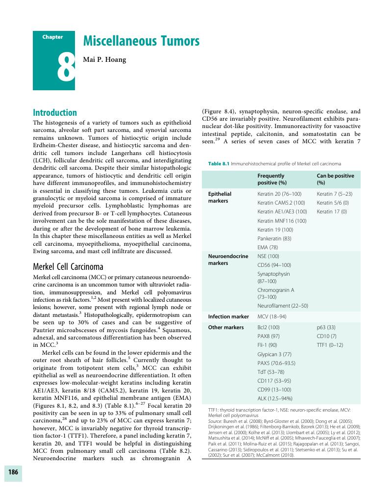
Image of the first page of this content. For PDF version, please use the ‘Save PDF’ preceeding this image.'
Type
Chapter
Information
Publisher: Cambridge University Press
Print publication year: 2017

Access options

Get access to the full version of this content by using one of the access options below. (Log in options will check for institutional or personal access. Content may require purchase if you do not have access.)

References

Penn, I, First, MR. Merkel’s cell carcinoma in organ recipients: Report of 41 cases. Transplantation 1999;68(11):1717–21.Google Scholar
Feng, H, Shuda, M, Chang, Y, et al. Clonal integration of a polyomavirus in human Merkel cell carcinoma. Science 2008;319(5866):1096–100.Google Scholar
Calder, KB, Smoller, BR. New insights into Merkel cell carcinoma. Adv Anat Pathol 2010;17(3):155–61.Google Scholar
Rocamora, A, Badia, N, Vives, R, et al. Epidermotropic primary neuroendocrine (Merkel cell) carcinoma of the skin with Pautrier-like microabscesses. Report of three cases and review of the literature. J Am Acad Dermatol 1987;16(6):1163–68.Google Scholar
Kim, D, Holbrook, K. The appearance, density and distribution of Merkel cells in human embryonic and fetal skin: Their relation to sweat gland and hair follicle development. J Invest Dermatol 1995;104(3):411–16.Google Scholar
Buresh, CJ, Oliai, BR, Miller, RT. Reactivity with TdT in Merkel cell carcinoma: A potential diagnostic pitfall. Am J Clin Pathol 2008;129(6):894–98.Google Scholar
Byrd-Gloster, AL, Khoor, A, Glass, LF, et al. Differential expression of thyroid trancription factor 1 in small cell lung carcinoma and Merkel cell tumor. Hum Pathol 2000;31(10):5862.CrossRefGoogle Scholar
Dong, HY, Liu, W, Cohen, P, Mahle, CE, Zhang, W. B-cell specific activation protein encoded by the PAX-5 gene is commonly expressed in merkel cell carcinoma and small cell carcinomas. Am J Surg Pathol 2005;29(5):687–92.Google Scholar
Drijkoningen, M, de Wolf-Peeters, C, van Limbergen, E, Desmet, V. Merkel cell tumor of the skin: An immunohistochemical study. Hum Pathol 1986;17(3):301–7.Google Scholar
Filtenborg-Barnkob, BE, Bzorek, M. Expression of anaplastic lymphoma kinase in Merkel cell carcinomas. Hum Pathol 2013;44(8):1656–64.Google Scholar
He, H, Fang, W, Liu, X, Weiss, LM, Chu, PG. Frequent expression of glypican-3 in Merkel cell carcinoma: An immunohistochemical study of 55 cases. Appl Immunohistochem Mol Morphol 2009;17(1):4046.CrossRefGoogle ScholarPubMed
Jensen, K, Kohler, S, Rouse, RV. Cytokeratin staining in Merkel cell carcinoma: An immunohistochemical study of cytokeratins 5/6, 7, 17, and 20. Appl Immunohistochem Mol Morpphol 2000;8(4):310–15.Google Scholar
Kolhe, R, Reid, MD, Lee, JR, Cohen, C, Ramalingam, P. Immunohistochemical expression of PAX5 and TdT by Merkel cell carcinoma and pulmonary small cell carcinoma: A potential diagnostic pitfall but useful discriminatory marker. Int J Clin Exp Pathol 2013;6(2):142–47.Google ScholarPubMed
Llombart, B, Monteagudo, C, Lopez-Guerrero, JA, et al. Clinicopapthological and immunohistochemical analysis of 20 cases of Merkel cell carcinoma in search of prognostic markers. Histopathology 2005;46(6):622–34.Google Scholar
Ly, TY, Walsh, NM, Pasternak, S. The spectrum of Merkel cell polyomavirus expression in Merkel cell carcinoma, in a variety of cutaneous neoplasms, and in neuroendocrine carcinomas from different anatomic sites. Hum Pathol 2012;43(4):557–66.Google Scholar
Matsushita, M, Nonaka, D, Iwasaki, T, et al. A new in situ hybridization and immunohistochemistry with a novel antibody to detect small T-antigen expressions of Merkel cell polyomavirus (MCPyV). Diagn Pathol 2014;9:65. doi: 10.1186/1746-1596-9-65.CrossRefGoogle ScholarPubMed
McNiff, JM, Cowper, SE, Lazova, R, Subtil, A, Glusac, EJ. CD56 staining in Merkel cell carcinoma and natural killer-cell lymphoma: Magic bullet, diagnostic pitfall, or both? J Cutan Pathol 2005;32(8):541–45.Google Scholar
Mhawech-Fauceglia, P, Saxena, R, Zhang, S, et al. Pax-5 immunoexpression in various types of benign and malignant tumours: A high-throughput tissue microarray analysis. J Clin Pathol 2007;60(6):709–14.CrossRefGoogle ScholarPubMed
Paik, JY, Hall, G, Clarkson, A, et al. Immunohistochemistry for Merkel cell polyomavirus is highly specific but not sensitive for the diagnosis of Merkel cell carcinoma in the Australian population. Hum Pathol 2011;42(1):1385–90.CrossRefGoogle Scholar
Molina-Ruiz, AM, Santonja, C, Rütten, A, et al. Immunohistochemistry in the diagnosis of cutaneous viral infections- part II: Cutaneous viral infections by parvoviruses, poxviruses, paramyxoviridae, picornaviridae, retroviruses and filoviruses. Am J Dermatopathol 2015;37(2):93106.CrossRefGoogle ScholarPubMed
Rajagopalan, A, Browning, D, Salama, S. CD99 expression in Merkel cell carcinoma: A case series with an unusual paranuclear dot-like staining pattern. J Cutan Pathol 2013;40(1):1924.Google Scholar
Sangoi, AR, Cassarino, DS. PAX-8 expression in primary and metastatic Merkel cell carcinoma: An immunohistochemical analysis. Am J Dermatopathol 2013;35(4):448–51.Google Scholar
Sidiropoulos, M, Hanna, W, Raphael, SJ, Ghorab, Z. Expression of TdT in Merkel cell carcinoma and small cell lung carcinoma. Am J Clin Pathol 2011;135(6):831–38.Google Scholar
Stetsenko, GY, Malekirad, J, Paulson, KG, et al. p63 expression in Merkel cell carcinoma predicts poorer survival yet may have limited clinical activity. Am J Clin Pathol 2013;140(6):838–44.Google Scholar
Su, LD, Fullen, DR, Lowe, L, et al. CD117 (kit receptor) expression in Merkel cell carcinoma. Am J Dermatopathol 2002;24(4):289–93.CrossRefGoogle ScholarPubMed
Sur, M, AlArdati, H, Ross, C, Alowami, S. TdT expression in Merkel cell carcinoma: Potential diagnostic pitfall with blastic hematological malignancies and expanded immunohistochemical analysis. Mod Pathol 2007;20(11):1113–20.Google Scholar
McCalmont, TH. Paranuclear dots of neurofilament reliably identify Merkel cell carcinoma. J Cutan Pathol 2010;37(8):821–23.Google Scholar
Hanly, AJ, Elgart, GW, Jorda, M, Smith, J, Nadji, M. Analysis of thyroid transcription factor-1 and cytokeratin 20 separates merkel cell carcinoma from small cell carcinoma of lung. J Cutan Pathol 2000; 27(3):118–20.Google Scholar
Smith, PD, Patterson, JW. Merkel cell carcinoma (neuroendocrine carcinoma of the skin). Am J Clin Pathol 2001;115(Suppl):S6878.Google Scholar
Calder, KB, Coplowitz, S, Schlauder, S, Morgan, MB. A case series and immunophenotypic analysis of CK20-/CK7+ primary neuroendocrine carcinoma of the skin. J Cutan Pathol 2007;34(12):918–23.Google Scholar
Shalin, SC, Cifarelli, CP, Suen, JY, Gao, L. Loss of cytokeratin 20 and acquisition of thyroid transcription factor-1 expression in a merkel cell carcinoma metastasis to the brain. Am J Dermatopathol 2014;36(11):904–6.Google Scholar
Zur Hausen, A, Rennspiess, D, Winnepenninckx, V, Speel, EJ, Kurz, AK. Early B-cell differentiation in Merkel cell carcinomas: Clues to cellular ancestry. Cancer Res 2013 Apr 10. doi: 10.1158/0008-5472.CAN-13-0616Google Scholar
Kutzner, H, Mentzel, T, Kaddu, S, et al. Cutaneous myoepithelioma: An under-recognized cutaneous neoplasm composed of myoepithelial cells. Am J Surg Pathol 2001;25(3):348–55.Google Scholar
Hornick, JL, Fletcher, CD. Myoepithelial tumors of soft tissue: A clinicopathologic and immunohistochemical study of 101 cases with evaluation of prognostic parameters. Am J Surg Pathol 2003;27(9):1183–96.Google Scholar
Jo, VY, Fletcher, CD. Myoepithelial neoplasms of soft tissue: An updated review of the clinicopathologic, immunophenotypic, and genetic features. Head Neck Pathol 2015;9(1):3238.Google Scholar
Gleason, BC, Fletcher, CD. Myoepithelial carcinoma of soft tissue in children: An aggressive neoplasm analyzed in a series of 29 cases. Am J Surg Pathol 2007;31(12):1813–24.Google Scholar
Folpe, AL, Schoolmeester, JK, McCluggage, WG, et al. SMARCB1-deficient vulvar neoplasms: A clinicopathologic, immunohistochemical, and molecular genetic study of 14 cases. Am J Surg Pathol. 2015;39(6):836–49.Google Scholar
Martins, C, Fonseca, I, Roque, L, et al. PLAG1 gene alterations in salivary gland pleomorphic adenoma and carcinoma ex-pleomorphic adenoma: A combined study using chromosome banding, in situ hybridization and immunocytochemistry. Mod Pathol 2005;18(8):1048–55.CrossRefGoogle ScholarPubMed
Antonescu, CR, Zhang, L, Chang, NE, et al. EWSR1-POU5F1 fusion in soft tissue myoepithelial tumors. A molecular analysis of sixty-six cases, including soft tissue, bone, and visceral lesions, showing common involvement of the EWSR1 gene. Genes Chromosomes Cancer 2010;49(12):1114–24.Google Scholar
Miettinen, M, Farnburg-Smith, JV, Virolainen, M, Shmookler, BM, Fetsch, JF. Epithelioid sarcoma: An immunohistochemical analysis of 112 classical and variant cases and a distinction of the differential diagnosis. Hum Pathol 1999;30(8):934–42.Google Scholar
Chase, DR, Enzinger, FM, Weiss, SW, Langloss, JM. Keratin in epithelioid sarcoma. An immunohistochemical study. Am J Surg Pathol 1984;8(6):435–41.Google Scholar
Kato, H, Hatori, M, Kokubun, S, et al. CA125 expression in epithelioid sarcoma. Jpn J Clin Oncol 2004;34(3):149–54.Google Scholar
Oda, Y, Tsuneyoshi, M. Extrarenal rhabdoid tumors of the soft tissue: Clinicopathological and molecular genetic review and distinction from other soft tissue sarcomas with rhabdoid features. Pathol Int 2006;56(6):287–95.Google Scholar
Biegel, JA, Tan, L, Zhang, F, et al. Alterations of the hSNF5/INI1 gene in central nervous system atypical teratoid/rhabdoid tumors and renal and extrarenal rhabdoid tumors. Clin Cancer Res 2002;8(11):3461–67.Google Scholar
Hornick, JL, Dal Cin, P, Fletcher, CD. Loss of INI1 expression is characteristic of both conventional and proximal-type epithelioid sarcoma. Am J Surg Pathol 2009;33(4):542–50.CrossRefGoogle ScholarPubMed
Kohashi, K, Yamada, Y, Hotokebuchi, Y, et al. ERG and SALL4 expression in SMARCB1/INI1-deficient tumors: A useful tool for distinguishing epithelioid sarcoma from malignant rhabdoid tumor. Hum Pathol 2015;46(2):225–30.Google Scholar
Hollmann, TJ, Hornick, JL. INI1-deficient tumors: Diagnostic features and molecular genetics. Am J Surg Pathol 2011;35(10):e47e63.Google Scholar
Kohashi, K, Oda, Y, Yamamoto, H, et al. Reduced expression of SMARCB1/INI1 protein in synovial sarcoma. Mod Pathol 2010;23(7):981–90.Google Scholar
Rossi, S, Orvieto, E, Furlanetto, A, et al. Utility of the immunohistochemical detection of FLI-1 expression in round cell and vascular neoplasm using a monoclonal antibody. Mod Pathol 2004;17(5):547–52.Google Scholar
Folpe, AL, Hill, CE, Parham, DM, et al. Immunohistochemical detection of FLI-1 protein expression – a study of 132 round cell tumors with emphasis on CD99-positive mimics of Ewing’s sarcoma/primitive neuroectodermal tumor. Am J Surg Pathol 2000;24(12):1657–62.Google Scholar
Tomlins, SA, Palanisamy, N, Brenner, JC, et al. Usefulness of a monoclonal ERG/FLI1 antibody for immunohistochemical discrimination of Ewing family tumors. Am J Clin Pathol 2013;139(6):771–79.Google Scholar
Mhawech-Fauceglia, P, Herrmann, F, Penetrante, R, et al. Diagnostic utility of FLI-1 monoclonal antibody and dual-colour, break-apart probe fluorescence in situ (FISH) analysis in Ewing’s sarcoma/primitive neuroectodermal tumour (EWS/ PNET): A comparative study with CD99 and FLI-1 polyclonal antibodies. Histopathology 2006;49(6):569–75.Google Scholar
Llombart-Bosch, A, Navarro, S. Immunohistochemical detection of EWS and FLI-1 proteins in Ewing sarcoma and primitive neuroectodermal tumors: Comparative analysis with CD99 (MIC-2) expression. Appl Immunohistochem Mol Morphol 2001;9(3):255–60.Google Scholar
Wang, WL, Patel, NR, Caragea, M, et al. Expression of ERG, an Ets family transcription factor, identifies ERG-rearranged Ewing sarcoma. Mod Pathol 2012;25(10):1378–83.CrossRefGoogle ScholarPubMed
Miettinen, M, Ekfors, T. Alveolar soft part sarcoma. Immunohistochemical evidence for muscle cell differentiation. Am J Clin Pathol 1990;93(1):3238.Google Scholar
Ladanyi, M, Lui, MY, Antonescu, CR, et al. The der(17)t(X; 17)(p11; q25) of human alveolar soft part sarcoma fuses the TFE3 transcription factor gene to ASPL, a novel gene at 17q25. Oncogene 2001;20(1):4857.Google Scholar
Argani, P, Lal, P, Hutchinson, B, et al. Aberrant nuclear immunoreactivity for TFE3 in neoplasms with TFE3 gene fusions: A sensitive and specific immunohistochemical assay. Am J Surg Pathol 2003;27(6):750–61.Google Scholar
Rekhi, B, Ingle, A, Agarwal, M, et al. Alveolar soft tissue sarcoma “revisited”: Clinicopathologic review of 47 cases from a tertiary cancer referral centre, including immunohistochemical expression of TFE3 in 22 cases and 21 other tumours. Pathology 2012;44(1):1117.Google Scholar
Williams, A, Bartle, G, Sumathi, VP, et al. Detection of ASPL/TFE3 fusion transcripts and the TFE3 antigen in formalin-fixed, paraffin-embedded tissue in a series of 18 cases of alveolar soft part sarcoma: Useful diagnostic tools in cases with unusual histological features. Virchows Arch 2011;458(3):291300.Google Scholar
Argani, P, Antonescu, CR, Couturier, J, et al. PRCC-TFE3 renal carcinomas: Morphologic, immunohistochemical, ultrastructural, and molecular analysis of an entity associated with the t(X;1)(p11.2;q21). Am J Surg Pathol 2002;26(12):1553–66.Google Scholar
Pang, LJ, Chang, B, Zou, H, et al. Alveolar soft part sarcoma: A bimarker diagnostic strategy using TFE3 immunoassay and ASPL-TFE3 fusion transcripts in paraffin-embedded tumor tissues. Diagn Mol Pathol 2008;17(4):245–52.Google Scholar
Tsuji, K, Ishikawa, Y, Imamura, T. Technique for differentiating alveolar soft part sarcoma from other tumors in paraffin-embedded tissue: Comparison of immunohistochemistry for TFE3 and CD147 and of reverse transcription polymerase chain reaction for ASPSCR1-TFE3 transcript. Hum Pathol 2012;43(3):356–63.Google Scholar
Miettinen, M, Limon, J, Niezabitowski, A, Lasota, J. Patterns of keratin polypeptides in 110 biphasic, monophasic, and poorly differentiated synovial sarcomas. Virchows Arch 2000;437(3):275–83.Google Scholar
Folpe, AL, Schmidt, RA, Chapman, D, Gown, AM. Poorly differentiated synovial sarcoma: Immunohistochemical distinction from primitive neuroectodermal tumors of high-grade malignant malignant peripheral nerve sheath tumors. Am J Surg Pathol 1998;22(6):673–82.Google Scholar
Kosemehmetoglu, K, Vrana, JA, Folpe, AL. TLE1 expression is not specific for synovial sarcoma: A whole section study of 163 soft tissue and bone neoplasms. Mod Pathol 2009;22(7):872–78.Google Scholar
Terry, J, Saito, T, Subramanian, S, et al. TLE1 as a diagnostic immunohistochemical marker for synovial sarcoma emergin from gene expression profiling studies. Am J Surg Pathol 2007;31(2):240–46.Google Scholar
Foo, WC, Cruise, MW, Wick, MR, Hornick, JL. Immunohistochemistry staining for TLE1 distinguishes synovial sarcoma from histologic mimics. Am J Clin Pathol 2011;135(6):839–44.Google Scholar
Jagdis, A, Rubin, BP, Tubbs, RR, Pacheco, M, Nielsen, TO. Prospective evaluation of TLE1 as a diagnostic immunohistochemical marker in synovial sarcoma. Am J Surg Pathol 2009;33(12):1743–51.Google Scholar
Valente, AL, Tull, J, Zhang, S. Specificity of TLE1 expression in unclassified high-grade sarcomas for the diagnosis of synovial sarcoma. Appl Immunohistochem Mol Morphol 2013;21(5):408–13.CrossRefGoogle ScholarPubMed
Rekhi, B, Basak, R, Desai, SB, Jambhekar, NA. Immunohistochemical validation of TLE1, a novel marker, for synovial sarcomas. Indian J Med Res 2012;136(5):766–75.Google Scholar
Paulli, M, Bergamaschi, G, Tonon, L, et al. Evidence for a polyclonal nature of the cell infiltrate in sinus histiocytosis with massive lymphadenopathy (Rosai-Dorfman disease). Br J Haematol 1995;91(2):415–18.Google Scholar
Miettinen, M, Paliakka, P, Haveri, P, Saxen, E. Sinus histiocytosis with massive lymphadenopathy. A nodal and extranodal proliferation of S-100 protein positive histiocytes? Am J Clin Pathol 1987;88(3):270–77.Google Scholar
Perrin, C, Michiels, JF, Lacour, JP, Chagnon, A, Fuzibet, JG. Sinus histiocytosis (Rosai-Dorfman disease) clinically limited to the skin. An immunohistochemical and ultrastructural study. J Cutan Pathol 1993;20(3):368–74.Google Scholar
Slone, SP, Fleming, DR, Buchino, JJ. Sinus histiocytosis with massive lymphadenopathy and Langerhans cell histiocytosis express the cellular adhesion molecule CD31. Arch Pathol Lab Med 2003;127(3):341–44.Google Scholar
Levine, PH, Jahan, N, Murari, P, Manak, M, Jaffe, ES. Detection of human herpes virus 6 in tissues involved by sinus histiocytosis with massive lymphadenopathy (Rosai-Dorfman disease). J Infect Dis 1992;166(2):291–95.Google Scholar
Mehraein, Y, Wagner, M, Remberger, K, et al. Parvovirus B19 detected in Rosai-Dorfman disease in nodal and extranodal manifestations. J Clin Pathol 2006;59(12):1320–26.Google Scholar
Al-Daraji, W, Anandan, A, Klassen-Fischer, M, et al. Soft tissue Rosai-Dorfman disease: 29 new lesions in 18 patients, with detection of polyomavirus antigen in 3 abdominal cases. Ann Diagn Pathol 2010;14(5):309–16.Google Scholar
Kuo, TT, Chen, TC, Lee, LY, Lu, PH. IgG4-positive plasma cells in cutaneous Rosai-Dorfman disease: An additional immunohistochemical feature and possible relationship to IgG4-related sclerosing disease. J Cutan Pathol 2009;36(10):1069–73.Google Scholar
Zhang, X, Hyjek, E, Vardiman, J. A subset of Rosai-Dorfman disease exhibits features of IgG4-related disease. Am J Clin Pathol 2013;139(5):622–32.CrossRefGoogle ScholarPubMed
Swerdlow, S, Campo, E, Pileri, SA, et al. The 2016 revision of the World Health Organization classification of lymphoid neoplasms. Blood 2016;127(20):2375–90.Google Scholar
Dalia, S, Jaglal, M, Chervenick, P, Cualing, H, Sokol, L. Clinicopathologic characteristics and outcomes of histiocytic and dendritic cell neoplasms: The Moffitt Cancer Center experience over the last twenty five years. Cancer 2014;6(4):2275–95.Google Scholar
Kapur, P, Erickson, C, Rakheja, D, Carder, KR, Hoang, MP. Congenital self-healing reticulohistiocytosis (Hashimoto-Pritzker disease): Ten-year experience at Dallas Children’s Medical Center. J Am Acad Dermatol 2007;56(2):290–94.CrossRefGoogle ScholarPubMed
Go, H, Jeon, YK, Huh, J, et al. Frequent detection of BRAFV600E mutation in histiocytic and dendritic cell neoplasms. Histopathology 2014;65(2):261–72.Google Scholar
Hornick, JL, Jaffe, ES, Fletcher, CD. Extranodal histiocytic sarcoma: Clinicopathologic analysis of 14 cases of a rare epithelioid malignancy. Am J Surg Pathol 2004;28(9):1133–44.Google Scholar
Pileri, SA, Grogan, TM, Harris, NL, et al. Tumours of histiocytes and accessory dendritic cells: An immunohistochemical approach to classification from the International Lymphoma Study Group based on 61 cases. Histopathology 2002;41(1):129.Google Scholar
Chan, JK, Fletcher, CD, Nayler, SJ, Cooper, K. Follicular dendritic cell sarcoma: Clinicopathologic analysis of 17 cases suggesting a malignant potential higher than currently recognized. Cancer 1997;79(2):294313.Google Scholar
Patsatsi, A, Kyriakou, A, Sotiriadis, D. Benign cephalic histiocytosis: Case report and review of the literature. Pediatr Dermatol 2014;31(5):547–50.Google Scholar
Gianotti, R, Alessi, E, Caputo, R. Benign cephalic histiocytosis: A distinct entity or a part of a wide spectrum of histiocytic proliferative disorders of children? A histopathological study. Am J Dermatopathol 1993;15(4):315–19.Google Scholar
Nguyen, NV, Prok, L, Burgos, A, Bruckner, AL. Hereditary progressive mucinous histiocytosis: New insights into a rare disease. Pediatr Dermatol 2015;32(6):e273–76.CrossRefGoogle ScholarPubMed
Schlegel, C, Metzler, G, Burgdorf, W, Schaller, M. Hereditary progressive mucinous histiocytosis: First report in a male patient. Acta Derm Venereol 2010;90(1):6567.Google Scholar
Sandell, RF, Carter, JM, Folpe, AL. Solitary (juvenile) xanthogranuloma: A comprehensive immunohistochemical study emphasizing recently developed markers of histiocytic lineage. Hum Pathol 2015;46(9):1390–97.Google Scholar
Nascimento, AG. A clinicopathologic and immunohistochemical comparative study of cutaneous and intramuscular forms of juvenile xanthogranuloma. Am J Surg Pathol 1997;21(6):645–52.Google Scholar
Kraus, MD, Haley, JC, Ruiz, R, et al. “Juvenile” xanthogranuloma. An immunophenotypic study with a reappraisal of histogenesis. Am J Dermatopathol 2001;23(2):104–11.Google Scholar
Sangueza, OP, Salmon, JK, White, CR Jr, Beckstead. Juvenile xanthogranuloma: A clinical, histopathologic and immunohistochemical study. J Cutan Pathol 1995;22(4):327–35.Google Scholar
Tomaszewski, MM, Lupton, GP. Unusual expression of S100 protein in histiocytic neoplasms. J Cutan Pathol 1998;25(3):129–35.Google Scholar
Barrow, MV, Holubar, K. Multicentric reticulohistiocytosis. A review of 33 patients. Medicine 1969;48(4):287305.Google Scholar
Nunnink, J, Krusinski, P, Yates, I. Multicentric reticulohistiocytosis and cancer: A case report and review of the literature. Med Pediatr Oncol 1985;13(5):272–79.Google Scholar
Oliver, GF, Umbert, I, Winkelmann, RK, Muller, SA. Reticulohistiocytoma cutis – review of 15 cases and an association with systemic vasculitis in two cases. Clin Exp Dermatol 1990;15(1):16.Google Scholar
Zelger, B, Cerio, R, Soyer, HP, et al. Reticulohistiocytoma and multicentric reticulohistiocytosis. Histopathologic and immunophenotypic distinct entities. Am J Dermatopathol 1994;16(6):577–84.Google Scholar
Perrin, C, Lacour, JP, Michiels, JF, Ortonne, JP. Reticulohistiocytomas versus multicentric reticulohistiocytosis. Am J Dermatopathol 1995;17(6):625–26.Google Scholar
Tashiro, A, Takeuchi, S, Nakahara, T, et al. Aberrant expression of CD10 in ground-glass-like multinucleated giant cells of multicentric reticulohistiocytosis. J Dermatol 2010;37(11):995–97.Google Scholar
Bakr, F, Webber, N, Fassihi, H, et al. Primary and secondary intralymphatic histiocytosis. J Am Acad Dermatol 2014;70(5):927–33.Google Scholar
Requena, L, El-Shabrawi-Caelen, L, Walsh, SN, et al. Intralymphatic histiocytosis. A clinicopathologic study of 16 cases. Am J Dermatopathol 2009;31(2):140–51.Google Scholar
Dumas, G, Prendki, V, Haroche, J, et al. Kikuchi-Fujimoto disease: Retrospective study of 91 cases and review of the literature. Medicine 2014;93(24):372–82.Google Scholar
Horny, HP, Metcalfe, DD, Bennett, JM, et al. Mastocytosis, In: Swerdlow, SH, Campo, E, Harris, NL, et al. (eds.) **WHO Classification of Tumours of Haematopoietic and Lymph Tissues, 4th edn. International Agency of Research and Development** IARC: Lyon, France; 2008, pp. 5463.Google Scholar
Horny, HP, Sillaber, C, Menke, D, et al. Diagnostic value of immunostaining for tryptase in patients with mastocytosis. Am J Surg Pathol 1998;22(9):1132–40.Google Scholar
Yang, F, Tran, TA, Carlson, JA, et al. Paraffin section immunophenotype of cutaneous and extracutaneous mast cell disease: Comparison to other hematopoietic neoplasms. Am J Surg Pathol 2000;24(5):703–9.Google Scholar
Hollman, TJ, Brenn, T, Hornick, JL. CD25 expression on cutaneous mast cells from adult patients presenting with urticaria pigmentosa is predictive of systemic mastocytosis. Am J Surg Pathol 2008;32(1):139–45.Google Scholar
Soilleux, EJ, Bowling, J, Hollowood, K. CD4 expression by mast cells in mastocytosis: A case report. J Clin Pathol 2009;62(6):564–66.Google Scholar
Sotlar, K, Cerny-Reiterer, S, Petat-Dutter, K, et al. Aberrant expression of CD30 in neoplastic mast cells in high-grade mastocytosis. Mod Pathol 2011;24(4):585–95.Google Scholar
Neiman, RS, Barcos, M, Berard, C, et al. Granulocytic sarcoma: A clinicopathologic study of 61 biopsied cases. Cancer 1981;48(6):1426–37.Google Scholar
Pileri, SA, Ascani, S, Cox, MC, et al. Myeloid sarcoma: Clinicopathologic, phenotypic and cytogenetic analysis of 92 adult patients. Leukemia 2007;21(2):340–50.Google Scholar
Cronin, DM, George, TI, Sundram, UN. An updated approach to the diagnosis of myeloid leukemia cutis. Am J Clin Pathol 2009;132(1):101–10.Google Scholar
Hurley, MY, Ghahramani, GK, Frisch, S, et al. Cutaneous myeloid sarcoma: Natural history and biology of an uncommon manifestation of acute myeloid leukemia. Acta Derm Venereol 2013;93(3):319–24.Google Scholar
Amador-Ortiz, C, Hurley, MY, Ghahramani, GK, et al. Use of classic and novel immunohistochemical markers in the diagnosis of cutaneous myeloid sarcoma. J Cutan Pathol 2011;38(12):945–53.Google Scholar
Benet, C, Gomes, A, Aguilar, C, et al. Histologic and immunohistologic characterization of skin localization of myeloid disorders a study of 173 cases. Am J Clin Pathol 2011;135(2):278–90.Google Scholar
Chang, CC, Eshoa, C, Kampalath, B, Shidham, VB, Perkins, S. Immunophenotypic profile of myeloid cells in granulocytic sarcoma by immunohistochemistry. Correlation with blast differentiation in bone marrow. Am J Clin Pathol 2000;114(5):807–11.Google Scholar
Lee, WJ, Moon, HR, Won, CH, et al. Precursor B- or T-lymphoblastic lymphoma presenting with cutaneous involvement: A series of 13 cases including 7 cases of cutaneous T-lymphoblastic lymphoma. J Am Acad Dermatol 2014;70(2):318–25.Google Scholar
Boccara, O, Laloum-Grynberg, E, Jeudy, G, et al. Cutaneous B-cell lymphoblastic lymphoma in children: A rare diagnosis. J Am Acad Dermatol 2012;66(1):5157.Google Scholar
Schmitt, IM, Manente, L, Di Matteo, A, et al. Lymphoblastic lymphoma of the pre-B phenotype with cutaneous presentation. Dermatology 1997;195(3):289–92.Google Scholar
Muljono, A, Graf, NS, Arbuckle, S. Primary cutaneous lymphoblastic lymphoma in children: A series of eight cases with review of the literature. Pathology 2009;41(3):223–28.Google Scholar
Najem, N, Zadeh, VB, Badawi, M, et al. Aleukemic leukemia cutis in a child preceding T-cell acute lymphoblastic leukemia. Pediatr Dermatol 2011;28(5):535–37.Google Scholar
Bonifazi, E, Caputo, R, Ceci, A, Meneghini, C. Congenital self-healing histiocytosis. Clinical, histologic, and ulstrastructural study. Arch Dermatol 1982;118(4):267–72.Google Scholar
Hashimoto, K, Takahashi, S, Lee, RG, Krull, EA. Congenital self-healing reticulohistiocytosis. Report of the seventh case with histochemical and ultrastructural studies. J Am Acad Dermatol 1984;11(3):447–54.Google Scholar
Melo, J, Catovsky, D, Galton, DAG. The relationship between chronic lymphocytic leukaemia and prolymphocytic leukaemia. I. Clinical and laboratory features of 300 patients and characterization of an intermediate group. Br J Haematol 1986;63(2):377–87.CrossRefGoogle ScholarPubMed
Menter, T, Dirnhofer, S, Tzankov, A. LEF1: A highly specific marker for the diagnosis of chronic lymphocytic B cell leukaemia/small lymphocytic B cell lymphoma. J Clin Pathol 2015;68(6):473–78.Google Scholar
Hartmann, K, Escribano, L, Grattan, C, et al. Cutaneous manifestations in patients with mastocytosis: Consensus report of the European Competence Network on Mastocytosis; the American Academy of Allergy, Asthmas & Immunology; and the European Academy of Allergology and Clinical Immunology. J Allergy Clin Immunol 2016;137(1):3545.Google Scholar
Severino, M, Chandesris, MO, Barete, S, et al. Telangiectasis macularis eruptiva perstans (TMEP): A form of cutaneous mastocytosis with potential systemic involvement. J Am Acad Dermatol 2016;74(5):885–91.Google Scholar

Save book to Kindle

To save this book to your Kindle, first ensure [email protected] is added to your Approved Personal Document E-mail List under your Personal Document Settings on the Manage Your Content and Devices page of your Amazon account. Then enter the ‘name’ part of your Kindle email address below. Find out more about saving to your Kindle.

Note you can select to save to either the @free.kindle.com or @kindle.com variations. ‘@free.kindle.com’ emails are free but can only be saved to your device when it is connected to wi-fi. ‘@kindle.com’ emails can be delivered even when you are not connected to wi-fi, but note that service fees apply.

Find out more about the Kindle Personal Document Service.

Available formats
×

Save book to Dropbox

To save content items to your account, please confirm that you agree to abide by our usage policies. If this is the first time you use this feature, you will be asked to authorise Cambridge Core to connect with your account. Find out more about saving content to Dropbox.

Available formats
×

Save book to Google Drive

To save content items to your account, please confirm that you agree to abide by our usage policies. If this is the first time you use this feature, you will be asked to authorise Cambridge Core to connect with your account. Find out more about saving content to Google Drive.

Available formats
×