Skip to main content Accessibility help
×
Hostname: page-component-78c5997874-dh8gc Total loading time: 0 Render date: 2024-11-05T03:22:29.386Z Has data issue: false hasContentIssue false

8 - Diffusion-weighted MRI of lymph nodes

Published online by Cambridge University Press:  10 November 2010
Bachir Taouli
Affiliation:
Mount Sinai School of Medicine, New York
Get access

Summary

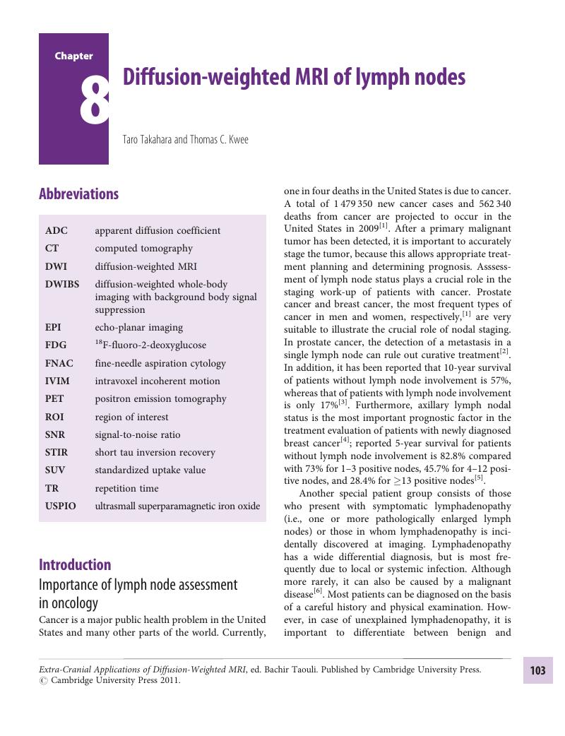
Image of the first page of this content. For PDF version, please use the ‘Save PDF’ preceeding this image.'
Type
Chapter
Information
Publisher: Cambridge University Press
Print publication year: 2010

Access options

Get access to the full version of this content by using one of the access options below. (Log in options will check for institutional or personal access. Content may require purchase if you do not have access.)

References

Jemal, A, Siegel, R, Ward, E, et al. Cancer statistics, 2009. CA Cancer J Clin 2009;59:225–49.CrossRefGoogle ScholarPubMed
Damber, JE, Aus, G. Prostate cancer. Lancet 2008;371:1710–21.CrossRefGoogle ScholarPubMed
Gervasi, , Mata, J, Easley, JD, et al. Prognostic significance of lymph nodal metastases in prostate cancer. J Urol 1989;142:332–6.CrossRefGoogle ScholarPubMed
Banerjee, M, George, J, Song, EY, Roy, A, Hryniuk, W. Tree-based model for breast cancer prognostication. J Clin Oncol 2004;22:2567–75.CrossRefGoogle ScholarPubMed
Fisher, B, Bauer, M, Wickerham, DL, and other NASAAB P investigators. Relation of number of positive axillary nodes to the prognosis of patients with primary breast cancer: an NSABP update. Cancer 1983;52:1551–7.3.0.CO;2-3>CrossRefGoogle Scholar
Ferrer, R. Lymphadenopathy: differential diagnosis and evaluation. Am Fam Physician 1998;58:1313–20.Google ScholarPubMed
Allaf, ME, Partin, AW, Carter, HB. The importance of pelvic lymph node dissection in men with clinically localized prostate cancer. Rev Urol 2006;8:112–19.Google ScholarPubMed
McLaughlin, SA, Wright, MJ, Morris, KT, et al. Prevalence of lymphedema in women with breast cancer 5 years after sentinel lymph node biopsy or axillary dissection: objective measurements. J Clin Oncol 2008;26:5213–19.CrossRefGoogle ScholarPubMed
Del Bianco, P, Zavagno, G, Burelli, P, et al. Morbidity comparison of sentinel lymph node biopsy versus conventional axillary lymph node dissection for breast cancer patients: results of the sentinella-GIVOM Italian randomised clinical trial. Eur J Surg Oncol 2008;34:508–13.CrossRefGoogle ScholarPubMed
Castelijns, JA, Brekel, MW. Imaging of lymphadenopathy in the neck. Eur Radiol 2002;12:727–38.CrossRefGoogle Scholar
Torabi, M, Aquino, SL, Harisinghani, MG. Current concepts in lymph node imaging. J Nucl Med 2004;45:1509–18.Google ScholarPubMed
Glazer, GM, Gross, BH, Quint, , et al. Normal mediastinal lymph nodes: number and size according to American Thoracic Society mapping. Am J Roentgenol 1985;144:261–5.CrossRefGoogle ScholarPubMed
Brekel, MW, Stel, HV, Castelijns, JA, et al. Cervical lymph node metastasis: assessment of radiologic criteria. Radiology 1990;177:379–84.CrossRefGoogle ScholarPubMed
Dorfman, RE, Alpern, MB, Gross, BH, Sandler, MA. Upper abdominal lymph nodes: criteria for normal size determined with CT. Radiology 1991;180:319–22.CrossRefGoogle ScholarPubMed
Schröder, W, Baldus, SE, Mönig, SP, et al. Lymph node staging of esophageal squamous cell carcinoma in patients with and without neoadjuvant radiochemotherapy: histomorphologic analysis. World J Surg 2002;26:584–7.CrossRefGoogle ScholarPubMed
Mönig, SP, Zirbes, TK, Schröder, W, et al. Staging of gastric cancer: correlation of lymph node size and metastatic infiltration. Am J Roentgenol 1999;173:365–7.CrossRefGoogle ScholarPubMed
Mönig, SP, Baldus, SE, Zirbes, TK, et al. Lymph node size and metastatic infiltration in colon cancer. Ann Surg Oncol 1999;6:579–81.CrossRefGoogle Scholar
Prenzel, KL, Mönig, SP, Sinning, JM, et al. Lymph node size and metastatic infiltration in non-small cell lung cancer. Chest 2003;123:463–7.CrossRefGoogle ScholarPubMed
Kyzas, PA, Evangelou, E, Denaxa-Kyza, D, Ioannidis, JP. F-fluorodeoxyglucose positron emission tomography to evaluate cervical node metastases in patients with head and neck squamous cell carcinoma: a meta-analysis. J Natl Cancer Inst 2008;100:712–20.CrossRefGoogle Scholar
Will, O, Purkayastha, S, Chan, C, et al. Diagnostic precision of nanoparticle-enhanced MRI for lymph-node metastases: a meta-analysis. Lancet Oncol 2006;7:52–60.CrossRefGoogle ScholarPubMed
Bondt, RB, Hoeberigs, MC, Nelemans, PJ, et al. Diagnostic accuracy and additional value of diffusion-weighted imaging for discrimination of malignant cervical lymph nodes in head and neck squamous cell carcinoma. Neuroradiology 2009;51:183–92.CrossRefGoogle ScholarPubMed
Padhani, AR, Liu, G, Koh, DM, et al. Diffusion-weighted magnetic resonance imaging as a cancer biomarker: consensus and recommendations. Neoplasia 2009;11:102–25.CrossRefGoogle ScholarPubMed
Glazer, GM, Orringer, MB, Chenevert, TL, et al. Mediastinal lymph nodes: relaxation time/pathologic correlation and implications in staging of lung cancer with MR imaging. Radiology 1988;168:429–31.CrossRefGoogle ScholarPubMed
Ranade, SS, Trivedi, PN, Bamane, VS. Mediastinal lymph nodes: relaxation time/pathologic correlation and implications in staging of lung cancer with MR imaging. Radiology 1990;174:284–5.CrossRefGoogle ScholarPubMed
Ioachim, HL, Medeiros, LJ. Ioachim's Lymph Node Pathology. Philadelphia, PA: Lippincott Williams and Wilkins; 2009.Google Scholar
Koh, DM, Collins, DJ. Diffusion-weighted MRI in the body: applications and challenges in oncology. Am J Roentgenol 2007;188:1622–35.CrossRefGoogle Scholar
Shiehmorteza, M, Sirlin, CB, Wolfson, T, et al. Effect of shot number on the calculated apparent diffusion coefficient in phantoms and in human liver in diffusion-weighted echo-planar imaging. J Magn Reson Imag 2009;30:547–53.CrossRefGoogle ScholarPubMed
Takahara, T, Imai, Y, Yamashita, T, et al. Diffusion weighted whole body imaging with background body signal suppression (DWIBS): technical improvement using free breathing, STIR and high resolution 3D display. Radiat Med 2004;22:275–82.Google ScholarPubMed
Bihan, D, Breton, E, Lallemand, D, et al. MR imaging of intravoxel incoherent motions: application to diffusion and perfusion in neurologic disorders. Radiology 1986;161:401–7.CrossRefGoogle ScholarPubMed
Nakai, G, Matsuki, M, Inada, Y, et al. Detection and evaluation of pelvic lymph nodes in patients with gynecologic malignancies using body diffusion-weighted magnetic resonance imaging. J Comput Assist Tomogr 2008;32:764–8.CrossRefGoogle ScholarPubMed
Weissleder, R, Elizondo, G, Wittenberg, J, et al. Ultrasmall superparamagnetic iron oxide: an intravenous contrast agent for assessing lymph nodes with MR imaging. Radiology 1990;175:494–8.CrossRefGoogle ScholarPubMed
Will, O, Purkayastha, S, Chan, C, et al. Diagnostic precision of nanoparticle-enhanced MRI for lymph-node metastases: a meta-analysis. Lancet Oncol 2006;7:52–60.CrossRefGoogle ScholarPubMed
Antoch, G, Bockisch, A. Combined PET/MRI: a new dimension in whole-body oncology imaging?Eur J Nucl Med Mol Imag 2009;36 (Suppl 1):S113–20.CrossRefGoogle ScholarPubMed
Sumi, M, Sakihama, N, Sumi, T, et al. Discrimination of metastatic cervical lymph nodes with diffusion-weighted MR imaging in patients with head and neck cancer. Am J Neuroradiol 2003;24:1627–34.Google ScholarPubMed
Sumi, M, Cauteren, M, Nakamura, T. MR microimaging of benign and malignant nodes in the neck. Am J Roentgenol 2006;186:749–57.CrossRefGoogle Scholar
Abdel Razek, AA, Soliman, NY, Elkhamary, S, Alsharaway, MK, Tawfik, A. Role of diffusion-weighted MR imaging in cervical lymphadenopathy. Eur Radiol 2006;16:1468–77.CrossRefGoogle ScholarPubMed
King, AD, Ahuja, AT, Yeung, DK, et al. Malignant cervical lymphadenopathy: diagnostic accuracy of diffusion-weighted MR imaging. Radiology 2007;245:806–13.CrossRefGoogle ScholarPubMed
Akduman, EI, Momtahen, AJ, Balci, NC, et al. Comparison between malignant and benign abdominal lymph nodes on diffusion-weighted imaging. Acad Radiol 2008;15:641–6.CrossRefGoogle ScholarPubMed
Holzapfel, K, Duetsch, S, Fauser, C, et al. Value of diffusion-weighted MR imaging in the differentiation between benign and malignant cervical lymph nodes. Eur J Radiol 2009;72:381–7.CrossRefGoogle ScholarPubMed
Sumi, M, Nakamura, T. Diagnostic importance of focal defects in the apparent diffusion coefficient-based differentiation between lymphoma and squamous cell carcinoma nodes in the neck. Eur Radiol 2009;19:975–81.CrossRefGoogle Scholar
Koşucu, P, Tekinbaş, C, Erol, M, et al. Mediastinal lymph nodes: assessment with diffusion-weighted MR imaging. J Magn Reson Imag 2009;30:292–7.CrossRefGoogle ScholarPubMed
Perrone, A, Guerrisi, P, Izzo, L, et al. Diffusion-weighted MRI in cervical lymph nodes: differentiation between benign and malignant lesions. Eur J Radiol 2009; doi: 10.1016/j.ejrad.2009.07.039.CrossRef
Tofts, PS, Lloyd, D, Clark, CA, et al. Test liquids for quantitative MRI measurements of self-diffusion coefficient in vivo. Magn Reson Med 2000;43:368–74.3.0.CO;2-B>CrossRefGoogle ScholarPubMed
Lin, G, Ho, KC, Wang, JJ, et al. Detection of lymph node metastasis in cervical and uterine cancers by diffusion-weighted magnetic resonance imaging at 3 T. J Magn Reson Imag 2008;28:128–35.CrossRefGoogle Scholar
Kim, JK, Kim, KA, Park, BW, Kim, N, Cho, KS. Feasibility of diffusion-weighted imaging in the differentiation of metastatic from nonmetastatic lymph nodes: early experience. J Magn Reson Imag 2008;28:714–19.CrossRefGoogle ScholarPubMed
Park, SO, Kim, JK, Kim, KA, et al. Relative apparent diffusion coefficient: determination of reference site and validation of benefit for detecting metastatic lymph nodes in uterine cervical cancer. J Magn Reson Imag 2009;29:383–90.CrossRefGoogle ScholarPubMed
Vandecaveye, V, Keyzer, F, Vander Poorten, V, et al. Head and neck squamous cell carcinoma: value of diffusion-weighted MR imaging for nodal staging. Radiology 2009;251:134–46.CrossRefGoogle ScholarPubMed
Sakurada, A, Takahara, T, Kwee, TC, et al. Diagnostic performance of diffusion-weighted magnetic resonance imaging in esophageal cancer. Eur Radiol 2009;19:1461–9.CrossRefGoogle ScholarPubMed
Yasui, O, Sato, M, Kamada, A. Diffusion-weighted imaging in the detection of lymph node metastasis in colorectal cancer. Tohoku J Exp Med 2009;218:177–83.CrossRefGoogle ScholarPubMed
Nomori, H, Mori, T, Ikeda, K, et al. Diffusion-weighted magnetic resonance imaging can be used in place of positron emission tomography for N staging of non-small cell lung cancer with fewer false-positive results. J Thorac Cardiovasc Surg 2008;135:816–22.CrossRefGoogle ScholarPubMed
Kwee, TC, Takahara, T, Luijten, PR, Nievelstein, RA. ADC measurements of lymph nodes: inter- and intra-observer reproducibility study and an overview of the literature. Eur J Radiol 2009; doi: 10.1016/j.ejrad.2009.03.026.CrossRef
Brenner, DJ, Hall, EJ. Computed tomography: an increasing source of radiation exposure. N Engl J Med 2007;357:2277–84.CrossRefGoogle ScholarPubMed
Namasivayam, S, Kalra, MK, Torres, WE, Small, WC. Adverse reactions to intravenous iodinated contrast media: a primer for radiologists. Emerg Radiol 2006;12:210–15.CrossRefGoogle ScholarPubMed
Kwee, TC, Quarles van Ufford, HM, Beek, FJ, et al. Whole-body MRI, including diffusion-weighted imaging, for the initial staging of malignant lymphoma: comparison to computed tomography. Investig Radiol 2009;44:683–90.CrossRefGoogle Scholar
Uto, T, Takehara, Y, Nakamura, Y, et al. Higher sensitivity and specificity for diffusion-weighted imaging of malignant lung lesions without apparent diffusion coefficient quantification. Radiology 2009;252:247–54.CrossRefGoogle ScholarPubMed
Thoeny, HC, Triantafyllou, M, Birkhaeuser, FD, et al. Combined ultrasmall superparamagnetic particles of iron oxide-enhanced and diffusion-weighted magnetic resonance imaging reliably detect pelvic lymph node metastases in normal-sized nodes of bladder and prostate cancer patients. Eur Urol 2009;55:761–9.CrossRefGoogle Scholar
Delfaut, EM, Beltran, J, Johnson, G, et al. Fat suppression in MR imaging: techniques and pitfalls. Radiographics 1999;19:373–82.CrossRefGoogle ScholarPubMed
Takahara, T, Hendrikse, J, Yamashita, T, et al. Diffusion-weighted MR neurography of the brachial plexus: feasibility study. Radiology 2008;249:653–60.CrossRefGoogle ScholarPubMed
Beaulieu, C. The basis of anisotropic water diffusion in the nervous system: a technical review. NMR Biomed 2002;15:435–55.CrossRefGoogle ScholarPubMed
Takahara, T, Hendrikse, J, Kwee, TC, et al. Diffusion-weighted MR neurography of the sacral plexus with unidirectional motion probing gradients. Eur Radiol 2009; doi: 10.1007/s00330-009-1665-2.CrossRef

Save book to Kindle

To save this book to your Kindle, first ensure [email protected] is added to your Approved Personal Document E-mail List under your Personal Document Settings on the Manage Your Content and Devices page of your Amazon account. Then enter the ‘name’ part of your Kindle email address below. Find out more about saving to your Kindle.

Note you can select to save to either the @free.kindle.com or @kindle.com variations. ‘@free.kindle.com’ emails are free but can only be saved to your device when it is connected to wi-fi. ‘@kindle.com’ emails can be delivered even when you are not connected to wi-fi, but note that service fees apply.

Find out more about the Kindle Personal Document Service.

Available formats
×

Save book to Dropbox

To save content items to your account, please confirm that you agree to abide by our usage policies. If this is the first time you use this feature, you will be asked to authorise Cambridge Core to connect with your account. Find out more about saving content to Dropbox.

Available formats
×

Save book to Google Drive

To save content items to your account, please confirm that you agree to abide by our usage policies. If this is the first time you use this feature, you will be asked to authorise Cambridge Core to connect with your account. Find out more about saving content to Google Drive.

Available formats
×