Skip to main content Accessibility help
×
Hostname: page-component-745bb68f8f-f46jp Total loading time: 0 Render date: 2025-01-27T12:34:39.336Z Has data issue: false hasContentIssue false

Section 2 - Non-clinical Assessment of Alzheimer’s Disease Candidate Drugs

Published online by Cambridge University Press:  03 March 2022
Jeffrey Cummings
Affiliation:
University of Nevada, Las Vegas
Jefferson Kinney
Affiliation:
University of Nevada, Las Vegas
Howard Fillit
Affiliation:
Alzheimer’s Drug Discovery Foundation
Get access

Summary

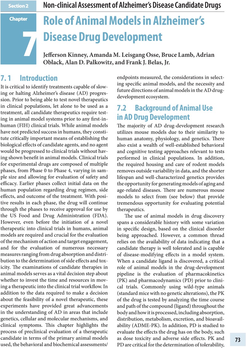
Image of the first page of this content. For PDF version, please use the ‘Save PDF’ preceeding this image.'
Type
Chapter
Information
Alzheimer's Disease Drug Development
Research and Development Ecosystem
, pp. 73 - 134
Publisher: Cambridge University Press
Print publication year: 2022

Access options

Get access to the full version of this content by using one of the access options below. (Log in options will check for institutional or personal access. Content may require purchase if you do not have access.)

References

References

Zhao, M, Lepak, AJ, Andes, DR. Animal models in the pharmacokinetic/pharmacodynamic evaluation of antimicrobial agents. Bioorg Med Chem 2016; 24: 6390–400.CrossRefGoogle ScholarPubMed
Ambrose, PG, Bhavnani, SM, Rubino, CM, et al. Pharmacokinetics–pharmacodynamics of antimicrobial therapy: it’s not just for mice anymore. Clin Infect Dis 2007; 44: 7986.Google Scholar
Fitten, LJ, Flood, JF, Baxter, CF, Tachiki, KH, Perryman, K. Long-term oral administration of memory-enhancing doses of tacrine in mice: a study of potential toxicity and side effects. J. Gerontol 1987; 42: 681–5.Google Scholar
Scearce-Levie, K, Sanchez, PE, Lewcock, JW. Leveraging preclinical models for the development of Alzheimer disease therapeutics. Nat Rev Drug Discov 2020; 19: 447–62.CrossRefGoogle ScholarPubMed
Fan, L, Mao, C, Hu, X, et al. New insights into the pathogenesis of Alzheimer’s disease. Front Neurol 2020; 10:1312.Google Scholar
Kinney, JW, Bemiller, SM, Murtishaw, AS, et al. Inflammation as a central mechanism in Alzheimer’s disease. Alzheimers Dement (N Y) 2018; 4: 575–90.Google ScholarPubMed
Schenk, D, Barbour, N, Dunn, W, et al. Immunization with amyloid-β attenuates Alzheimer-disease-like pathology in the PDAPP mouse. Nature 1999; 400: 173.CrossRefGoogle ScholarPubMed
Orgogozo, J-M, Gilman, S, Dartigues, J-F, et al. Subacute meningoencephalitis in a subset of patients with AD after Abeta42 immunization. Neurology 2003; 61: 4654.Google Scholar
Nicoll, JAR, Wilkinson, D, Holmes, C, et al. Neuropathology of human Alzheimer disease after immunization with amyloid-β peptide: a case report. Nat Med 2003; 9: 448–52.Google Scholar
Fandos, N, Pérez-Grijalba, V, Pesini, P, et al. Plasma amyloid β42/40 ratios as biomarkers for amyloid β cerebral deposition in cognitively normal individuals. Alzheimers Dement (Amst) 2017; 8: 179–87.Google Scholar
Moore, BD, Martin, J, de Mena, L, et al. Short Aβ peptides attenuate Aβ42 toxicity in vivo. J Exp Med 2018; 215: 283301.CrossRefGoogle ScholarPubMed
Salazar, A, Leisgang, A, Ortiz, A, Kinney, J. Dementia insights: what do animal models of Alzheimer’s disease tell us? Pract Neurol 2019;July/August:23–34.Google Scholar
Janelidze, S, Stomrud E, Smith R, et al. Cerebrospinal fluid p-tau217 performs better than p-tau181 as a biomarker of Alzheimer’s disease. Nat Commun 2020; 11: 1683.Google Scholar
Thijssen, EH, La Joie R, Wolf A, et al. Diagnostic value of plasma phosphorylated tau181 in Alzheimer’s disease and frontotemporal lobar degeneration. Nat Med 2020; 26: 387–97.CrossRefGoogle ScholarPubMed
Van Dam, D, De Deyn, PP. Animal models in the drug discovery pipeline for Alzheimer’s disease. Br J Pharmacol 2011; 164: 1285–300.Google Scholar
Cummings, J, Lee, G, Ritter, A, Sabbagh, M, Zhong, K. Alzheimer’s disease drug development pipeline: 2020. Alzheimers Dement (N Y) 2020; 6: e12050.Google Scholar
Sterniczuk, R, Antle, MC, LaFerla, FM, Dyck, RH. Characterization of the 3×Tg-AD mouse model of Alzheimer’s disease: part 2. Behavioral and cognitive changes. Brain Res 2010; 1348: 149–55.Google Scholar
Martinez-Coria, H, Green, KN, Billings, LM, et al. Memantine improves cognition and reduces Alzheimer’s-like neuropathology in transgenic mice. Am J Pathol 2010; 176: 870–80.CrossRefGoogle ScholarPubMed
Fiebich, BL, Batista, CRA, Saliba, SW, Yousif, NM, de Oliveira, ACP . Role of microglia TLRs in neurodegeneration. Front Cell Neurosci 2018; 12 : 329.CrossRefGoogle ScholarPubMed
Zhao, J, Bi, W, Xiao, S, et al. Neuroinflammation induced by lipopolysaccharide causes cognitive impairment in mice. Sci Rep 2019; 9: 5790.Google Scholar
Weintraub, MK, Kranjac, D, Eimerbrink, MJ, et al. Peripheral administration of poly I:C leads to increased hippocampal amyloid-beta and cognitive deficits in a non-transgenic mouse. Behav Brain Res 2014; 266: 183–7.CrossRefGoogle Scholar
Walker, DG, Tang, TM, Lue, L-F. Increased expression of toll-like receptor 3, an anti-viral signaling molecule, and related genes in Alzheimer’s disease brains. Exp Neurol 2018; 309: 91106.CrossRefGoogle ScholarPubMed
Alzheimer’s Association. 2020 Alzheimer’s disease facts and figures. Alzheimers Dement 2020; 16: 391460.Google Scholar
Yeoman, M, Scutt, G, Faragher, R. Insights into CNS ageing from animal models of senescence. Nat Rev Neurosci 2012; 13: 435–45.Google Scholar
Morley, JE, Farr, SA, Kuma, VB, Armbrecht, HJ. The SAMP8 mouse: a model to develop therapeutic interventions for Alzheimer’s disease. Curr Pharm Des 2012; 18: 1123–30.Google Scholar
Carroll, JC, Roasrio, ER, Chang, L, et al. Progesterone and estrogen regulate Alzheimer-like neuropathology in female 3×Tg-AD mice. J Neurosci 2007; 27: 13357–65.Google Scholar
Palm, R, Chang, J, Blair, J, et al. Down-regulation of serum gonadotropins but not estrogen replacement improves cognition in aged-ovariectomized 3×Tg AD female mice. J Neurochem 2014; 130: 115–25.Google Scholar
Rolo, AP, Palmeira, CM. Diabetes and mitochondrial function: role of hyperglycemia and oxidative stress. Toxicol Appl Pharmacol 2006; 212: 167–78.CrossRefGoogle ScholarPubMed
Murtishaw, AS, Hearney CF, Bolton MM, et al. Intermittent streptozotocin administration induces behavioral and pathological features relevant to Alzheimer’s disease and vascular dementia. Neuropharmacology 2018; 137: 164–77.Google Scholar
Candeias, E, Duarte, AI, Carvalho, C, et al. The impairment of insulin signaling in Alzheimer’s disease. IUBMB Life 2012; 64: 951–7.CrossRefGoogle ScholarPubMed
Dineley, KT, Jahrling, JB, Denner, L. Insulin resistance in Alzheimer’s disease. Neurobiol Dis 2014; 72: 92103.CrossRefGoogle ScholarPubMed
Lee, Y, Kim, Y-H, Park, SJ, et al. Insulin/IGF signaling-related gene expression in the brain of a sporadic Alzheimer’s disease monkey model induced by intracerebroventricular injection of streptozotocin. J Alzheimers Dis 2014; 38: 251–67.Google Scholar
Yeo, H-G, Lee, Y, Jeon, CY, et al. Characterization of cerebral damage in a monkey model of Alzheimer’s disease induced by intracerebroventricular injection of streptozotocin. J Alzheimers Dis 2015; 46: 9891005.Google Scholar
Bauzon, J, Lee, G, Cummings, J. Repurposed agents in the Alzheimer’s disease drug development pipeline. Alzheimers Res Ther 2020; 12: 98.Google Scholar
Knight, EM, Martins, VA, Gümüsgöz, S, Allan, SM, Lawrence, CB. High-fat diet-induced memory impairment in triple-transgenic Alzheimer’s disease (3×TgAD) mice is independent of changes in amyloid and tau pathology. Neurobiol Aging 2014; 35: 1821–32.CrossRefGoogle Scholar
Bracko, O, Vinarcsik, LK, Cruz, Hernández JC, et al. High fat diet worsens Alzheimer’s disease-related behavioral abnormalities and neuropathology in APP/PS1 mice, but not by synergistically decreasing cerebral blood flow. Sci Rep 2020; 10: 9884.Google Scholar
Rollins, CPE, Gallion, D, Kong, V, et al. Contributions of a high-fat diet to Alzheimer’s disease-related decline: A longitudinal behavioural and structural neuroimaging study in mouse models. Neuroimage Clin 2019; 21: 101606.CrossRefGoogle ScholarPubMed
Bhat, NR, Thirumangalakudi, L. Increased tau phosphorylation and impaired brain insulin/IGF signaling in mice fed a high fat/high cholesterol diet. J Alzheimers Dis 2013; 36: 781–9.CrossRefGoogle ScholarPubMed
Busquets, O, Ettcheto, M, Pallàs, M, et al. Long-term exposition to a high fat diet favors the appearance of β-amyloid depositions in the brain of C57BL/6 J mice. A potential model of sporadic Alzheimer’s disease. Mech Ageing Dev 2017; 162: 3845.CrossRefGoogle Scholar
Pugazhenthi, S, Qin, L, Reddy, PH. Common neurodegenerative pathways in obesity, diabetes, and Alzheimer’s disease. Biochim Biophys Acta 2017; 1863: 1037–45.Google ScholarPubMed
Prüßing, K, Voig, A, Schulz, JB. Drosophila melanogaster as a model organism for Alzheimer’s disease. Mol Neurodegener 2013; 8: 35.CrossRefGoogle Scholar
Tan, FHP, Azzam, G. Drosophila melanogaster: Deciphering Alzheimer’s disease. Malays J Med Sci 2017; 24: 620.Google ScholarPubMed
Drummond, E, Wisniewski, T. Alzheimer’s disease: experimental models and reality. Acta Neuropathol 2017; 133: 155–75.CrossRefGoogle Scholar
Callahan, PM, Hutchings, EJ, Kille, NJ, Chapman, JM, Terry, AV. Positive allosteric modulator of α7 nicotinic-acetylcholine receptors, PNU-120596 augments the effects of donepezil on learning and memory in aged rodents and non-human primates. Neuropharmacology 2013; 67: 201–12.CrossRefGoogle ScholarPubMed
Vardigan, JD, Cannon, CE, Puri, V, et al. Improved cognition without adverse effects: novel M1 muscarinic potentiator compares favorably to donepezil and xanomeline in rhesus monkey. Psychopharmacology (Berl) 2015; 232: 1859–66.Google Scholar
Link, CD. Invertebrate models of Alzheimer’s disease. Genes Brain Behav 2005; 4: 147–56.CrossRefGoogle ScholarPubMed
Tse, FL, Laplanche, R. Absorption, metabolism, and disposition of [14 C]SDZ ENA 713, an acetylcholinesterase inhibitor, in minipigs following oral, intravenous, and dermal administration. Pharm Res 1998; 15: 1614–20.Google Scholar
Wang, D. Tumor necrosis factor-alpha alters electrophysiological properties of rabbit hippocampal neurons. J Alzheimers Dis 2019; 68: 1257–71.Google Scholar
Brody, DL, Holtzman, DM. Morris water maze search strategy analysis in PDAPP mice before and after experimental traumatic brain injury. Exp Neurol 2006; 197: 330–40.CrossRefGoogle ScholarPubMed
Li, D, Huang, Y, Cheng, B, et al. Streptozotocin induces mild cognitive impairment at appropriate doses in mice as determined by long-term potentiation and the Morris water maze. J Alzheimers Dis 2016; 54: 8998.Google Scholar
Sadowski, M, Pankiewicz, J, Scholtzova, H, et al. Amyloid-β deposition is associated with decreased hippocampal glucose metabolism and spatial memory impairment in APP/PS1 mice. J Neuropathol Exp Neurol 2004; 63: 418–28.CrossRefGoogle ScholarPubMed
Xu, H, Rösler, TW, Carlsson, T, et al. Memory deficits correlate with tau and spine pathology in P301S MAPT transgenic mice. Neuropathol Appl Neurobiol 2014; 40: 833–43.CrossRefGoogle ScholarPubMed
Possin, KL, Kramer, JH, Finkbeine, S, et al. Cross-species translation of the Morris maze for Alzheimer’s disease. J Clin Invest 2016; 126: 779–83.CrossRefGoogle ScholarPubMed
Dodart, J-C, Bales, KR, Gannon, KS, et al. Immunization reverses memory deficits without reducing brain Aβ burden in Alzheimer’s disease model. Nat Neurosci 2002; 5: 452–7.CrossRefGoogle ScholarPubMed
Shen, L, Hang, B, Geng, Y, et al. Amelioration of cognitive impairments in APPswe/PS1dE9 mice is associated with metabolites alteration induced by total salvianolic acid. PLoS One 2017; 12: e0174763.Google Scholar
Barbeau, E, Didic, M, Tramoni, E, et al. Evaluation of visual recognition memory in MCI patients. Neurology 2004; 62: 1317–22.Google Scholar
Didic, M, Felician, O, Barbeau, E, et al. Impaired visual recognition memory predicts Alzheimer’s disease in amnestic mild cognitive impairment. Dement Geriatr Cogn Disord 2013; 35: 291–9.Google Scholar
Zhang, R, Xue, G, Wang, S, et al. Novel object recognition as a facile behavior test for evaluating drug effects in AβPP/PS1 Alzheimer’s disease mouse model. J Alzheimers Dis 2012; 31: 801–12.Google Scholar
Koola, MM. Galantamine–memantine combination in the treatment of Alzheimer’s disease and beyond. Psychiatry Res 2020; 293: 113409.Google Scholar
Wu, Z, Zhao, L, Chen, X, Cheng, X, Zhang, Y. Galantamine attenuates amyloid-β deposition and astrocyte activation in APP/PS1 transgenic mice. Exp Gerontol 2015; 72: 244–50.CrossRefGoogle ScholarPubMed
Puzzo, D, Lee, L, Palmeri, A, Calabrese, G, Arancio, O. Behavioral assays with mouse models of Alzheimer’s disease: practical considerations and guidelines. Biochem Pharmacol 2014; 88: 450–67.CrossRefGoogle ScholarPubMed
Chen, Y, Shi, G-W, Liang, Z-M, et al. Resveratrol improves cognition and decreases amyloid plaque formation in Tg6799 mice. Mol Med Rep 2019; 19: 3783–90.Google Scholar
Guerreiro, R, Wojtas, A, Bras, J, et al. TREM2 variants in Alzheimer’s disease. N Engl J Med 2013; 368: 117–27.Google Scholar
Lannfelt, L, Relkin, NR, Siemers, ER. Amyloid-ß-directed immunotherapy for Alzheimer’s disease. J Intern Med 2014; 275: 284–95.CrossRefGoogle ScholarPubMed
De Strooper, B, Vassar, R, Golde, T. The secretases: enzymes with therapeutic potential in Alzheimer disease. Nat Rev Neurol 2010; 6: 99107.CrossRefGoogle ScholarPubMed
Cummings, JL, Morstorf, T, Zhong, K. Alzheimer’s disease drug-development pipeline: few candidates, frequent failures. Alzheimers Res Ther 2014; 6: 37.Google Scholar
Bales, KR. The value and limitations of transgenic mouse models used in drug discovery for Alzheimer’s disease: an update. Expert Opin Drug Discov 2012; 7: 281–97.CrossRefGoogle ScholarPubMed
Shineman, DW, Basi, GS, Bizon, JL, et al. Accelerating drug discovery for Alzheimer’s disease: best practices for preclinical animal studies. Alzheimers Res Ther 2011; 3: 28.CrossRefGoogle ScholarPubMed
Preuss, C, Pandey, R, Piazza, E, et al. A novel systems biology approach to evaluate mouse models of late-onset Alzheimer’s disease. Mol Neurodegener 2020; 15: 67.Google Scholar
Wan, Y-W, Al-Ouran, R, Mangleburg, CG, et al. Meta-analysis of the Alzheimer’s disease human brain transcriptome and functional dissection in mouse models. Cell Reports 2020; 32: 107908.CrossRefGoogle ScholarPubMed
Hayden, KM, Jones, RN, Zimmer, C, et al. Factor structure of the National Alzheimer’s Coordinating Centers uniform dataset neuropsychological battery: an evaluation of invariance between and within groups over time. Alzheimer Dis Assoc Disord 2011; 25: 128–37.Google Scholar
Hansen, DV, Hanson, JE, Sheng, M. Microglia in Alzheimer’s disease. J Cell Biol 2018; 217: 459–72.CrossRefGoogle ScholarPubMed

References

Tanzi, RE, Bertram, L. Twenty years of the Alzheimer’s disease amyloid hypothesis: a genetic perspective. Cell 2005; 120: 545–55.Google Scholar
Coric, V, van Dyck, CH, Salloway, S, et al. Safety and tolerability of the gamma-secretase inhibitor avagacestat in a phase 2 study of mild to moderate Alzheimer disease. Arch Neurol 2012; 69: 1430–40.CrossRefGoogle Scholar
Fleisher, AS, Raman, R, Siemers, ER, et al. Phase 2 safety trial targeting amyloid beta production with a gamma-secretase inhibitor in Alzheimer disease. Arch Neurol 2008; 65: 1031–8.Google Scholar
Miles, LA, Crespi, GA, Doughty, L, Parker, MW. Bapineuzumab captures the N-terminus of the Alzheimer’s disease amyloid-beta peptide in a helical conformation. Sci Rep 2013; 3: 1302.CrossRefGoogle ScholarPubMed
Salloway, S, Sperling, R, Fox, NC, et al. Two phase 3 trials of bapineuzumab in mild-to-moderate Alzheimer’s disease. N Engl J Med 2014; 370: 322–33.Google Scholar
Sevigny, J, Chiao, P, Bussière, T, et al. The antibody aducanumab reduces Aβ plaques in Alzheimer’s disease. Nature 2016; 537: 50–6.Google Scholar
Iwatsubo, T, Odaka, A, Suzuki, N, et al. Visualization of A beta 42(43)and A beta 40 in senile plaques with end-specific A beta monoclonals: evidence that an initially deposited species is A beta 42(43). Neuron 1994; 13: 4553.Google Scholar
Kumar-Singh, S, Theuns, J, Van Broeck, B, et al. Mean age-of-onset of familial alzheimer disease caused by presenilin mutations correlates with both increased Abeta42 and decreased Abeta40. Hum Mutat 2006; 27: 686–95.Google Scholar
Alzheimer’s Association Expert Advisory Workgroup on NAPA. Workgroup on NAPA’s scientific agenda for a national initiative on Alzheimer’s disease. Alzheimers Dement 2012; 8: 357–71.Google Scholar
Shi, Y, Inoue, H, Wu, JC, Yamanaka, S. Induced pluripotent stem cell technology: a decade of progress. Nat Rev Drug Discov 2017; 16: 115–30.Google Scholar
Khurana, V, Tardiff, DF, Chung, CY, Lindquist, S. Toward stem cell-based phenotypic screens for neurodegenerative diseases. Nat Rev Neurol 2015; 11: 339–50.Google Scholar
Yagi, T, Ito, D, Nihei, Y, Ishihara, T, Suzuki, N. N88S seipin mutant transgenic mice develop features of seipinopathy/BSCL2-related motor neuron disease via endoplasmic reticulum stress. Hum Mol Genet 2011; 20: 3831–40.Google Scholar
Liu, Q, Waltz, S, Woodruff, G, et al. Effect of potent gamma-secretase modulator in human neurons derived from multiple presenilin 1-induced pluripotent stem cell mutant carriers. JAMA Neurol 2014; 71: 1481–9.Google Scholar
Kounnas, MZ, Danks, AM, Cheng, S, et al. Modulation of gamma-secretase reduces beta-amyloid deposition in a transgenic mouse model of Alzheimer’s disease. Neuron 2010; 67: 769–80.Google Scholar
van der Kant, R, Langness, VF, Herrera, CM, et al. Cholesterol metabolism is a druggable axis that independently regulates tau and amyloid-β in iPSC-derived Alzheimer’s disease neurons. Cell Stem Cell 2019; 24: 363–75.e9.Google Scholar
Israel, MA, Yuan, SH, Bardy, C, et al. Probing sporadic and familial Alzheimer’s disease using induced pluripotent stem cells. Nature 2012; 482: 216–20.Google Scholar
Kondo, T, Imamura, K, Funayama, M, et al. iPSC-based compound screening and in vitro trials identify a synergistic anti-amyloid β combination for Alzheimer’s disease. Cell Rep 2017; 21: 2304–12.CrossRefGoogle ScholarPubMed
Shi, Y, Kirwan, P, Smith, J, et al. A human stem cell model of early Alzheimer’s disease pathology in Down syndrome. Sci Transl Med 2012; 4: 124ra29.Google Scholar
Brownjohn, PW, Smith, J, Portelius, E, et al. Phenotypic screening identifies modulators of amyloid precursor protein processing in human stem cell models of Alzheimer’s disease. Stem Cell Rep 2017; 8: 870–82.Google Scholar
Kukar, TL, Ladd, TB, Bann, MA, et al. Substrate-targeting γ-secretase modulators. Nature 2008; 453: 925–9.Google Scholar
Weggen, S, Eriksen, JL, Das, P, et al. A subset of NSAIDs lower amyloidogenic Abeta42 independently of cyclooxygenase activity. Nature 2001; 414: 212–16.Google Scholar
Crump, CJ, Johnson, DS, Li, Y-M. Development and mechanism of γ-secretase modulators for Alzheimer’s disease. Biochemistry 2013; 52: 3197–216.Google Scholar
Wagner, SL, Rynearson, KD, Duddy, SK, et al. Pharmacological and toxicological properties of the potent oral gamma-secretase modulator BPN-15606. J Pharmacol Exp Ther 2017; 362: 3144.Google Scholar
Wagner, SL, Zhang, C, Cheng, S, et al. Soluble gamma-secretase modulators selectively inhibit the production of the 42-amino acid amyloid beta peptide variant and augment the production of multiple carboxy-truncated amyloid beta species. Biochemistry 2014; 53: 702–13.CrossRefGoogle ScholarPubMed
Rynearson, KD, Buckle, RN, Herr, RJ, et al. Design and synthesis of novel methoxypyridine-derived gamma-secretase modulators. Bioorg Med Chem 2020; 28: 115734.CrossRefGoogle ScholarPubMed
Rynearson, KD, Buckle, RN, Barnes, KD, et al. Design and synthesis of aminothiazole modulators of the gamma-secretase enzyme. Bioorg Med Chem Lett 2016; 26: 3928–37.CrossRefGoogle ScholarPubMed
Sunderland, T, Linker, G, Mirza, N, et al. Decreased beta-amyloid1–42 and increased tau levels in cerebrospinal fluid of patients with Alzheimer disease. JAMA 2003; 289: 2094–103.Google Scholar
Hardy, JA, Higgins, GA. Alzheimer’s disease: the amyloid cascade hypothesis. Science 1992; 256: 184–5.Google Scholar
Gilman, S, Koller, M, Black, RS, et al. Clinical effects of Abeta immunization (AN1792) in patients with AD in an interrupted trial. Neurology 2005; 64: 1553–62.CrossRefGoogle Scholar
Wagner, SL, Tanzi, RE, Mobley, WC, Galasko, D. Potential use of gamma-secretase modulators in the treatment of Alzheimer disease. Arch Neurol 2012; 69: 1255–8.Google Scholar
Potter, R, Patterson, BW, Elbert, DL, et al. Increased in vivo amyloid-beta42 production, exchange, and loss in presenilin mutation carriers. Sci Transl Med 2013; 5: 189ra77.CrossRefGoogle ScholarPubMed
Kretner, B, Fukumori, A, Gutsmiedl, A, et al. Attenuated Aβ42 responses to low potency γ-secretase modulators can be overcome for many pathogenic presenilin mutants by second-generation compounds. J Biol Chem 2011; 286: 15240–51.Google Scholar
Koch, P, Tamboli, IY, Mertens, J, et al. Presenilin-1 L166P mutant human pluripotent stem cell-derived neurons exhibit partial loss of γ-secretase activity in endogenous amyloid-β generation. Am J Pathol 2012; 180: 2404–16.Google Scholar
Yuan, SH, Martin, J, Elia, J, et al. Cell-surface marker signatures for the isolation of neural stem cells, glia and neurons derived from human pluripotent stem cells. PLoS One 2011; 6: e17540.Google Scholar
Rynearson, KD, Ponnusamym, M, Prikhodko, O, et al. Preclinical validation of a potent γ-secretase modulator for Alzheimer’s disease prevention. J Exp Med 2021; 218: e20202560.Google Scholar
Caldwell, AB, Liu, Q, Schroth, GP, et al. Dedifferentiation and neuronal repression define familial Alzheimer’s disease.Sci Adv 2020; 6: eaba5933.Google Scholar
Antonell, A, Lladó, A, Altirriba, J, et al. A preliminary study of the whole-genome expression profile of sporadic and monogenic early-onset Alzheimer’s disease. Neurobiol Aging 2013; 34: 1772–8.Google Scholar

References

Polanco, JC, Li, C, Bodea, LG, et al. Amyloid-beta and tau complexity: towards improved biomarkers and targeted therapies. Nat Rev Neurol 2018; 14: 2239.Google Scholar
Gotz, J, Bodea, LG, Goedert, M. Rodent models for Alzheimer disease. Nat Rev Neurosci 2018; 19: 583–98.Google Scholar
Grieb, P. Intracerebroventricular streptozotocin injections as a model of Alzheimer’s disease: in search of a relevant mechanism. Mol Neurobiol 2016; 53: 1741–52.Google Scholar
Kamat, PK, Rai, S, Nath, C. Okadaic acid induced neurotoxicity: an emerging tool to study Alzheimer’s disease pathology. Neurotoxicology 2013; 37: 163–72.Google Scholar
Kim, HY, Lee, DK, Chung, BR, Kim, HV, Kim, Y. Intracerebroventricular injection of amyloid-beta peptides in normal mice to acutely induce Alzheimer-like cognitive deficits. J Vis Exp 2016; 109: 53308.Google Scholar
de Calignon, A, Polydoro, M, Suarez-Calvet, M, et al. Propagation of tau pathology in a model of early Alzheimer’s disease. Neuron 2012; 73: 685–97.Google Scholar
Braak, H, Braak, E. Neuropathological stageing of Alzheimer-related changes. Acta Neuropathol 1991; 82: 239–59.Google Scholar
Hardy, J, Selkoe, DJ. The amyloid hypothesis of Alzheimer’s disease: progress and problems on the road to therapeutics. Science 2002; 297: 353–6.Google Scholar
Leon, WC, Canneva, F, Partridge, V, et al. A novel transgenic rat model with a full Alzheimer’s-like amyloid pathology displays pre-plaque intracellular amyloid-beta-associated cognitive impairment. J Alzheimers Dis 2010; 20: 113–26.Google Scholar
Cohen, RM, Rezai-Zadeh, K, Weitz, TM, et al. A transgenic Alzheimer rat with plaques, tau pathology, behavioral impairment, oligomeric Abeta, and frank neuronal loss. J Neurosci 2013; 33: 6245–56.CrossRefGoogle ScholarPubMed
Malcolm, JC, Breuillaud, L, Do Carmo, S, et al. Neuropathological changes and cognitive deficits in rats transgenic for human mutant tau recapitulate human tauopathy. Neurobiol Dis 2019; 127: 323–38.Google Scholar
Do Carmo, S, Cuello, AC. Modeling Alzheimer’s disease in transgenic rats. Mol Neurodegener 2013; 8: 37.Google Scholar
Zimmer, ER, Parent, MJ, Cuello, AC, Gauthier, S, Rosa-Neto, P. MicroPET imaging and transgenic models: a blueprint for Alzheimer’s disease clinical research. Trends Neurosci 2014; 37: 629–41.Google Scholar
Kuang, E, Wan, Q, Li, X, et al. ER stress triggers apoptosis induced by Nogo-B/ASY overexpression. Exp Cell Res 2006; 312: 1983–8.Google Scholar
Saito, T, Matsuba, Y, Mihira, N, et al. Single APP knock-in mouse models of Alzheimer’s disease. Nat Neurosci 2014; 17: 661–3.Google Scholar
Bard, F, Cannon, C, Barbour, R, et al. Peripherally administered antibodies against amyloid beta-peptide enter the central nervous system and reduce pathology in a mouse model of Alzheimer disease. Nat Med 2000; 6: 916–19.Google Scholar
DeMattos, RB, Bales, KR, Cummins, DJ, et al. Peripheral anti-A beta antibody alters CNS and plasma A beta clearance and decreases brain Abeta burden in a mouse model of Alzheimer’s disease. Proc Natl Acad Sci USA 2001; 98: 8850–5.Google Scholar
Bohrmann, B, Baumann, K, Benz, J, et al. Gantenerumab: a novel human anti-A beta antibody demonstrates sustained cerebral amyloid-beta binding and elicits cell-mediated removal of human amyloid-beta. J Alzheimers Dis 2012; 28: 4969.Google Scholar
Lord, A, Gumucio, A, Englund, H, et al. An amyloid-beta protofibril-selective antibody prevents amyloid formation in a mouse model of Alzheimer’s disease. Neurobiol Dis 2009; 36: 425–34.Google Scholar
Sevigny, J, Chiao, P, Bussiere, T, et al. The antibody aducanumab reduces Abeta plaques in Alzheimer’s disease. Nature 2016; 537: 50–6.Google Scholar
Cummings, JL, Morstorf, T, Zhong, K. Alzheimer’s disease drug-development pipeline: few candidates, frequent failures. Alzheimers Res Ther 2014; 6: 37.Google Scholar
Jack, CR Jr., Bennett, DA, Blennow, K, et al. NIA-AA Research Framework: toward a biological definition of Alzheimer’s disease. Alzheimers Dement 2018; 14: 535–62.CrossRefGoogle Scholar
Alzheimer’s Association. 2019 Alzheimer’s disease facts and figures. Alzheimers Dement 2019; 15: 321–87.Google Scholar
Amadoru, S, Dore, V, McLean, CA, et al. Comparison of amyloid PET measured in centiloid units with neuropathological findings in Alzheimer’s disease. Alzheimers Res Ther 2020; 12: 22.Google Scholar
Maeda, J, Ji, B, Irie, T, et al. Longitudinal, quantitative assessment of amyloid, neuroinflammation, and anti-amyloid treatment in a living mouse model of Alzheimer’s disease enabled by positron emission tomography. J Neurosci 2007; 27: 10957–68.Google Scholar
Maeda, J, Zhang, MR, Okauchi, T, et al. In vivo positron emission tomographic imaging of glial responses to amyloid-beta and tau pathologies in mouse models of Alzheimer’s disease and related disorders. J Neurosci 2011; 31: 4720–30.Google Scholar
Snellman, A, Lopez-Picon, FR, Rokka, J, et al. Longitudinal amyloid imaging in mouse brain with 11C-PIB: comparison of APP23, Tg2576, and APPswe-PS1dE9 mouse models of Alzheimer disease. J Nucl Med 2013; 54: 1434–41.Google Scholar
Snellman, A, Rokka, J, Lopez-Picon, FR, et al. Applicability of [(11)C]PiB micro-PET imaging for in vivo follow-up of anti-amyloid treatment effects in APP23 mouse model. Neurobiol Aging 2017; 57: 8494.Google Scholar
Snellman, A, Rokka, J, Lopez-Picon, FR, et al. In vivo PET imaging of beta-amyloid deposition in mouse models of Alzheimer’s disease with a high specific activity PET imaging agent [(18)F]flutemetamol. EJNMMI Res 2014; 4: 37.Google Scholar
Toyama, H, Ye, D, Ichise, M, et al. PET imaging of brain with the beta-amyloid probe, [11C]6-OH-BTA-1, in a transgenic mouse model of Alzheimer’s disease. Eur J Nucl Med Mol Imaging 2005; 32: 593600.Google Scholar
Kuntner, C, Kesner, AL, Bauer, M, et al. Limitations of small animal PET imaging with [18F]FDDNP and FDG for quantitative studies in a transgenic mouse model of Alzheimer’s disease. Mol Imaging Biol 2009; 11: 236–40.Google Scholar
Poisnel, G, Dhilly, M, Moustie, O, et al. PET imaging with [18 F]AV-45 in an APP/PS1-21 murine model of amyloid plaque deposition. Neurobiol Aging 2012; 33: 2561–71.CrossRefGoogle Scholar
Brendel, M, Jaworska, A, Griessinger, E, et al. Cross-sectional comparison of small animal [18F]-florbetaben amyloid-PET between transgenic AD mouse models. PLoS One 2015; 10: e0116678.Google Scholar
Brendel, M, Jaworska, A, Overhoff, F, et al. Efficacy of chronic BACE1 inhibition in PS2APP mice depends on the regional Abeta deposition rate and plaque burden at treatment initiation. Theranostics 2018; 8: 4957–68.Google Scholar
Parent, MJ, Zimmer, ER, Shin, M, et al. Multimodal imaging in rat model recapitulates Alzheimer’s disease biomarkers abnormalities. J Neurosci 2017; 37: 12263–71.Google Scholar
Zimmer, ER, Leuzy, A, Gauthier, S, Rosa-Neto, P. Developments in tau PET imaging. Can J Neurol Sci 2014; 41: 547–53.Google Scholar
Leuzy, A, Chiotis, K, Lemoine, L, et al. Tau PET imaging in neurodegenerative tauopathies: still a challenge. Mol Psychiatry 2019; 24: 1112–34.Google Scholar
Fodero-Tavoletti, MT, Okamura, N, Furumoto, S, et al. 18F-THK523: a novel in vivo tau imaging ligand for Alzheimer’s disease. Brain 2011; 134: 1089–100.Google Scholar
Maruyama, M, Shimada, H, Suhara, T, et al. Imaging of tau pathology in a tauopathy mouse model and in Alzheimer patients compared to normal controls. Neuron 2013; 79: 1094–108.Google Scholar
Ishikawa, A, Tokunaga, M, Maeda, J, et al. In vivo visualization of tau accumulation, microglial activation, and brain atrophy in a mouse model of tauopathy rTg4510. J Alzheimers Dis 2018; 61: 1037–52.Google Scholar
Ni, R, Ji, B, Ono, M, et al. Comparative in vitro and in vivo quantifications of pathologic tau deposits and their association with neurodegeneration in tauopathy mouse models. J Nucl Med 2018; 59: 960–6.Google Scholar
Brendel, M, Yousefi, BH, Blume, T, et al. Comparison of (18)F-T807 and (18)F-THK5117 PET in a mouse model of tau pathology. Front Aging Neurosci 2018; 10: 174.Google Scholar
Hattori, N, Huang, SC, Wu, HM, et al. Acute changes in regional cerebral (18)F-FDG kinetics in patients with traumatic brain injury. J Nucl Med 2004; 45: 775–83.Google Scholar
Zimmer, ER, Parent, MJ, Souza, DG, et al. [(18)F]FDG PET signal is driven by astroglial glutamate transport. Nat Neurosci 2017; 20: 393–5.Google Scholar
Stoessl, AJ. Glucose utilization: still in the synapse. Nat Neurosci 2017; 20: 382–4.Google Scholar
Silverman, DH. Brain 18F-FDG PET in the diagnosis of neurodegenerative dementias: comparison with perfusion SPECT and with clinical evaluations lacking nuclear imaging. J Nucl Med 2004; 45: 594607.Google Scholar
Bohnen, NI, Djang, DS, Herholz, K, Anzai, Y, Minoshima, S. Effectiveness and safety of 18F-FDG PET in the evaluation of dementia: a review of the recent literature. J Nucl Med 2012; 53: 5971.Google Scholar
Mosconi, L, Tsui, WH, Herholz, K, et al. Multicenter standardized 18F-FDG PET diagnosis of mild cognitive impairment, Alzheimer’s disease, and other dementias. J Nucl Med 2008; 49: 390–8.Google Scholar
Chételat, G, Arbizu, J, Barthel, H, et al. Amyloid-PET and 18F-FDG-PET in the diagnostic investigation of Alzheimer’s disease and other dementias. Lancet Neurol 2020; 19: 951–62.Google Scholar
Luo, F, Rustay, NR, Ebert, U, et al. Characterization of 7- and 19-month-old Tg2576 mice using multimodal in vivo imaging: limitations as a translatable model of Alzheimer’s disease. Neurobiol Aging 2012; 33: 933–44.Google Scholar
Martin-Moreno, AM, Brera, B, Spuch, C, et al. Prolonged oral cannabinoid administration prevents neuroinflammation, lowers beta-amyloid levels and improves cognitive performance in Tg APP 2576 mice. J Neuroinflamm 2012; 9: 8.Google Scholar
Heneka, MT, Ramanathan, M, Jacobs, AH, et al. Locus ceruleus degeneration promotes Alzheimer pathogenesis in amyloid precursor protein 23 transgenic mice. J Neurosci 2006; 26: 1343–54.Google Scholar
Poisnel, G, Herard, AS, El Tannir El Tayara, N, et al. Increased regional cerebral glucose uptake in an APP/PS1 model of Alzheimer’s disease.Neurobiol Aging 2012; 33: 19952005.Google Scholar
Lu, XY, Huang, S, Chen, QB, et al. Metformin ameliorates Abeta pathology by insulin-degrading enzyme in a transgenic mouse model of Alzheimer’s disease. Oxid Med Cell Longev 2020; 2020: 2315106.Google Scholar
Sung, KK, Jang, DP, Lee, S, et al. Neural responses in rat brain during acute immobilization stress: a [F-18]FDG micro PET imaging study. Neuroimage 2009; 44: 1074–80.CrossRefGoogle Scholar
Toyama, H, Ichise, M, Liow, J-S, et al. Evaluation of anesthesia effects on [18F]FDG uptake in mouse brain and heart using small animal PET. Nucl Med Biol 2004; 31: 251–6.Google Scholar
Ottoy, J, Verhaeghe, J, Niemantsverdriet, E, et al. Validation of the semiquantitative static SUVR method for (18)F-AV45 PET by pharmacokinetic modeling with an arterial input function. J Nucl Med 2017; 58: 1483–9.Google Scholar
Price, JC, Klunk, WE, Lopresti, BJ, et al. Kinetic modeling of amyloid binding in humans using PET imaging and Pittsburgh Compound-B. J Cereb Blood Flow Metab 2005; 25: 1528–47.Google Scholar
Buckner, RL, Snyder, AZ, Shannon, BJ, et al. Molecular, structural, and functional characterization of Alzheimer’s disease: evidence for a relationship between default activity, amyloid, and memory. J Neurosci 2005; 25: 7709–17.Google Scholar
Lu, H, Zou, Q, Gu, H, et al. Rat brains also have a default mode network. Proc Natl Acad Sci USA 2012; 109: 3979–84.Google Scholar
Jack, CR Jr., Wiste, HJ, Vemuri, P, et al. Brain beta-amyloid measures and magnetic resonance imaging atrophy both predict time-to-progression from mild cognitive impairment to Alzheimer’s disease. Brain 2010; 133: 3336–48.Google Scholar
Terry, RD, Masliah, E, Salmon, DP, et al. Physical basis of cognitive alterations in Alzheimer’s disease: synapse loss is the major correlate of cognitive impairment. Ann Neurol 1991; 30: 572–80.Google Scholar
Jagust, W. Imaging the evolution and pathophysiology of Alzheimer disease. Nat Rev Neurosci 2018; 19: 687700.Google Scholar
Ottoy, J, Niemantsverdriet, E, Verhaeghe, J, et al. Association of short-term cognitive decline and MCI-to-AD dementia conversion with CSF, MRI, amyloid- and (18)F-FDG-PET imaging. Neuroimage Clin 2019; 22: 101771.Google Scholar
Heggland, I, Storkaas, IS, Soligard, HT, Kobro-Flatmoen, A, Witter, MP. Stereological estimation of neuron number and plaque load in the hippocampal region of a transgenic rat model of Alzheimer’s disease. Eur J Neurosci 2015; 41: 1245–62.Google Scholar
Macdonald, IR, DeBay, DR, Reid, GA, et al. Early detection of cerebral glucose uptake changes in the 5×FAD mouse. Curr Alzheimer Res 2014; 11: 450–60.Google Scholar
Yoshiyama, Y, Higuchi, M, Zhang, B, et al. Synapse loss and microglial activation precede tangles in a P301S tauopathy mouse model. Neuron 2007; 53: 337–51.Google Scholar
Chiquita, S, Ribeiro, M, Castelhano, J, et al. A longitudinal multimodal in vivo molecular imaging study of the 3×Tg-AD mouse model shows progressive early hippocampal and taurine loss. Hum Mol Genet 2019; 28: 2174–88.Google Scholar
Kang, MS, Aliaga, AA, Shin, M, et al. Amyloid-beta modulates the association between neurofilament light chain and brain atrophy in Alzheimer’s disease. Mol Psychiatry 2020;https://doi.org/10.1038/s41380-020-0818-1.Google Scholar
Vincent, JL, Patel, GH, Fox, MD, et al. Intrinsic functional architecture in the anaesthetized monkey brain. Nature 2007; 447: 83–6.CrossRefGoogle ScholarPubMed
Rilling, JK, Barks, SK, Parr, LA, et al. A comparison of resting-state brain activity in humans and chimpanzees. Proc Natl Acad Sci USA 2007; 104: 17146–51.Google Scholar
Greicius, MD, Srivastava, G, Reiss, AL, Menon, V. Default-mode network activity distinguishes Alzheimer’s disease from healthy aging: evidence from functional MRI. Proc Natl Acad Sci USA 2004; 101: 4637–42.Google Scholar
Buckner, RL, Sepulcre, J, Talukdar, T, et al. Cortical hubs revealed by intrinsic functional connectivity: mapping, assessment of stability, and relation to Alzheimer’s disease. J Neurosci 2009; 29: 1860–73.Google Scholar
Sheline, YI, Raichle, ME. Resting state functional connectivity in preclinical Alzheimer’s disease. Biol Psychiatry 2013; 74: 340–7.Google Scholar
Wang, L, Zang, Y, He, Y, et al. Changes in hippocampal connectivity in the early stages of Alzheimer’s disease: evidence from resting state fMRI. Neuroimage 2006; 31: 496504.Google Scholar
Huijbers, W, Mormino, EC, Schultz, AP, et al. Amyloid-beta deposition in mild cognitive impairment is associated with increased hippocampal activity, atrophy and clinical progression. Brain 2015; 138: 1023–35.Google Scholar
Salloway, S, Sperling, R, Gilman, S, et al. A Phase 2 multiple ascending dose trial of bapineuzumab in mild to moderate Alzheimer disease. Neurology 2009; 73: 2061–70.Google Scholar
Sperling, RA, Jack, CR Jr., Black, SE, et al. Amyloid-related imaging abnormalities in amyloid-modifying therapeutic trials: recommendations from the Alzheimer’s Association Research Roundtable Workgroup. Alzheimers Dement 2011; 7: 367–85.Google Scholar
Luo, F, Rustay, NR, Seifert, T, et al. Magnetic resonance imaging detection and time course of cerebral microhemorrhages during passive immunotherapy in living amyloid precursor protein transgenic mice. J Pharmacol Exp Ther 2010; 335: 580–8.Google Scholar
Constantinides, C, Murphy, K. Molecular and integrative physiological effects of isoflurane anesthesia: the paradigm of cardiovascular studies in rodents using magnetic resonance imaging. Front Cardiovasc Med 2016; 3: 23.Google Scholar
Stenroos, P, Paasonen, J, Salo, RA, et al. Awake rat brain functional magnetic resonance imaging using standard radio frequency coils and a 3D printed restraint kit. Front Neurosci 2018; 12: 548.Google Scholar
Paasonen, J, Stenroos, P, Salo, RA, Kiviniemi, V, Grohn, O. Functional connectivity under six anesthesia protocols and the awake condition in rat brain. Neuroimage 2018; 172: 920.Google Scholar
Wu, TL, Mishra, A, Wang, F, et al. Effects of isoflurane anesthesia on resting-state fMRI signals and functional connectivity within primary somatosensory cortex of monkeys. Brain Behav 2016; 6: e00591.Google Scholar
Ferris, CF, Smerkers, B, Kulkarni, P, et al. Functional magnetic resonance imaging in awake animals. Rev Neurosci 2011; 22: 665–74.Google Scholar
Dopfel, D, Zhang, N. Mapping stress networks using functional magnetic resonance imaging in awake animals. Neurobiol Stress 2018; 9: 251–63.Google Scholar
Low, LA, Bauer, LC, Pitcher, MH, Bushnell, MC. Restraint training for awake functional brain scanning of rodents can cause long-lasting changes in pain and stress responses. Pain 2016; 157: 1761–72.Google Scholar
Mizuma, H, Shukuri, M, Hayashi, T, Watanabe, Y, Onoe, H. Establishment of in vivo brain imaging method in conscious mice. J Nucl Med 2010; 51: 1068–75.CrossRefGoogle ScholarPubMed
Alstrup, AK, Smith, DF. Anaesthesia for positron emission tomography scanning of animal brains. Lab Anim 2013; 47: 12–18.CrossRefGoogle ScholarPubMed
Momosaki, S, Hatano, K, Kawasumi, Y, et al. Rat-PET study without anesthesia: anesthetics modify the dopamine D1 receptor binding in rat brain. Synapse 2004; 54: 207–13.Google Scholar
Kyme, AZ, Zhou, VW, Meikle, SR, Fulton, RR. Real-time 3D motion tracking for small animal brain PET. Phys Med Biol 2008; 53: 2651–66.CrossRefGoogle ScholarPubMed
Schulz, D, Southekal, S, Junnarkar, SS, et al. Simultaneous assessment of rodent behavior and neurochemistry using a miniature positron emission tomograph. Nat Methods 2011; 8: 347–52.CrossRefGoogle ScholarPubMed
Miranda, A, Kang, MS, Blinder, S, et al. PET imaging of freely moving interacting rats. Neuroimage 2019; 191: 560–7.Google Scholar
Miranda, A, Staelens, S, Stroobants, S, Verhaeghe, J. Fast and accurate rat head motion tracking with point sources for awake brain PET. IEEE Trans Med Imaging 2017; 36: 1573–82.Google Scholar
Nakamura, A, Kaneko, N, Villemagne, VL, et al. High performance plasma amyloid-beta biomarkers for Alzheimer’s disease. Nature 2018; 554: 249–54.Google Scholar
Janelidze, S, Mattsson, N, Palmqvist, S, et al. Plasma p-tau181 in Alzheimer’s disease: relationship to other biomarkers, differential diagnosis, neuropathology and longitudinal progression to Alzheimer’s dementia. Nat Med 2020; 26: 379–86.Google Scholar
Niemantsverdriet, E, Ottoy, J, Somers, C, et al. The cerebrospinal fluid Abeta1-42/Abeta1-40 ratio improves concordance with amyloid-PET for diagnosing Alzheimer’s disease in a clinical setting. J Alzheimers Dis 2017; 60: 561–76.Google Scholar
Hansson, O, Zetterberg, H, Buchhave, P, et al. Prediction of Alzheimer’s disease using the CSF Abeta42/Abeta40 ratio in patients with mild cognitive impairment. Dement Geriatr Cogn Disord 2007; 23: 316–20.Google Scholar
Barten, DM, Cadelina, GW, Hoque, N, et al. Tau transgenic mice as models for cerebrospinal fluid tau biomarkers. J Alzheimers Dis 2011; 24: 127–41.Google Scholar
Acker, CM, Forest, SK, Zinkowski, R, Davies, P, d’Abramo, C. Sensitive quantitative assays for tau and phospho-tau in transgenic mouse models. Neurobiol Aging 2013; 34: 338–50.Google Scholar
Preische, O, Schultz, SA, Apel, A, et al. Serum neurofilament dynamics predicts neurodegeneration and clinical progression in presymptomatic Alzheimer’s disease. Nat Med 2019; 25: 277–83.Google Scholar
Bacioglu, M, Maia, LF, Preische, O, et al. Neurofilament light chain in blood and CSF as marker of disease progression in mouse models and in neurodegenerative diseases. Neuron 2016; 91: 5666.Google Scholar
Kang, MS, Shin, M, Ottoy, J, et al. Preclinical in vivo longitudinal assessment of KG207-M as a disease-modifying Alzheimer’s disease therapeutic. J Cereb Blood Flow Metab August 2021; doi: 10.1177/0271678X211035625.Google Scholar
Dirnagl, U, Przesdzing, I, Kurreck, C, Major, S. A laboratory critical incident and error reporting system for experimental biomedicine. PLoS Biol 2016; 14: e2000705.Google Scholar

References

Hallmans, G, Vaught, JB. Best practices for establishing a biobank. In Methods in Biobanking, Dillner, J (ed.). New York: Humana Press; 2011: 241–60.Google Scholar
Betsou, F, Lehmann, S, Ashton, G, et al. Standard preanalytical coding for biospecimens: defining the sample preanalytical code. Cancer Epidemiol Prev Biomark 2010; 19: 1004–11.Google Scholar
Dillner, J (ed.). Methods in Biobanking. New York: Humana Press; 2011.Google Scholar
Lehmann, S, Guadagni, F, Mooreet, H, et al. Standard preanalytical coding for biospecimens: review and implementation of the Sample PREanalytical Code (SPREC). Biopreserv Biobank 2012; 10: 366–74.Google Scholar
McQueen, MJ, Keys, JL, Bamford, K, Hall, K. The challenge of establishing, growing and sustaining a large biobank: a personal perspective. Clin Biochem 2014; 47: 239–44.Google Scholar
Cummings, J, Lee, G, Ritter, A, Sabbagh, M, Zhong, K. Alzheimer’s disease drug development pipeline: 2020. Alzheimers Dement (N Y) 2020; 6: e12050.Google Scholar
Cummings, J. The role of biomarkers in Alzheimer’s disease drug development. In Reviews on Biomarker Studies in Psychiatric and Neurodegenerative Disorders, Guest, PC (ed.). New York: Springer; 2019: 2961.Google Scholar
Jack, CR, Vemuri, P, Wistet, HJ, et al. Evidence for ordering of Alzheimer’s disease biomarkers. Arch Neurol 2011; 68: 1526–35.Google Scholar
Ashton, NJ, Leuzy, A, Lim, YM, et al. Increased plasma neurofilament light chain concentration correlates with severity of post-mortem neurofibrillary tangle pathology and neurodegeneration. Acta Neuropathol Commun 2019; 7: 5.Google Scholar
Mattsson, N, Andreasson, U, Zetterberg, H, Blennow, K. Association of plasma neurofilament light with neurodegeneration in patients with Alzheimer disease. JAMA Neurol 2017; 74: 557–66.Google Scholar
Mattsson, N, Cullen, NC, Andreasson, U, Zetterberg, H, Blennow, K. Association between longitudinal plasma neurofilament light and neurodegeneration in patients with Alzheimer disease. JAMA Neurol 2019; 76: 791–9.Google Scholar
Pereira, JB, Westman, E, Hansson, O. Association between cerebrospinal fluid and plasma neurodegeneration biomarkers with brain atrophy in Alzheimer’s disease. Neurobiol Aging 2017; 58: 1429.Google Scholar
Preische, O, Schultz, SA, Apel, A, et al. Serum neurofilament dynamics predicts neurodegeneration and clinical progression in presymptomatic Alzheimer’s disease. Nat Med 2019; 25: 277–83.Google Scholar
Rojas, JC, Karydas, A, Bang, J, et al. Plasma neurofilament light chain predicts progression in progressive supranuclear palsy. Ann Clin Transl Neurol 2016; 3: 216–25.Google Scholar
Sánchez-Valle, R, Heslegrave, A, Foiani, MS, et al. Serum neurofilament light levels correlate with severity measures and neurodegeneration markers in autosomal dominant Alzheimer’s disease. Alzheimers Res Ther 2018; 10: 113.Google Scholar
Janelidze, S, Hertze, J, Zetterberg, H, et al. Cerebrospinal fluid neurogranin and YKL-40 as biomarkers of Alzheimer’s disease. Ann Clin Transl Neurol 2015; 3: 1220.Google Scholar
Liu, W, Lin, H, He, X, et al. Neurogranin as a cognitive biomarker in cerebrospinal fluid and blood exosomes for Alzheimer’s disease and mild cognitive impairment. Transl Psychiatry 2020; 10: 19.Google Scholar
Portelius, E, Olsson, B, Hoglund, K, et al. Cerebrospinal fluid neurogranin concentration in neurodegeneration: relation to clinical phenotypes and neuropathology. Acta Neuropathol 2018; 136: 363–76.Google Scholar
Wellington, H, Paterson, RW, Portelius, E, et al. Increased CSF neurogranin concentration is specific to Alzheimer disease. Neurology 2016; 86: 829–35.Google Scholar
Liu, G, Sun, J-Y, Xu, M, Yang, X-Y, Sun, B-L. SORL1 variants show different association with early-onset and late-onset Alzheimer’s disease risk. J Alzheimers Dis 2017; 58: 1121–8.Google Scholar
Nicolas, G, Charbonnier, C, Wallon, D, et al. SORL1 rare variants: a major risk factor for familial early-onset Alzheimer’s disease. Mol Psychiatry 2016; 21: 831–6.Google Scholar
Pottier, C, Hannequin, D, Coutant, S, et al. High frequency of potentially pathogenic SORL1 mutations in autosomal dominant early-onset Alzheimer disease. Mol Psychiatry 2012; 17: 875–9.Google Scholar
Rogaeva, E, Meng, Y, Lee, JH, et al. The neuronal sortilin-related receptor SORL1 is genetically associated with Alzheimer’s disease. Nat Genet 2007; 39: 168–77.Google Scholar
Thonberg, H, Chiang, H-H, Lilius, L, Forsell, C. Identification and description of three families with familial Alzheimer disease that segregate variants in the SORL1 gene. Acta Neuropathol Commun 2017; 5: 43.Google Scholar
Verheijen, J, Van den Bossche, T, van der Zee, J, et al. A comprehensive study of the genetic impact of rare variants in SORL1 in European early-onset Alzheimer’s disease. Acta Neuropathol 2016; 132: 213–24.Google Scholar
Wen, Y, Miyashita, A, Kitamura, N, et al. SORL1 is genetically associated with neuropathologically characterized late-onset Alzheimer’s disease. J Alzheimers Dis 2013; 35: 387–94.Google Scholar
Kester, MI, Teunissen, CE, Sutphen, C, et al. Cerebrospinal fluid VILIP-1 and YKL-40, candidate biomarkers to diagnose, predict and monitor Alzheimer’s disease in a memory clinic cohort. Alzheimers Res Ther 2015; 7: 59.Google Scholar
Lee, J, Blennow, K, Andreasen, N, et al. The brain injury biomarker VLP-1 is increased in the cerebrospinal fluid of Alzheimer disease patients. Clin Chem 2008; 54: 1617–23.Google Scholar
Tarawneh, R, D’Angelo, G, Macy, E, et al. Visinin-like protein-1: diagnostic and prognostic biomarker in Alzheimer disease. Ann Neurol 2011; 70: 274–85.Google Scholar
Kinney, JW, Bemiller, SM, Murtishaw, AS, et al. Inflammation as a central mechanism in Alzheimer’s disease. Alzheimers Dement Transl Res Clin Interv 2018; 4: 575–90.Google Scholar
Moatamed, NA. Biobanking of urine samples. In Biobanking: Methods and Protocols, Yong, WH (ed.). New York: Springer; 2019: 115–24.Google Scholar
Peña-Bautista, C, Vigor, C, Galano, J-M, et al. New screening approach for Alzheimer’s disease risk assessment from urine lipid peroxidation compounds. Sci Rep 2019; 9: 14244.Google Scholar
Yao, F, Hong, X, Li, S, et al. Urine-based biomarkers for Alzheimer’s disease identified through coupling computational and experimental methods. J Alzheimers Dis 2018; 65: 421–31.Google Scholar
Boston, PF, Gopalkaje, K, Manning, L, Middleton, L, Loxley, M. Developing a simple laboratory test for Alzheimer’s disease: measuring acetylcholinesterase in saliva: a pilot study. Int J Geriatr Psychiatry 2008; 23: 439–40.Google Scholar
Sayer, R, Law, E, Connelly, PJ, Breen, KC. Association of a salivary acetylcholinesterase with Alzheimer’s disease and response to cholinesterase inhibitors. Clin Biochem 2004; 37: 98104.Google Scholar
Bermejo-Pareja, F, Antequera, D, Vargas, T, Molina, JA, Carro, E. Saliva levels of Abeta1–42 as potential biomarker of Alzheimer’s disease: a pilot study. BMC Neurol 2010; 10: 108.Google Scholar
Lee, M, Guo, JP, Kennedy, K, McGeer, EG, McGeer, PL. A method for diagnosing Alzheimer’s disease based on salivary amyloid-β protein 42 levels. J Alzheimers Dis 2017; 55: 1175–82.Google Scholar
Tsuruoka, M, Hara, J, Hirayama, A, et al. Capillary electrophoresis-mass spectrometry-based metabolome analysis of serum and saliva from neurodegenerative dementia patients. Electrophoresis 2013; 34: 2865–72.Google Scholar
Shi, M, Sui, Y-T, Peskind, ER, et al. Salivary tau species are potential biomarkers of Alzheimer disease. J. Alzheimers Dis 2011; 27: 299305.Google Scholar
Carro, E, Bartolomé, F, Bermejo-Pareja, F, et al. Early diagnosis of mild cognitive impairment and Alzheimer’s disease based on salivary lactoferrin. Alzheimers Dement Diagn Assess Dis Monit 2017; 8: 131–8.Google Scholar
Takahashi, K, Yamanaka, S. Induction of pluripotent stem cells from mouse embryonic and adult fibroblast cultures by defined factors. Cell 2006; 126: 663–76.Google Scholar
Ooi, L, Sidhu, K, Poljak, A, et al. Induced pluripotent stem cells as tools for disease modelling and drug discovery in Alzheimer’s disease. J. Neural Transm (Vienna) 2013; 120: 103–11.Google Scholar
Takahashi, K, Tanabe, K, Ohnuki, M, et al. Induction of pluripotent stem cells from adult human fibroblasts by defined factors. Cell 2007; 131: 861–72.Google Scholar
Yu, J, Vodyanik, MA, Smuga-Otto, K, et al. Induced pluripotent stem cell lines derived from human somatic cells. Science 2007; 318: 1917–20.Google Scholar
Yahata, N, Asai, M, Kitaoka, S, et al. Anti-Aβ drug screening platform using human iPS cell-derived neurons for the treatment of Alzheimer’s disease. PLoS One 2011; 6: e25788.Google Scholar
Yagi, T, Ito, D, Okada, Y, et al. Modeling familial Alzheimer’s disease with induced pluripotent stem cells. Hum Mol Genet 2011; 20: 4530–9.Google Scholar
Hossini, AM, Megges, M, Prigione, A, et al. Induced pluripotent stem cell-derived neuronal cells from a sporadic Alzheimer’s disease donor as a model for investigating AD-associated gene regulatory networks. BMC Genomics 2015; 16: 84.Google Scholar
Majolo, F, Marinowic, DR, Machado, DC, Da Costa, JC. Important advances in Alzheimer’s disease from the use of induced pluripotent stem cells. J Biomed Sci 2019; 26: 15.Google Scholar
Yang, J, Li, S, He, X-B, Cheng, C, Le, W. Induced pluripotent stem cells in Alzheimer’s disease: applications for disease modeling and cell-replacement therapy. Mol Neurodegener 2016; 11: 39Google Scholar
Zhang, R, Zhang, L, Xie, X. iPSCs and small molecules: a reciprocal effort towards better approaches for drug discovery. Acta Pharmacol Sin 2013; 34: 765–76.Google Scholar
Dragunow, M. The adult human brain in preclinical drug development. Nat Rev Drug Discov 2008; 7: 659–66.Google Scholar
Israel, MA, Yuan, SH, Bardy, C, et al. Probing sporadic and familial Alzheimer’s disease using induced pluripotent stem cells. Nature 2012; 482: 216–20.Google Scholar
Kondo, T, Asai, M, Tsukita, K, et al. Modeling Alzheimer’s disease with iPSCs reveals stress phenotypes associated with intracellular Aβ and differential drug responsiveness. Cell Stem Cell 2013; 12: 487–96.Google Scholar
Cryan, JF, O’Riordan, KJ, Cowan, CSM, et al. The microbiota-gut–brain axis. Physiol Rev 2019; 99: 18772013.Google Scholar
Gareau, MG. Microbiota–gut–brain axis and cognitive function. In Microbial Endocrinology: The Microbiota–Gut–Brain Axis in Health and Disease, Lyte, M, Cryan, JF (eds.). New York: Springer; 2014: 357–71.Google Scholar
Quigley, EMM. Microbiota–brain–gut axis and neurodegenerative diseases. Curr Neurol Neurosci Rep 2017; 17: 94.Google Scholar
Calsolaro, V, Edison, P. Neuroinflammation in Alzheimer’s disease: current evidence and future directions. Alzheimers Dement 2016; 12: 719–32.Google Scholar
Jia, W, Rajani, C, Kaddurah-Daouk, R, Li, H. Expert insights: the potential role of the gut microbiome–bile acid–brain axis in the development and progression of Alzheimer’s disease and hepatic encephalopathy. Med Res Rev 2020; 40: 1496–507.Google Scholar
Angelucci, F, Cechova, K, Amlerov, J, Hort, J. Antibiotics, gut microbiota, and Alzheimer’s disease. J Neuroinflamm 2019; 16: 108.Google Scholar
Liu, S, Gao, J, Zhu, M, Liu, K, Zhang, H-L. Gut microbiota and dysbiosis in Alzheimer’s disease: implications for pathogenesis and treatment. Mol Neurobiol 2020; 57: 5026–43.Google Scholar
He, Y, Li, B, Sun, D, Chen, S. Gut microbiota: implications in Alzheimer’s disease. J Clin Med 2020; 9: 2042.Google Scholar
Seo, D-O, Holtzman, DM. Gut microbiota: from the forgotten organ to a potential key player in the pathology of Alzheimer’s disease. J Gerontol Ser A 2020; 75: 1232–41.Google Scholar
Syed, YY. Sodium oligomannate: first approval. Drugs 2020; 80: 441–4.Google Scholar
Wang, X, Sun, G, Geng, M, et al. Sodium oligomannate therapeutically remodels gut microbiota and suppresses gut bacterial amino acids: shaped neuroinflammation to inhibit Alzheimer’s disease progression. Cell Res 2019; 29: 787803.Google Scholar
Gilman, S, Koller, M, Black, RS, et al. Clinical effects of Abeta immunization (AN1792) in patients with AD in an interrupted trial. Neurology 2005; 64: 1553–62.Google Scholar
Nicoll, JAR, Buckland, GR, Harrison, CH, et al. Persistent neuropathological effects 14 years following amyloid-β immunization in Alzheimer’s disease. Brain J Neurol 2019; 142: 2113–26.Google Scholar
Castle, MJ, Baltanás, FC, Kovacs, I, et al. Postmortem analysis in a clinical trial of AAV2-NGF gene therapy for Alzheimer’s disease identifies a need for improved vector delivery. Hum Gene Ther 2020; 31: 415–22.Google Scholar
Halliday, GM, Shepherd, CE, McCann, H, et al. Effect of anti-inflammatory medications on neuropathological findings in Alzheimer disease. Arch Neurol 2000; 57: 831–6.Google Scholar
Beeri, MS, Schmeidler, J, Lesser, GT, et al. Corticosteroids, but not NSAIDs, are associated with less Alzheimer neuropathology. Neurobiol Aging 2012; 33: 1258–64.Google Scholar
Sparks, DL, Sabbagh, M, Connor, D, et al. Statin therapy in Alzheimer’s disease. Acta Neurol Scand Suppl 2006; 185: 7886.Google Scholar
Li, G, Larson, EB, Sonnen, JA, et al. Statin therapy is associated with reduced neuropathologic changes of Alzheimer disease. Neurology 2007; 69: 878–85.Google Scholar
Cummings, JL, Ringman, J, Vinters, HV. Neuropathologic correlates of trial-related instruments for Alzheimer’s disease. Am J Neurodegener Dis 2014; 3: 45–9.Google Scholar
Clark, CM, Pontecorvo, MJ, Beach, TG, et al. Cerebral PET with florbetapir compared with neuropathology at autopsy for detection of neuritic amyloid-β plaques: a prospective cohort study. Lancet Neurol 2012; 11: 669–78.Google Scholar
Doré, V, Bullich, S, Rowe, CC, et al. Comparison of 18F-florbetaben quantification results using the standard Centiloid, MR-based, and MR-less CapAIBL® approaches: validation against histopathology. Alzheimers Dement 2019; 15: 807–16.Google Scholar
Thal, DR, Beach, TG, Zanette, M, et al. Estimation of amyloid distribution by [18F]flutemetamol PET predicts the neuropathological phase of amyloid β-protein deposition. Acta Neuropathol 2018; 136: 557–67.Google Scholar
Fleisher, AS, Pontecorvo, MJ, Devous, MD, Sr., et al. Positron emission tomography imaging with [18F]flortaucipir and postmortem assessment of Alzheimer disease neuropathologic changes. JAMA Neurol 2020; 77: 829–39.Google Scholar
Apostolova, LG, Zarow, C, Biado, K, et al. Relationship between hippocampal atrophy and neuropathology markers: a 7 T MRI validation study of the EADC-ADNI Harmonized Hippocampal Segmentation Protocol. Alzheimers Dement 2015; 11: 139–50.Google Scholar
Strozyk, D, Blennow, K, White, L, Launer, LJ. CSF Aβ 42 levels correlate with amyloid-neuropathology in a population-based autopsy study. Neurology 2003; 60: 652–6.Google Scholar
Seeburger, JL, Holder, DJ, Combrinck, M, et al. Cerebrospinal fluid biomarkers distinguish postmortem-confirmed Alzheimer’s disease from other dementias and healthy controls in the OPTIMA cohort. J Alzheimers Dis 2015; 44: 525–39.Google Scholar
Horgan, RP, Kenny, LC. ‘Omic’ technologies: genomics, transcriptomics, proteomics and metabolomics. Obstet Gynaecol 2011; 13: 189–95.Google Scholar
Wenk, MR. The emerging field of lipidomics. Nat Rev Drug Discov 2005; 4: 594.Google Scholar
Astarita, G, Piomelli, D. Towards a whole-body systems [multi-organ] lipidomics in Alzheimer’s disease. Prostaglandins Leukot Essent Fatty Acids 2011; 85: 197203.Google Scholar
Gatz, M, Pedersen, NL, Berg, S, et al. Heritability for Alzheimer’s disease: the study of dementia in Swedish twins. J Gerontol Ser A 1997; 52: M117–25.Google Scholar
Gatz, M, Reynolds, CA, Fratiglioni, L, et al. Role of genes and environments for explaining Alzheimer disease. Arch Gen Psychiatry 2006; 63: 168.Google Scholar
Andrews, SJ, Fulton-Howard, B, Goate, A. Interpretation of risk loci from genome-wide association studies of Alzheimer’s disease. Lancet Neurol 2020; 19: 326–35.Google Scholar
Coon, KD, Myers, AJ, Craig, DW, et al. A high-density whole-genome association study reveals that APoE is the major susceptibility gene for sporadic late-onset Alzheimer’s disease. J Clin Psychiatry 2007; 68: 613–18.Google Scholar
Grupe, A, Abraham, R, Li, Y, et al. Evidence for novel susceptibility genes for late-onset Alzheimer’s disease from a genome-wide association study of putative functional variants. Hum Mol Genet 2007; 16: 865–73.Google Scholar
Raghavan, N, Tosto, G. Genetics of Alzheimer’s disease: the importance of polygenic and epistatic components. Curr Neurol Neurosci Rep 2018; 17: 78Google Scholar
Guerreiro, R, Wojtas, A, Bras, J, et al. TREM2 variants in Alzheimer’s disease. N Engl J Med 2013; 368: 117–27.Google Scholar
Jonsson, T, Stefansson, H, Steinberg, S, et al. Variant of TREM2 associated with the risk of Alzheimer’s disease. N Engl J Med 2013; 368: 107–16.Google Scholar
Courtney, E, Kornfeld, S, Janitz, K, Janitz, M. Transcriptome profiling in neurodegenerative disease. J Neurosci Methods 2010; 193: 189202.Google Scholar
Costa, V, Angelini, C, De Feis, I, Ciccodicola, A. Uncovering the complexity of transcriptomes with RNA-seq. J Biomed Biotechnol 2010; DOI: http://doi.org/10.1155/2010/853916.Google Scholar
Burgos, K, Malenica, I, Metpally, R, et al. Profiles of extracellular miRNA in cerebrospinal fluid and serum from patients with Alzheimer’s and Parkinson’s diseases correlate with disease status and features of pathology. PLoS One 2014; 9: e94839.Google Scholar
Magistri, M, Velmeshev, D, Makhmutova, M, Faghihi, MA. Transcriptomics profiling of Alzheimer’s disease reveal neurovascular defects, altered amyloid-β homeostasis, and deregulated expression of long noncoding RNAs. J Alzheimers Dis 2015; 48: 647–65.Google Scholar
Mills, JD, Nalpathamkalam, T, Jacobs, HIL, et al. RNA-seq analysis of the parietal cortex in Alzheimer’s disease reveals alternatively spliced isoforms related to lipid metabolism. Neurosci Lett 2013; 536: 90–5.Google Scholar
Mills, JD, Janitz, M. Alternative splicing of mRNA in the molecular pathology of neurodegenerative diseases. Neurobiol Aging 2012; 33: 1012.e1124.Google Scholar
Twine, NA, Janitz, K, Wilkins, MR, Janitz, M. Whole transcriptome sequencing reveals gene expression and splicing differences in brain regions affected by Alzheimer’s disease. PLoS One 2011; 6: e16266.Google Scholar
Wu, Y, Xu, J, Xu, J, et al. Lower serum levels of miR-29c-3p and miR-19b-3p as biomarkers for Alzheimer’s disease. Tohoku J Exp Med 2017; 242: 129–36.Google Scholar
Qin, J, Xu, Q. Functions and application of exosomes. Acta Pol Pharm 2014; 71: 537–43.Google Scholar
Yuyama, K, Igarashi, Y. Exosomes as carriers of Alzheimer’s amyloid-ß. Front Neurosci 2017; 11;DOI: http://doi.org/10.3389/fnins.2017.00229.Google Scholar
Vella, LJ, Sharples, RA, Nisbet, RM, Cappai, R, Hill, AF. The role of exosomes in the processing of proteins associated with neurodegenerative diseases. Eur Biophys J 2008; 37: 323–32.Google Scholar
Johnstone, RM, Adam, M, Hammond, JR, Orr, L, Turbide, C. Vesicle formation during reticulocyte maturation. Association of plasma membrane activities with released vesicles (exosomes). J Biol Chem 1987; 262: 9412–20.Google Scholar
Malm, T, Loppi, S, Kanninen, KM. Exosomes in Alzheimer’s disease. Neurochem Int 2016; 97: 193–9.Google Scholar
Théry, C, Ostrowski, M, Segura, E. Membrane vesicles as conveyors of immune responses. Nat Rev Immunol 2009; 9: 581.Google Scholar
Joshi, P, Turola, E, Ruiz, A, et al. Microglia convert aggregated amyloid-β into neurotoxic forms through the shedding of microvesicles. Cell Death Differ 2014; 21: 582–93.Google Scholar
Chivet, M, Hemming, F, Pernet-Gallay, K, Fraboulet, S, Sadoul, R. Emerging role of neuronal exosomes in the central nervous system. Front Physiol 2012; 3: 145.Google Scholar
Colombo, E, Borgiani, B, Verderio, C, Furlan, R. Microvesicles: novel biomarkers for neurological disorders. Front Physiol 2012; 3: 63.Google Scholar
Verderio, C, Muzio, L, Turola, E, et al. Myeloid microvesicles are a marker and therapeutic target for neuroinflammation. Ann Neurol 2012; 72: 610–24.Google Scholar
Raposo, G, Stoorvogel, W. Extracellular vesicles: exosomes, microvesicles, and friends. J Cell Biol 2013; 200: 373–83.Google Scholar
Sato, Y, Suzuki, I, Nakamura, T, et al. Identification of a new plasma biomarker of Alzheimer’s disease using metabolomics technology. J Lipid Res 2012; 53: 567–76.Google Scholar
Wilcoxen, KM, Uehara, T, Myint, KT, Sato, Y, Oda, Y. Practical metabolomics in drug discovery. Expert Opin Drug Discov 2010; 5: 249–63.Google Scholar
Graham, SF, Chevallier, OP, Elliott, CT, et al. Untargeted metabolomic analysis of human plasma indicates differentially affected polyamine and L-arginine metabolism in mild cognitive impairment subjects converting to Alzheimer’s disease. PLoS One 2015; 10: https://doi.org/10.1371/journal.pone.0119452.Google Scholar
Kaddurah-Daouk, R, Zhu, H, Sharma, S, et al. Alterations in metabolic pathways and networks in Alzheimer’s disease. Transl Psychiatry 2013; 3: e244.Google Scholar
Kori, M, Aydın, B, Unal, S, Arga, KY, Kazan, D. Metabolic biomarkers and neurodegeneration: a pathway enrichment analysis of Alzheimer’s disease, Parkinson’s disease, and amyotrophic lateral sclerosis. OMICS J Integr Biol 2016; 20: 645–61.Google Scholar
Mousavi, M, Jonsson, P, Antti, H, et al. Serum metabolomic biomarkers of dementia. Dement Geriatr Cogn Disord Extra 2014; 4: 252–62.Google Scholar
Toledo, JB, Arnold, M, Kastenmüller, G, et al. Metabolic network failures in Alzheimer’s disease: a biochemical road map. Alzheimers Dement 2017; 13: 965–84.Google Scholar
Trushina, E, Mielke, MM. Recent advances in the application of metabolomics to Alzheimer’s disease. Biochim Biophys Acta 2014; 1842: 1232–9.Google Scholar
Voyle, N, Kim, M, Proitsi, P, et al. Blood metabolite markers of neocortical amyloid-β burden: discovery and enrichment using candidate proteins. Transl Psychiatry 2016; 6: e719.Google Scholar
Orešič, M, Hyötyläinen, T, Herukka, S-K, et al. Metabolome in progression to Alzheimer’s disease. Transl Psychiatry 2011; 1: e57.Google Scholar
Cui, Y, Liu, X, Wang, M, et al. Lysophosphatidylcholine and amide as metabolites for detecting Alzheimer disease using ultrahigh-performance liquid chromatography–quadrupole time-of-flight mass spectrometry–based metabonomics. J Neuropathol Exp Neurol 2014; 73: 954–63.Google Scholar
Zhou, M, Haque, RU, Dammer, EB, et al. Targeted mass spectrometry to quantify brain-derived cerebrospinal fluid biomarkers in Alzheimer’s disease. Clin Proteomics 2020; 17: 19.Google Scholar
Sapkota, S, Huan, T, Tran, T, et al. Metabolomics analyses of salivary samples discriminate normal aging, mild cognitive impairment, and Alzheimer’s disease groups and produce biomarkers predictive of neurocognitive performance. Alzheimers Dement 2015; 11: P654.Google Scholar
Liang, Q, Liu, H, Zhang, T, et al. Metabolomics-based screening of salivary biomarkers for early diagnosis of Alzheimer’s disease. RSC Adv 2015; 5: 96074–9.Google Scholar

Save book to Kindle

To save this book to your Kindle, first ensure [email protected] is added to your Approved Personal Document E-mail List under your Personal Document Settings on the Manage Your Content and Devices page of your Amazon account. Then enter the ‘name’ part of your Kindle email address below. Find out more about saving to your Kindle.

Note you can select to save to either the @free.kindle.com or @kindle.com variations. ‘@free.kindle.com’ emails are free but can only be saved to your device when it is connected to wi-fi. ‘@kindle.com’ emails can be delivered even when you are not connected to wi-fi, but note that service fees apply.

Find out more about the Kindle Personal Document Service.

Available formats
×

Save book to Dropbox

To save content items to your account, please confirm that you agree to abide by our usage policies. If this is the first time you use this feature, you will be asked to authorise Cambridge Core to connect with your account. Find out more about saving content to Dropbox.

Available formats
×

Save book to Google Drive

To save content items to your account, please confirm that you agree to abide by our usage policies. If this is the first time you use this feature, you will be asked to authorise Cambridge Core to connect with your account. Find out more about saving content to Google Drive.

Available formats
×