It is a riddle, wrapped in a mystery, inside an enigma; but perhaps there is a key.
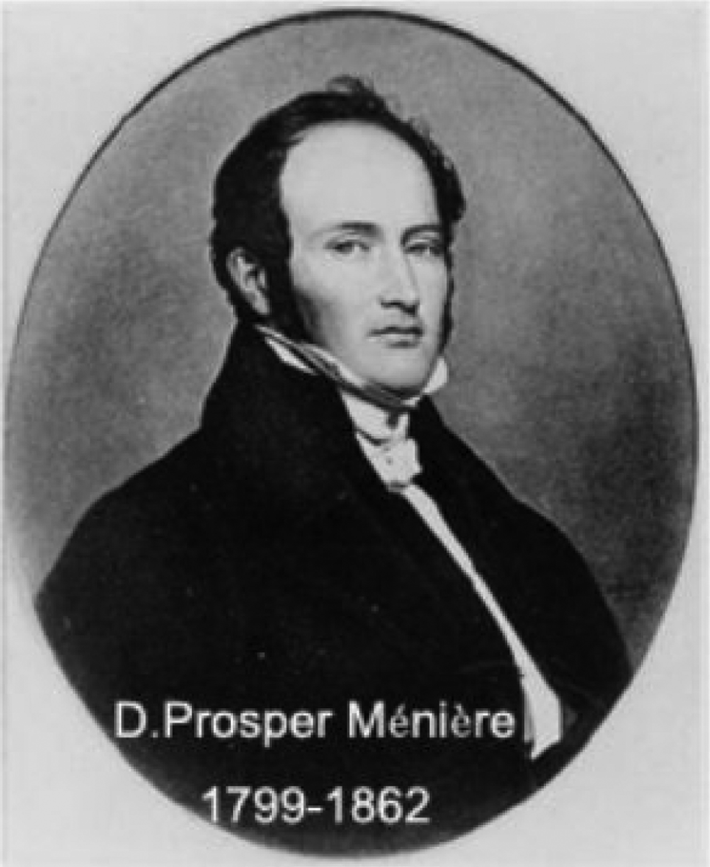
Churchill's wartime comment on the Soviet Union (British Broadcasting Corporation broadcast, London, 1 October 1939) could equally be applied to what is now termed Ménière's disease. Even the acute or grave accents required for the correct spelling of Prosper's surname still exercise many a linguist and are disputed (Figure 1). Most of us will now accept that there is a curious and quite distinct disorder of the inner ear, with some very characteristic features. The progress is not always uniform, but normally starts as a unilateral and fluctuating, possibly low frequency, cochlear deafness, with recruitment. The most obvious additional feature is the paroxysmal vertigo that accompanies this, and which is, arguably, the only symptom we can influence. Finally, there is tinnitus and a sense of fullness, which in the early stages of the disease frequently convince the non-expert to believe that this is a eustachian tube problem.

Fig. 1 Prosper Ménière.
The Archives of The Journal of Laryngology and Otology nicely illustrate how our forebears tried to make sense of this illness and influence its natural history. What emerges is a confusion about the definition, underlying pathology and treatment that persists to this day.
The short review that follows concentrates on some key articles from the last century and a half of publications in the Journal, following the same format as in our earlier review of the pathology and surgery of otosclerosis.Reference Flood and Kenyon1 As previously, the online review also contains direct electronic links to the archived material quoted.
Introduction
The eponym attached to the symptom complex of deafness, tinnitus and vertigo is so well established that it is curious to read of the difficulty in tracing Ménière's original description. Dan M'Kenzie, in Reference M'Kenzie1924, sought out the primary evidence and found contradictory reports, written in a style that did not always appeal to him.Reference M'Kenzie2
‘Ménière's original case’, D M'Kenzie (Reference M'Kenzie1924)
So stealthily has the story (I had nearly written the “myth”) of Ménière's original case insinuated itself into our ontological literature, that we are all in the habit of looking upon its data, its traditional data, as proved, and have little or no hesitation in basing theories and explanations of various kinds upon them. But it is well to refer occasionally to original sources, and a few months ago I made an attempt to get at Ménière's own account. The result of my efforts was a surprise.
Politzer, whose sources of information are always carefully furnished, refers us to the Gazette Medicale de Paris of 1861, and thither I turned, expecting my quest to be soon completed. But the search turned out to be by no means so easy as I had anticipated.
Ménière manifests a tendency to sandwich remarks into his paragraphs just as they occur to him, whether they are strictly germane to the subject in hand, or not. There is little of the French love for order and arrangement in Ménière's style…
I give a literal translation of Ménière's account, preserving the original punctuation:— “I have already spoken, a long time ago, of a young girl, who, having travelled by night in winter on the outside of a diligence, when she was at a catamenial period, had, in consequence of a considerable cold, complete and sudden deafness. Having been received into the service of M. Chomel she manifested as her chief symptom continual vertigo, the slightest effort to move produced vomiting, and death followed on the fifth day. The necropsy showed that the cerebrum, cerebellum and spinal cord were absolutely exempt from any alteration, but as the patient had become suddenly deaf after having always had perfect hearing, I removed the temporals in order to examine with care what could be the cause of this complete deafness, so rapidly supervening. The sole lesion I found was the semicircular canals filled with a red plastic material, a sort of bloody exudate, of which scarcely any traces were perceived in the vestibule, and which did not exist in the cochlea. The most attentive search has enabled me to establish with all the precision desirable that the semicircular canals were the only parts of the labyrinth which showed abnormality, and this consisted, as I have said, in the presence of a plastic lymph replacing the liquid of Cotugno.”
It is clear that our current desired levels of evidence were not required to recommend treatment in the early days of the journal. Dr Labit provides a typical example of this. The statement that follows was announced by Dr Labit to the French Society of Otology and Laryngology, in the year that WG Grace scored his one-hundredth century.3
‘Ménière's vertigo’, M Labit (1895)
This affection arises generally from sudden exudation or haemorrhage of the labyrinth, and the author has thought that the benefits of subcutaneous injections of pilocarpine, seen in pleuritic or pleural effusion, may be extended to this affection. He quotes three cases of Ménière's vertigo successfully treated by this means.
There was clearly some muddled thinking on vertigo in the 1890s, but also doubt about the fame of Ménière. The idea that ossicular removal might be of benefit was subsequently mooted, and the notions of gastric and nasal vertigo, which certainly seem beyond the province or indeed experience of the modern neuro-otologist, were discussed. The good Drs White and Burnett, who are quoted below in an article by Dundas Grant, seem to lack an evidence base for their recommendations, but nonetheless seem happy to discuss these notions.Reference Dundas4 They may well have also experienced the 70 per cent rate of remission of symptoms, as seen with every subsequent intervention (or even the natural history).
‘Otology’, J Dundas Grant (Reference Dundas1894)
A discussion on vertigo took place in the Medical Society of Virginia. (“Med. Record,” Oct. 8, 1892.) Dr. Brady considered the gastric form the most common. Dr. Bedford Brown insisted on the necessity for examination of the urine for albumen, casts and sugar whenever the tendency to vertigo is marked and persistent, adding that he had never seen a case of chronic nephritis or diabetes mellitus without more or less vertigo. Dr. Joseph White believed that all cases of so-called “nasal vertigo” were really aural vertigo. C. H. Burnett (American Otol. Soc.—“Med. Record,” July 29, 1893) disputes the claim of Ménière to the honour of having aural vertigo named after him. He ascribes it to middle-ear disease, and advises surgical treatment—the removal of the incus.
Emile Ménière, son of Prosper, featured regularly in transcripts of proceedings in the early days of the journal, and clearly did not share the doubts about his illustrious name.Reference Ménière, Ménière and Dundas5 He was certain that the middle ear was not involved, but was equally honest that he had no miracle cure to offer.
‘Case of complete deafness occurring in the course of leucocythaemia’, E Ménière (Reference Ménière, Ménière and Dundas1895)
Addressing the Paris Society of Laryngology, Otology and Rhinology, Emile Ménière stated:
Madame X., aged forty-three, suffering from leucocythaemia, was considered to be in a desperate condition six months ago. One day, during a period of general amelioration, in the month of August, the patient experienced violent tinnitus, vertigo, and nausea. The right ear became completely deaf in the space of a few hours, the left ear was affected to a considerable extent, at the same time maintaining a very slight degree of hearing power, which continuously became less and less. On examination of the ear, no visible lesion was observed either recent or of old standing. The functional examination confirmed the fact of her having complete deafness on the right side, and almost complete on the left. In view of the small number of cases published, one case, although incomplete, possesses considerable interest. Dr. Ménière has seen four such in his practice, but without having the opportunity of making a post-mortem examination. He draws attention to the fact that the symptoms which presented themselves at the commencement were those of Ménière's complexus, and that they occurred during a period of amelioration as regards the leucocythaemia. According to other authors, in the cases that have been observed there has generally existed some lesion of the middle ear, but in this case nothing of the kind could be detected. No method of treatment produced any result.
Responding to the presidential address of G Stoker to The British Laryngological, Rhinological, And Otological Association, Dundas Grant tried to distinguish the symptom complex from what we now recognise as the primary condition.Reference Dundas and Stoker6 He further proposed some holistic measures to treat Ménière's symptoms rather than the disease itself. ‘Sound’ red wine, leeches and the application of cold to the head are more reminiscent of the eighteenth century than the nineteenth century, but may prove to be as efficacious as any claimed vasodilator, diuretic or labyrinthine sedative.
‘Discussion on the treatment of Ménière's symptoms’, J Dundas Grant (Reference Dundas and Stoker1895)
Ménière's symptoms may depend on —
A. Disease affecting the auditory nerve directly:
(a) In its intra-cranial course.
(b) In its labyrinthine distribution.
B. Disease affecting the auditory nerve indirectly from accessions of increase of tympanic pressure due to —
(a) Catarrhal indrawing of the membrane.
(b) Pressure of granulations on the stapes.
(c) Unopposed over-action of the tensor, from —
1. Antero-inferior perforation.
2. Paralysis of the stapedius.
C. Hyperaesthesia sensoria et acoustica.
Thus in congestive conditions we may resort to the application of cold to the head, derivation towards the feet, intestines, skin, and kidneys, abstraction of blood either generally by venesection in the arm or locally, by leeching over the tragus or mastoid, or by free incision or even excision of the inferior turbinated body; lowering of the diet and avoidance of excitement; further, by compression of the vertebral arteries and the administration of bromide of potassium or hydrobromic acid.
In anaemic forms we have to put an end to any direct loss of blood or other source of weakness, and to restore at the same time the qualities of the vital fluid and the tone of the depressed nervous system. Hence we administer suitable food, judicious stimulation by means of spirits or sound red wine, and iron in any of the most assimilable forms…
Of vertigo arising from indirect irritation of the labyrinth, little need here be said, if we accept the unquestionable evidence, notably that of Prof. Gelle, that the most typical Ménière's symptoms may arise from disease of the middle ear, affecting the stapedio-vestibular articulation. So striking is the pathological evidence, that Gradenigo, in Schwartze's handbook, gives to this condition the appellation of “Ménière's disease,” rather than to the labyrinthine affection to which Ménière himself attributed the symptoms.
In reply to Dr. Law's query as to the desirability of extraction of the stapes for Ménière's symptoms, I have no experience to offer, but I have found in the few cases in which patients have come under my care on account of vertigo that milder means have sufficed. Moreover, in some of the reported cases of stapedectomy the operation has been credited with setting up vertigo. Extraction of the incus or section of its long process is probably the most justifiable surgical procedure.
In response comes a statement that appeals more to modern thinking on this disease process.Reference Dundas and Stoker6
Dr. MACNAUGHTON-JONES: …I consider that, in discussing Ménière's disease, we ought to confine ourselves strictly to those intra-labyrinthine causes which are most in our minds when dealing with direct pressure on the nervous elements in the labyrinth, increased tension in its arteries, or an effusion, due to recent inflammation or otherwise. We need not go through the various causes in the middle ear which may produce these symptoms.
It is obvious that the earliest writers were lumping all hearing loss associated with vertigo under an umbrella term of Ménière's disorder. This led to the enthusiasm for insufflation of many a eustachian tube, or gouging of a carious mastoid. One hundred years later, the Journal was still calling for clarity in diagnosis and definition.Reference Beasley and Jones7
‘Menière's disease: evolution of a definition’, N J P Beasley & N S Jones (Reference Beasley and Jones1996)
In 1861 Prosper Menière separated patients with episodic vertigo, hearing loss and tinnitus from a group previously described as having apoplectiform cerebral congestion. He suggested the cause was disease within the semicircular canals (Menière, 1861). Over the years it became apparent that within this group there were a number of patients with characteristic signs and symptoms and in 1938 a pathological correlate was found in the form of endolymphatic hydrops. Descriptions such as Menière's ‘disease’, Menière's ‘syndrome’ and Menière's ‘symptom complex’ led to a confusing array of terms for this condition and monitoring of treatment results became difficult. In response to this in 1972 the American Academy of Ophthalmology and Otolaryngology Committee on Hearing and Equilibrium published a clear definition of Menière's disease and criteria for the reporting of treatment results, it was updated in 1985 and again in 1995. We describe the changes that have taken place as the definition of Menière's disease has evolved.
There does seem a modern consensus that Ménière's disease is a peripheral disorder of the audiovestibular system, which may have something to do with homeostasis of the endolymphatic system, and any evaluation of the treatment for it is bedevilled by its natural history: a tendency to spontaneous remission (or an undue susceptibility to a placebo effect?).
As early as 1896, Professor Gruber of Vienna, in an address to the Austrian Otological Society, had already suggested that the endolymphatic sac might be implicated. Indeed, he suggested a hydrops-like condition, and called for the distinction between the disease and the symptom complex.Reference Prof8
‘On Morbus Ménièrei’, Prof. Gruber (Reference Prof1896)
The speaker recommended giving up the term “Ménière's symptoms”, but retaining that of “Ménière's disease” for the conditions to which Ménière applied it, namely, primary labyrinthine affections, haemorrhagic or exudative, with vertigo, subjective noises, and deafness, apart from pyrexial inflammations of the labyrinth.
When the so-called Ménière's symptoms arise from disease of other parts of the organs of hearing, they should be mentioned among the accidental complications, and not as the main term in the formal diagnosis of the case. He directed attention to the varying anatomical formation of the saccus endolymphaticus, and of the aqueductus vestibuli, and suggested that when they are exceptionally small the outflow of endolymph might be readily interfered with, and abnormal tension, such as to produce vertigo, readily induced.
Again, long before any histological evidence of hydrops, comes the suggestion that a glaucoma-like condition of the inner ear might be responsible.Reference Mackenzie9
‘Remarks on the nature, diagnosis, prognosis and treatment of aural vertigo’, S Mackenzie (Reference Mackenzie1894)
These remarks are introduced by reference to a pronounced case of aural vertigo in a man aged fifty, beginning with vertigo, vomiting, and deafness.
…the author inclines to the opinion that the seat of the lesion is to be found in the semicircular canals, and that this (the lesion) is of an irritative character producing its effects so long as there is no absolute atrophy of the auditory nerve. It is pointed out that Buzzard and Dalby suggest a bulbar origin for some of these cases of aural vertigo. As to the nature of the lesion it is observed that in few cases indeed has coarse disease been demonstrated in the semicircular canals. There are causes direct and indirect which may irritate the nerve terminations of the former, e.g., haemorrhage, syphilis of the latter, e.g., otitis media. The theory of Spear that there is a condition in the labyrinth resembling glaucoma, as well as Knapp's suggestion that Ménière's disease is an idiopathic, serous exudative otitis interna, are referred to. Seeing that aural vertigo occurs in the latter part of life, degenerative changes in the local blood vessels is probable, and Gower's statement that it is associated with gout (in the labyrinthine membrane) is supported.
When tinnitus, vertigo, nausea, and vomiting occur together, suspicion as to an aural origin ought to be entertained. In the majority of these cases a certain degree of deafness is appreciable. The treatment consists in the recumbent position during an attack and bromide of potassium in fair doses. Lithium may be given in gouty cases. Counter-irritation and attention to the ear affection. The inter-paroxysmal treatment consists in the administration of quinine. Pilocarpin is used by Field. Mercury is useful to keep down arterial tension, and is beneficial when given during the premonitory symptoms of an attack.
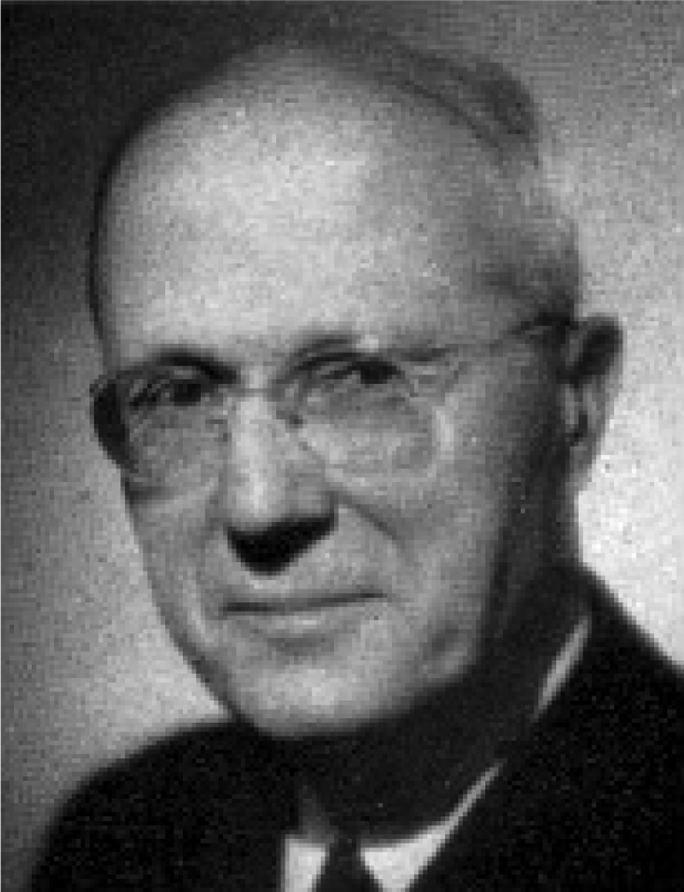
Georges Portmann (Figure 2) was a regular contributor to the Journal. He experimented on the role of the autonomic nervous system in the homeostasis of intralabyrinthine fluids.Reference Portmann10

Fig. 2 Georges Portmann.
‘Vasomotor affections of the internal ear’, G Portmann (Reference Portmann1928)
We have recently made a series of experiments on the action of the sympathetic in regard to the labyrinth. These experiments have been either pericarotid sympathectomy or injections of vasomotor drugs (stimulators or inhibitors of the sympathetic; stimulators or inhibitors of the parasympathetic). Our study has dealt also with the circulatory labyrinthine troubles caused by the compression of the vertebral artery or of the “common carotid artery,” or by peripheral vasodilatation (warm bath test).
These different experiments, of which the results agree with those obtained by Terracol (perivascular sympathectomy, section of the cervical sympathetic, injection of suprarenin into the superior cervical ganglion), allow of our bringing forward some conclusions on the vasomotor affections of the internal ear.
For a long time the action of the sympathetic on the labyrinthine circulation on the one hand, and on the secretion of the endolymphatic fluid on the other, has impressed otologists. It is astonishing that the study of it has not been more developed, for, in 1904, Ferreri pointed out certain affections of the internal ear in connection with some sympathetic lesions and spoke of “Glaucoma,” a term used again more recently by Aboulker and myself, to designate the hypertension of the endolymphatic fluid.
The opportunity to review temporal bone findings was later exploited by Hallpike and Cairns in 1938, in a lengthy and well-illustrated article, of which the below is a very small segment. Both patients mentioned below had succumbed to raised intracranial pressure and coning, after surgery to section the VIII nerve.Reference Hallpike and Cairns11
‘Observations on the pathology of Ménière's syndrome’, C S Hallpike & H Cairns (Reference Hallpike and Cairns1938)
It is the purpose of this paper to describe—it is believed, for the first time—the pathological changes in the temporal bones of two cases of Ménière's syndrome. In both death occurred shortly after operation for section of the VIIIth nerve.
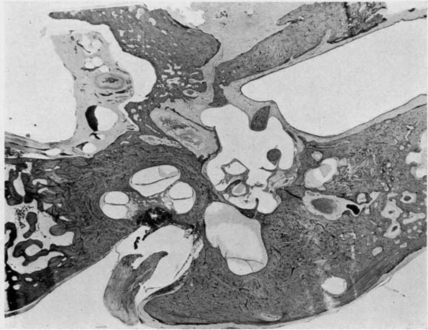
The vestibule.—There is a gross dilatation of the endolymph spaces, in particular of the saccule [Figure 3]. At the level of the stapes footplate the saccule extends around the lateral aspect of the utricle to enclose it on its posterior aspect and extends into the inner extremity of the lateral semi-circular canal. The perilymph cistern has been obliterated.
The opening of the saccule into the ductus endolymphaticus contains a dense reddish staining coagulum.
There is a gross dilatation of the endolymph system which affects chiefly the scala media of the cochlea and the saccule.
…additional emphasis must accordingly be thrown upon the condition found in and around the saccus endolymphaticus in all of the three temporal bones which have been examined. It will be recalled that in all of these the soft perisaccular connective tissue, identified by Guild as the normal absorption area of the endolymph, was found to be absent. It might well be that the general dilatation of the endolymph system in these cases was due to hypersecretion and had led to the obliteration by pressure of the soft perisaccular tissue. But this seems to be rendered unlikely by the fact that in the unaffected ear of the first case the same absence of this perisaccular tissue is to be seen. The suggestion is therefore made that the endolymphatic dilatation in these cases is due, at any rate in part, to some failure in the normal absorptive mechanism; and that morphological evidence of this failure is provided by the absence from these cases of the normal absorption area of perisaccular connective tissue.
…it seems possible to explain the attacks as being due to rapidly initiated bouts of asphyxia of the labyrinthine end-organs, brought about by extremely rapid rises of fluid pressure, in response to relatively very small volume increases in the endolymph. Upon the basis of this hypothesis it is supposed that there exists a chronic condition of lowered function of the affected labyrinth due to increased endolymph pressure and resulting anoxaemia of its end-organs. This condition fluctuates in a manner typical of the clinical course of the disease, and may proceed during the course of the attack to complete paralysis of the affected labyrinth.

Fig. 3 ‘Case I. Left temporal bone. Low power view showing chronic inflammatory changes within the tympanum, and gross dilatation of the endolymph spaces of the vestibule and the cochlea.’Reference Hallpike and Cairns11
We should now turn to attempts to treat the disorder. Urban Pritchard, in 1900, was clearly an enthusiast for the diagnosis of the disease, as it at least offered a benign prognosis.Reference Pritchard12 John M Graham has memorably remarked that ‘Ménière's disease is easy to diagnose. Some people do it all the time’ (L Flood, personal communication).
‘Vertigo of Ménière’, U Pritchard (Reference Pritchard1900)
It was truly a grand discovery in medical science which has enabled us to differentiate the symptoms of that disease, now associated with the name of Ménière, from those far more serious ones which generally indicate disease of the brain. With proper care the physician is now able to diagnose this condition with precision in perhaps ninety-nine cases out of every hundred that are brought before him; and yet, through lack of a little special knowledge, many a medical man has mistaken this Ménière's vertigo either for mere biliousness on the one hand, or for grave brain trouble on the other. How often have some of us heard from patients suffering from Ménière's disease, “Oh, but Dr. — told me that I must make my will and settle my affairs, as this will prove fatal sooner or later; and now you smile, and say that I cannot die from it”!
…we have to confess that our knowledge of the subject has not advanced so very much since Dr. Ménière introduced it to the profession now some forty years ago (1861).
Our actual knowledge of the causes of this condition is still very vague. We are satisfied that it is an affection of the semicircular canals, that it is probably due to congestion of, or extravasation of blood into, those canals; but we want to go further. True, this explains readily enough the reason for the apoplectiform variety of the disease, where there is sudden severe vertigo, vomiting, and tinnitus, accompanied with more or less complete loss of hearing; but it only partially explains the epileptiform condition.
…acute inflammation such as occasionally occurs in the course of mumps or in tertiary syphilis, sometimes produce[s] this condition. More rarely the cause is traumatic; thus, I have seen it brought about by a smart blow behind the ear from a golf-ball, which, I need hardly explain, is a small, very hard ball driven with considerable force.
Gout and sunstroke are sometimes the exciting causes, but our knowledge on this point is very imperfect. Occasionally, though not nearly as frequently as is commonly taught, this form of Ménière's vertigo is associated with middle-ear catarrh.
When it occurs in the course of chronic middle-ear catarrh the treatment is that of the catarrh itself, and in some of these cases the relief given by inflation by means of the catheter or by Politzer's bag is very marked.
In intercranial [sic] lesions the Ménière's symptoms are only of secondary importance as compared with the gravity of the lesion itself; but bromides and hydrobromic acid will often afford great relief.
In those somewhat rare cases which are associated with middle-ear catarrh, the ordinary treatment for that condition is of much service, and it is in these cases that some improvement in the hearing power may be hoped for. But I deprecate the indiscriminate and forcible inflation, by means of the catheter or of Politzer's method, in most cases of epileptiform; indeed, sometimes where hyperaesthesia of the nerve exists positive harm may result therefrom.
Few of us would, I imagine, dare to recommend bicycling as a treatment for patients suffering from Ménière's vertigo, but I have a case of a lady who resorts to bicycling when the giddiness comes on, as she invariably finds it is arrested by this form of exercise. I shall be glad to know if others have met with a similar experience.
Pritchard goes on to describe a truly remarkable outcome for a woman with a chronically discharging post-aural fistula, vertigo and hearing loss. Today, we would suspect a labyrinthine fistula; in 1900, syphilis or tuberculosis was as likely. He clearly felt that a golden age of surgery in the labyrinth was dawning, but he may have been a little premature!
…by one brilliant achievement Mr. Charles Ballance has opened up a vista of glorious possibilities.
E. M—, aged 54, admitted to St. Thomas's Hospital, February 5, 1900.
The petrous was removed till no further sign of pus was discoverable. In doing this the semicircular canals were, in part, destroyed, and the back of the vestibule opened. Clear fluid escaped. The cavity in the petrous at the conclusion of the operation was about 5/8 inch in depth, and in size would have taken half a Barcelona nut[*]. It was repeatedly swabbed out with absolute phenol. For several days the patient was very sick and giddy.
March 20: Healing has been complete for some days, and the patient can run down the ward. There is no vertigo, and the tinnitus has ceased. The hearing has returned in a marvellous way. Three weeks after the operation the watch could be heard at 6 feet, and a whisper at, at least, 23 feet distance.
With such a case before us are we not justified in looking forward with sanguine expectation to the assistance which shall be rendered by surgery in the near future, whereby vertigo, tinnitus, and deafness in many a labyrinthine case may be overcome?
Of a truth, gentlemen, we may indeed say that in recent years Aural Surgery has advanced by leaps and bounds. How much further will she bring us along the road which leads to victory? By what paths will she make her way? These things we know not. Only Imagination can foreshadow the answer, and only Time himself can prove the truth of her prophecy.
*Our readers will surely know that the Barcelona nut is a hazelnut-like product of the Spanish filbert tree; the authors of this review did not, however.
Parry, surgeon to the Victoria Infirmary and to the Royal Hospital for Sick Children in Glasgow, reported the sectioning of the VII cranial nerve, undismayed by previous reports universally resulting in death!Reference Parry13 Few twenty-first century surgeons will not wince at his experience with the facial nerve, but, equally, few will fail to be impressed by his measures to tackle the result.
‘A case of tinnitus and vertigo treated by division of the auditory nerve’, R H Parry (Reference Parry1904)
S. D—, aged thirty, engineer, was recommended for treatment in February, 1902, by Dr. Jones of Glasgow, who in his letter expressed a hope that something might be done by operation to relieve his condition, as all other forms of treatment had failed.
The patient stated that he had enjoyed excellent health until six years ago, when one day he was suddenly seized with giddiness while he was lighting his pipe. He was not far from his house at the time and managed to stagger to it. While the attack was on him everything seemed to be turning round. During the next six months he had several such attacks, and he had finally to give up work for two years. The giddiness became more or less constant, affecting him more particularly when walking. He improved somewhat towards the end of that period, and was able to resume work and to remain at it for two years.
A year ago he was again obliged to give up work owing to the frequency of the attacks of vertigo. He complained of noises in the left ear, but only occasionally in the right. He had always been partially deaf in [the] left ear, from which there had been a discharge until two years ago. There is no history of alcoholism or syphilis. During the three weeks in which he was under observation before operation his suffering remained as great as before admission; and it was made absolutely clear to us that he had in no way exaggerated his misery. He expressed anxiety to be relieved of the pain in the back of his head and the vertigo, which affected him both at rest and while walking about in the ward.
The fact that a mastoid operation had not been carried out suggested that, in the opinion of the several medical men who had seen him, the seat of the trouble lay deeper, and that a simple mastoidectoniy would not bring about the desired result. On the value of the removal of the semicircular canals no opinion could be offered, nor at the time (two years ago) could any information be obtained as to the probable effect of such a procedure.
The dura mater was separated from the surface of the petrous portion as far as the promontory caused by the superior semicircular canal. On removing the roof of the tympanic cavity an excellent view was obtained of that space, and it is interesting to note that it was possible by that method to determine the presence of pus or granulation tissue in the middle ear. More bone was removed in the direction of the mastoid antrum, but as no evidence of active disease was found either here or in the middle ear the fear that a septic focus had been left en route was set at rest.
The roof of the internal auditory meatus was next removed, when the seventh and eighth nerves were easily recognised. The eighth nerve was drawn aside and could have been divided then, but owing, perhaps, to the fact that no untoward accident had occurred and to the desire to sever completely the vestibular and cochlear divisions, more bone was removed; but unfortunately, the detached portion proved to be the commencement of the Fallopian aqueduct, and in the withdrawal of it the seventh nerve was torn. This regrettable, and, it may be added, avoidable accident, permitted of a very thorough examination of the nerves in that opening. The plug was removed from the middle ear and iodoform gauze introduced in its place through the external meatus.
For fully a month he was kept at rest, although in the third week he was most anxious to get up, declaring that he felt quite equal to it.
When allowed to move about again he still complained of giddiness and the noises in the head. The general opinion was that he was better, although, in his anxiety for a complete cure, we found it difficult to get him to admit it.
At the end of that period he was induced to undergo a second operation in order to overcome the facial paralysis. The spinal accessory nerve was divided and attached to the facial close to the stylo-mastoid foramen. He has no voluntary power over the facial muscles; they move in association with the movements of the shoulder.
The present is, as far as can be ascertained, the first in which the auditory nerve has been divided in the living subject. Two cases have been reported recently, but both ended fatally.
The advent of sac surgery held the promise of treating the underlying disorder of endolymphatic hydrops using a hearing conserving approach. This surgery will always be associated with the name of Portmann, and it remains, arguably, the only surgery that holds any hope of treating the underlying pathological disorder of Ménière's disease. Extracts from his article, published in 1927, suggest an unusual experience on draining of the sac.Reference Portmann14 The extracts also indicate that Portmann was a stoical surgeon, who could tackle live surgery before illustrious gatherings.
‘The saccus endolymphaticus and an operation for draining the same for the relief of vertigo’, G Portmann (Reference Portmann1927)
The contribution to the therapeutics of vertigo which forms the subject of this work establishes the practical result of a series of researches on comparative anatomy, human anatomy and physiology of the saccus endolymphaticus, which I have pursued incessantly for the last eight years.
To continue our comparison, as in glaucoma the ophthalmologists puncture the cornea in order to suppress the intraocular hypertension, capable of destroying for ever the value of the eye; so it seems logical in some cases of serous labyrinthinitis, when medical treatment has failed, to make a decompression of the internal ear by the removal of the excess of endolymphatic fluid.
In a word, it is necessary to search for the saccus through the mastoid in the area of a triangle formed above, by a line corresponding to the floor of the antrum, in front by the aqueductus Fallopii, behind by the lateral sinus.
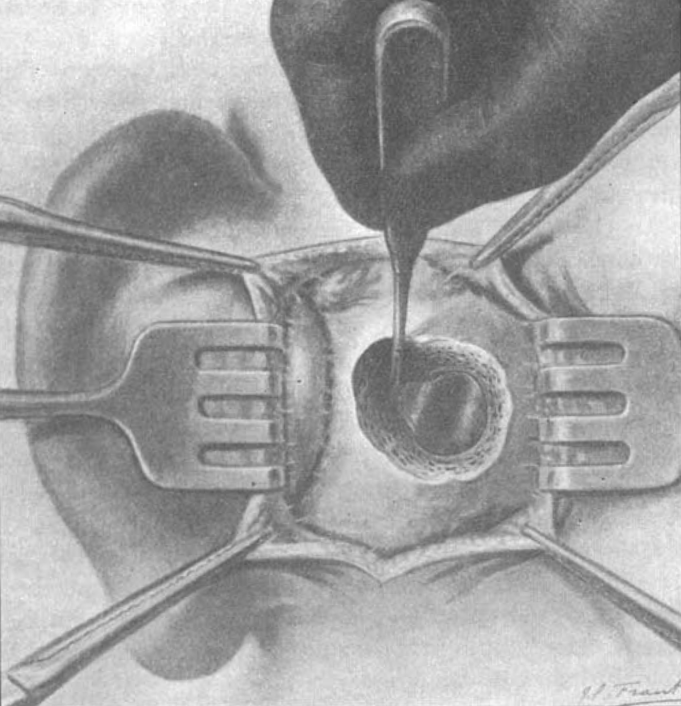
Fourth step.—The petrous wall of the saccus endolymphaticus is freed by removing the greater part of the fossa endolymphatica. By the help of a very fine needle, mounted on a Luer syringe, one makes an exploratory puncture of the saccus in contact with the zone of adhesion [Figure 4]. The operator, who feels perfectly the sensation of penetrating the little cavity of the saccus, then makes, with a paracentesis knife, an incision from 2 to 3 mm. long into the saccus. One or two drops of fluid, like spring water, flow from the opening of the wound.
I have on several occasions practised this operative intervention on the living most successfully, and quite recently (10th October 1926) I operated (1) at Groningen, before the International Oto-Rhino-Laryngological College, and (2) at Rome (23rd October 1926) before the Italian Congress of Oto-Neuro-Oculists, on two typical cases of vertigo, cured by opening of the saccus endolymphaticus. The severity of the vertigo prevented the unhappy patients who were attacked by it from doing any kind of work.

Fig. 4 ‘By the help of a very fine needle, mounted on a Luer syringe, one makes an exploratory puncture of the saccus in contact with the zone of adhesion.’14
In a memorable address to the Royal Society of Medicine, Professor Alan Kerr tackled the doubts that many felt (but few dared to express) surrounding the management, and even the underlying pathology, of Ménière's disease.Reference Kerr15 As in the fable of ‘The Emperor's New Clothes’, we believe what we are told, we see what others tell us they see, and it is a brave individual who then sticks his, or her, head over the parapet and contradicts dogma. Those of us who were privileged to be there that day will never forget sitting spellbound in that lecture theatre as he spoke. This is also one of the most thought provoking papers ever published in the Journal and worth reading in full. What follows, as with all the articles cited here, is a ‘taster’.
‘Emotional investments in surgical decision making’, A G Kerr (Reference Kerr2002)
Pathology
First, what is the pathology? We know what the inner ear looks like at post-mortem examination but we still do not know why this happens or how it gets to this end situation. There is hydrops but we also know that at post-mortem many temporal bones show hydrops in people who have never complained of the symptoms of Ménière's disease.
There have been many reports of fibrosis in the peri-saccular tissue, altered glycoprotein metabolism and immune-mediated and viral aetiologies but none has produced any clear cut evidence so that we still are uncertain about the pathological mechanisms.
Prognosis
Secondly what is the prognosis? Sadly we still do not really know the natural history of this condition. There are studies of cohorts of patients but these tend to be unrepresentative of the totality of Ménière's disease patients in that they are collected by those who are interested in this condition and are biased by tertiary referrals.
Proficiency
Thirdly, [do] we need to consider the proficiency of any particular treatment? Apart from a very small minority of enthusiasts, most of whom have considerable emotional investment in this area, there are no claims that any drugs or procedures have any effect on the hearing in the long term. However, let me raise a doubt in your mind. Isn't it odd, in this condition where the hearing and vertigo occur together that many treatments that are said to improve the course of one, the vertigo, should fail to have any significant effect on the other, the hearing? In other words, is the alleged effect on the vertigo really the result of the treatment?
Probably the most common operation for Ménière's disease is some form of endolymphatic sac surgery. This is sometimes done without conviction on the basis that ‘one has got to do something’. Discussion of sac surgery often generates a lot of emotion and confusion.
Illustrations of sac surgery procedures show Silastic® sheets, fine capillary tubes or tubes with one-way valves, with the common objective of draining endolymph. But can they? Firstly, the utriculo-endolymphatic valve may not be patent. Secondly, the endolymphatic duct may be blocked or even virtually non-existent. However, they are at their most fantastic, (as in fantasy), when we consider the nature of the sac which is alveolar. It is more like a tiny lung than a tiny gall bladder. So, just where are these drains going? It is more likely that they are causing damage than draining.
The sceptical neuro-otologist could, at worst, invite professional ruin, as he predicted. Meetings of the Royal Society of Medicine are rarely reduced to helpless laughter (the whole presentation is worth reading in full), but Kerr showed how this expressed doubt regarding efficacy could develop, until:
‘Patients used to come a long way to see me and expected me to do something. I did not. Therefore I am no longer an expert in Ménière's disease because I have lost most of my referrals.’
Sadly, I am pessimistic about this. Even with the advent of tightly controlled evidence-based medicine, we are still going to see doubtful operations being performed on patients with Ménière's disease. It might be possible to control these in other conditions but Ménière's disease fluctuates too much, sac surgery is probably an easy way to procrastinate and there are too many vested interests ready to exploit this.
After decades of doubt as to the proposed underlying disorder, endolymphatic hydrops may finally be dispelled by advances in imaging. Magnetic resonance imaging (MRI) scans showing a distended scala media may prove far more convincing than a negative summating potential on electrocochleography. Doubting Thomas was told ‘blessed are those who have not seen and yet have believed’ (John 20:29). Recent studies published in the Journal suggest we may yet be able to see the hydrops, as has long been proposed, in vivo and in real time.Reference Hornibrook, Coates, Goh, Gourley and Bird16, Reference Fang, Chen, Gu, Liu, Zhang and Cao17
‘Magnetic resonance imaging for Ménière's disease: correlation with tone burst electrocochleography’, J Hornibrook et al. (Reference Hornibrook, Coates, Goh, Gourley and Bird2012)
The newly developed use of magnetic resonance imaging of the human inner ear, on a 3 Tesla scanner with intratympanically administered gadolinium, can now reliably distinguish perilymph from endolymph and visually confirm the presence or absence of endolymphatic hydrops. Transtympanic tone burst electrocochleography is an established, and under-utilised evoked response electrophysiological test for hydrops, but it relies on a symptom score to indicate the likelihood of hydrops being present. The current diagnostic criteria for Ménière's disease make no allowance for any in vivo test, making diagnostic errors likely. In this small pilot study of three patients undergoing tone burst electrocochleography, subsequent magnetic resonance imaging confirmed or excluded the hydrops that the electrocochleography predicted.
Magnetic resonance imaging of the inner ear is an exciting new technique. In addition to its use in Ménière's disease, it has been used to study idiopathic sudden SNHL [sensorineural hearing loss] and fluctuating hearing loss without vertigo. It should contribute to our understanding of these inner ear disorders. In Ménière's disease, it could be the in vivo ‘proof’ of hydrops, and the investigation by which electrophysiological tests are validated. Magnetic resonance imaging of the inner ear could contribute to a more precise clinical diagnosis of Ménière's disease than is currently possible, in particular to distinguish it from other causes of recurrent vertigo.
Of course, it could not be that simple. It is all too easy to ‘see’ changes that we seek, either when hunting a perilymph fistula at tympanotomy or when looking for a dehiscent superior semicircular canal on computed tomography. We need an objective scoring system it seems.
‘A new magnetic resonance imaging scoring system for perilymphatic space appearance after intratympanic gadolinium injection, and its clinical application’, Z M Fang et al. (Reference Fang, Chen, Gu, Liu, Zhang and Cao2012)
The derived scoring model was highly accurate for diagnosing Ménière's disease and delayed endolymphatic hydrops. Two magnetic resonance imaging scoring methods for the perilymphatic space were proposed for the diagnosis of endolymphatic hydrops: a pre-1 value (a new variable that predicts individual probability) of more than 0.3982299, or a sum of all labyrinth component scores of less than 14.5.
It is salutary to consider that the most evidence-based interventions for Ménière's disease still depend on ablation of vestibular function, whether nerve section, or exploiting the ototoxicity of gentamicin. There is little suggestion that anything we do influences the natural history for cochlear function, and all research is bedevilled by the fluctuating pattern of the disease and that placebo response.
Conclusion
We hope that readers will not be discouraged and have been inspired by the attempts of our ancestors in otology, as recorded in the Journal Archives, to make some sense of the disorder. As in many other areas of our speciality, we still do not understand Ménière's related problems, and we still need to do something about that lack of understanding.


