Hostname: page-component-cd9895bd7-q99xh Total loading time: 0 Render date: 2024-12-22T15:24:51.829Z Has data issue: false hasContentIssue false

Amoebiasis in Iran: a systematic review and meta-analysis

Published online by Cambridge University Press:  11 July 2018

Ali Haghighi
Affiliation:
Department of Parasitology and Mycology, School of Medicine, Shahid Beheshti University of Medical Science, Tehran, Iran
Seyed Mohammad Riahi
Affiliation:
Department of Epidemiology, School of Public Health, Student's committee research, Shahid Beheshti University of Medical Sciences, Tehran, Iran Faculty of Health, Birjand University of Medical Sciences, Birjand, Iran
Ali Taghipour
Affiliation:
Department of Parasitology and Mycology, School of Medicine, Shahid Beheshti University of Medical Science, Tehran, Iran
Adel Spotin
Affiliation:
Infectious and Tropical Diseases Research Center, Tabriz University of Medical Sciences, Tabriz, Iran Student Research Committee, Tabriz University of Medical Sciences, Tabriz, Iran
Mostafa Javanian
Affiliation:
Infectious Diseases and Tropical Medicine Research Center, Health Research Institute, Babol University of Medical Sciences, Babol, I.R. Iran
Mohsen Mohammadi
Affiliation:
Non-Communicable Pediatric Diseases Research Center, Health Research Institute, Babol University of Medical Sciences, Babol, I.R. Iran
Mohammadreza Esmaeili Dooki
Affiliation:
Non-Communicable Pediatric Diseases Research Center, Health Research Institute, Babol University of Medical Sciences, Babol, I.R. Iran
Ali Rostami*
Affiliation:
Infectious Diseases and Tropical Medicine Research Center, Health Research Institute, Babol University of Medical Sciences, Babol, I.R. Iran Immunoregulation Research Center, Health Research Institute, Babol University of Medical Sciences, Babol, I.R. Iran
*
Author for correspondence: Ali Rostami, E-mail: [email protected], [email protected]
Rights & Permissions [Opens in a new window]

Abstract

A comprehensive meta-analysis study was performed to estimate the reliable national prevalence and molecular epidemiology of amoebiasis in Iran. Nine English and Persian databases were searched to achieve the relevant studies. Pooled estimates were generated and meta-regression was performed. We identified 71 eligible articles involving 330 930 subjects from 25 provinces to be included in the final analysis. Moreover, 17 studies compromising 462 polymerase chain reaction (PCR)-positive isolates performed molecular analysis to inter-species differentiation. The pooled prevalence of Entamoeba infection among Iranian population was about 1% (95% CI 0.8–2.0%). Moreover, regarding Human Development Index (HDI), a higher prevalence was observed in undeveloped provinces. Out of 462 PCR-positive isolates, 83% (95% CI 69–94%) and 12% (95% CI 3–24%) were Entamoeba dispar, Entamoeba histolytica, respectively. In subgroup analysis based on molecular results, in general, population prevalence of Entamoeba dispar and E. histolytica were 91% (95% CI 80–99%) and 7%, (95% CI 0–19%), respectively, while prevalence of these species in patients with gastrointestinal disorders were 75% (95% CI 45–96%) and 18% (95% CI 1–43%), respectively. Our findings indicate the low burden of amoebiasis in Iran. E. dispar, that is mostly non-pathogenic, was identified as most prevalent species. Nevertheless, we suggest more public health interventions in areas with lower HDI.

Type
Review
Copyright
Copyright © Cambridge University Press 2018 

Introduction

The genus Entamoeba is constituted by cosmopolitan parasites belonging to the phylum Amoebozoa with world distribution. This genus contains seven species: Entamoeba histolytica, Entamoeba dispar, Entamoeba moshkovskii, Entamoeba coli, Entamoeba poleki, Entamoeba bangladeshi and Entamoeba hartmanni [Reference Fotedar1, Reference Royer2]. Among these, the first three (E. histolytica, E. dispar and E. moshkovskii) species are morphologically identical and considered as Entamoeba complex. Of those, only E. histolytica is the causative agent of amoebiasis, a global expanded gastrointestinal disease [Reference Haque3, Reference Ximénez4]. Although, some recent studies suggested that E. moshkovskii could have potential pathogenic effect in human [Reference Fotedar5, Reference Shimokawa6], E. dispar is still considered commensal organism [Reference Ali, Clark and Petri7].

Amebic infection is most prevalent in developing countries located in tropical and subtropical zones and its prevalence is associated with climatic conditions, sanitary and socio-economic status [Reference Nowak, Mastalska and Loster8]. It is estimated that about 50 million people were affected by invasive amoebiasis, resulting in up to 100 000 deaths per annum [Reference Haque3]. In a comprehensive global burden of disease study 1990–2010, Murray et al. (2013) reported 32 persons per 100 000 (95% confidence interval 25–41) disability-adjusted life years for amoebiasis [Reference Murray9].

Due to different biochemical, genetic and pathogenic features of Entamoeba complex, the differentiation of three aforementioned species is avery important issue in the effective clinical management of patients. For example, an infection with non-pathogenic species could mistakenly be diagnosed as E. histolytica infection and patient be unnecessarily treated with metronidazole that is the drug of choice for invasive amoebiasis, but not effective for the non-invasive species [Reference Al-Areeqi10, Reference Wolfe11].

Iran is one of the largest developing countries in Middle-East area with highly diverse geography, climatic and sociodemographic conditions. According to the Statistical Centre of Iran, the number of its total population is 80.28 million and approximately one-third of the people live below the national poverty line [12]. During the past years, several microscopy-based studies have investigated the prevalence of amoebiasis in different population groups, although discriminating studies between species (using molecular methods) are relatively few. According to the results of these studies, amoebiasis should be considered as a public health problem in Iran. Nevertheless, there is no comprehensive study showing the reliable status of amoebiasis at the national level. In this study, we performed a systematic review and meta-analysis to achieve an overview regarding the prevalence of amoebiasis and/or Entamoeba complex in Iranian people and also identify the different species circulating among of them.

Methods

Search strategy and study selection

This systematic review and meta-analysis study was implemented in accordance with the Preferred Reporting Items for Systematic Reviews and Meta-Analyses (PRISMA) guidelines [Reference Moher13]. To assess the prevalence of Entamoeba complex in Iran, relevant studies were searched from five English language databases (PubMed, Web of Science, Science Direct, Scopus and Google Scholar) and four Persian language databases (Scientific Information Database, Iran-Medex, Iran Doc and MagIran) from 1 January 1995 to 30 September 2017. This study was performed using the following keywords: ‘Iran’, ‘Islamic Republic of Iran’, ‘intestinal parasite’ ‘amoebiasis’, ‘Entamoeba’, ‘Entamoeba histolytica’, ‘Entamoeba dispar’, ‘Entamoeba moshkovskii’, ‘E. histolytica’, ‘E. dispar’, ‘E. moshkovskii’, ‘Entamoeba complex’, ‘epidemiology’, ‘frequency’, ‘prevalence’ ‘molecular epidemiology’ and ‘PCR’ alone or combined together with ‘OR’ and/or ‘AND’. Reference lists of retrieved articles were explored for additional studies. We restricted our search to human subjects. After duplicate removal, the initial title and abstract screening were performed by two independent researchers (A.R. and A.T.). Only peer-reviewed original observational studies reporting the prevalence of Entamoeba complex using stool examination were included. Serological studies, conference papers, reviews and letters or correspondences were excluded.

Data extraction and study quality assessment

A data extraction form in an Excel sheet was designed by three investigators, A.R., A.T. and A.H. Full-text review was performed for all the selected included papers by two independent researchers (A.R. and A.T.) and information were extracted and sorted for the following variables: the first author's last name, publication year, implementation year, name of study region, design of study, type of studied population, mean age or age range of studied population, total sample size, number of infected subjects, number of E. histolytica or E. dispar or E. moshkovskii, in molecular studies. In cases of disagreement in data extraction, the consensus was achieved through discussion with a third researcher (A.H.). In order to evaluate the quality assessment of included studies, we used the JBI (Joanna Briggs Institute) Prevalence Critical Appraisal Tool [Reference Munn14]. We divided included studies to five population sub-groups based on types of participants recruited: (1) general population, (2) children, (3) immunocompromised patients, (4) patients with gastrointestinal disorders and (5) mentally retarded patients. In this classification, transplanted individuals, HIV positive patients, individuals undergoing hemodialysis or chemotherapy and individuals taking immunosuppressive drugs were considered as immunocompromised patients. Moreover, to evaluate the impact of poverty on the prevalence of infection, studied provinces were divided into developed, relatively developed and undeveloped areas according to Human Development Index (HDI) [Reference Sabermahani15].

Data synthesis and statistical analysis

For the meta-analysis, we applied a random effects model to calculate pooled prevalence estimates with 95% confidence intervals using metaprop command in Stata software. Freeman-Tukey double arcsine transformation and score confidence intervals were applied to calculate the pooled prevalence in raw cell counts, for the individual studies [Reference Freeman and Tukey16Reference Nyaga, Arbyn and Aerts18]. Heterogeneity between studies was assessed using the I 2 measure and the Cochran Q-statistic and an I 2 value above 75% indicates high heterogeneity [Reference Riahi and Mokhayeri19]. To address the sources of heterogeneity we separately performed meta-regression and subgroup analyses. Meta-regression was used for some predictors such as geographical latitude/longitude of different provinces and implementation years of studies. In addition, subgroup analysis was used for (HDI and type of participants. Assessing publication bias in prevalence studies is not routine and logical, because the main aim in these studies was only the estimate of prevalence and these studies did not examine the association between exposure an outcome (i.e. odds ratio, relative risk, etc.). Therefore, the extent of reported prevalence has no effect on the publication and in these studies, there was no publication bias. In all statistical analyses, the significance level was considered as P value < 0.05 and meta-analysis was done by using STATA version 13 (STATA Corp., College Station, Texas).

Retrieving sequence and phylogenetic tree

To show a cladistic relationship between the populations of Entamoeba spp. a maximum likelihood haplotype tree was drawn by MEGA 5.05 software. The sequences generated at 18S small subunit ribosomal RNA (18S rRNA) gene of E. histolytica (Accession nos: KX528457-KX528462) and E. moshkoskii (Accession no: AB520687) were directly retrieved from the GenBank database for FASTA format. The topology of the constructed tree was supported by bootstrap values of higher than 60%. Entamoeba bovis was considered as an out-group branch (Accession no: FN666250).

Results

Study characteristics

Our systematic literature search yielded 945 studies, of which 862 had not eligibility to be included in the quantitative analysis based on inclusion and exclusion criteria. A flowchart illustrating of the study selection process is depicted in Figure 1. Finally, a total of 71 studies involving 330 937 Iranian people were included in the meta-analysis. Among these, 36 studies (n = 237 117) were in general population, 17 (n = 64 471) in patients with gastrointestinal disorders, eight (n = 27 398) in children only, six studies (n = 910) in immunocompromised patients and four (n = 1041) in mentally retarded patients. The studies were performed in all geographical area of Iran. The majority of studies have cross-sectional design and few (some studies related with immunocompromised and mentally retarded patients) have a case-control design. Main characteristics of the included studies have been embedded in Table 1. Among the included studies, 17 (containing 462 polymerase chain reaction (PCR)-positive isolates) performed molecular analysis on Entamoeba complex isolates for inter-species differentiation (Table 2).
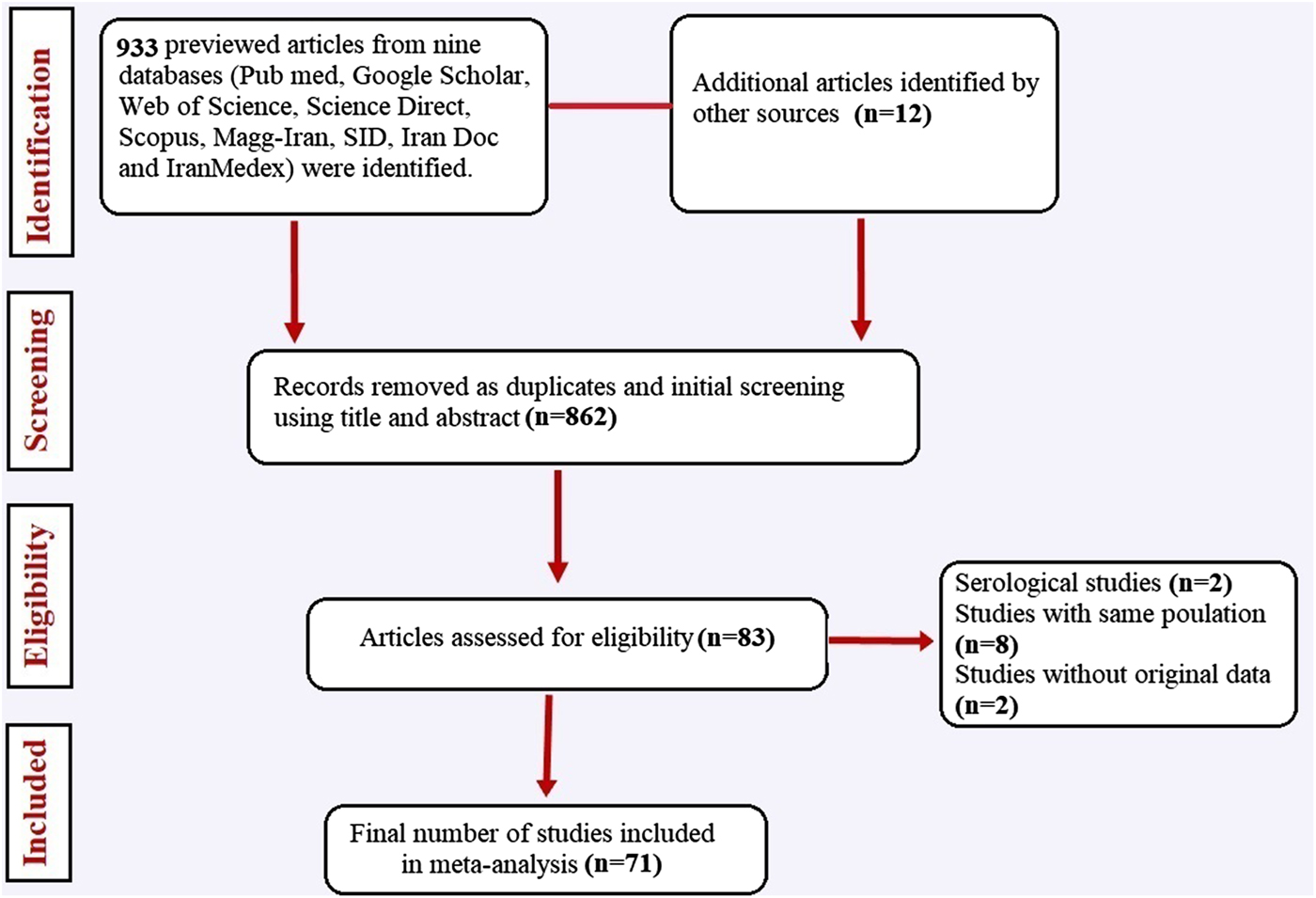
Fig. 1. Flow chart of the study selection process showing inclusion and exclusion of studies identified.

Table 1. Main characteristics of selected studies reporting the prevalence of Entamoeba complex in Iran
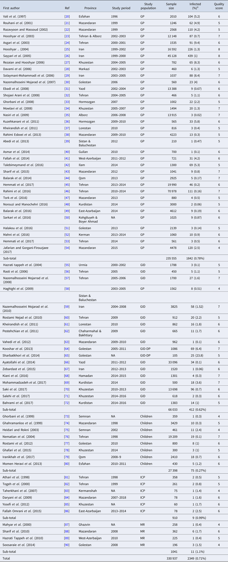
GP, general population; MR, mentally retarded patients; ICP, immunocompromised patients; GID, patients with gastrointestinal disorders; GID-DP, diarrheic patients with gastrointestinal disorders.

Studies are listed in order of year published.

a The percentages presented in this table are crude.

Table 2. Main characteristics of studies reporting molecular distinguish of Entamoeba complex

a The percentages presented in this table are crude.

Results of meta-analysis on prevalence using microscopic results

The pooled prevalence of Entamoeba infection among Iranian general population from 1995 to 2017 was 1% (95% CI 0.8–2.0%), however, there was significant heterogeneity in this meta-analysis (I 2 = 98.65%, P < 0.001) (Fig. 2 and Supplementary Fig. S1). A similar prevalence was observed among patients with gastrointestinal disorders, immunocompromised patients and mentally retarded patients 1% (95% CI 0.8–2.0%). The slowly lower prevalence was observed among children 1% (95% CI 0.0–1.0%) (Fig. 2). As shown in Figure 3 and Supplementary Figure S2, meta-regression analysis on implementation year demonstrated that it is a crucial source of heterogeneity (tau2 deceased from 0.0002 to 0) and prevalence of Entamoeba infection reduced during the time (1995–2017) (b = 0.009 P = 0.001 R 2 = 100%). Subgroup analysis regarding HDI demonstrated that prevalence of Entamoeba infection is higher in undeveloped provinces (2%, 95% CI 1–4%) compared with relatively developed (1%, 95% CI 1–3%) or developed provinces (1%, 95% CI 0–1%) (Supplementary Fig. S3). Meta-regression on the prevalence of Entamoeba infection in different provinces according to geographical latitude/longitude have not yielded any significant results (data not shown).
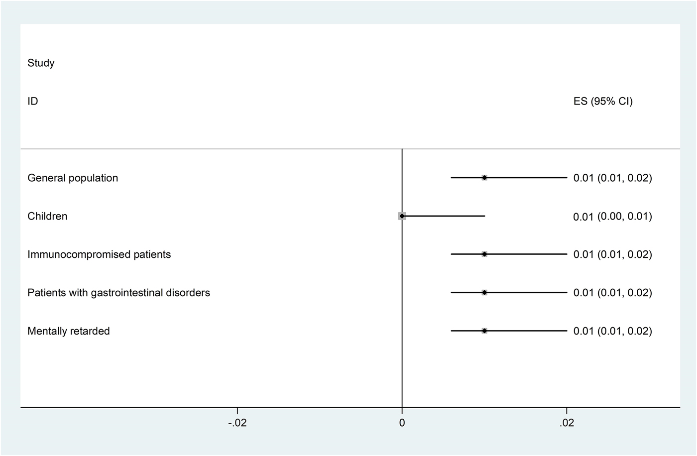
Fig. 2. Forest plot for random-effects meta-analysis on prevalence Entamoeba infection in different groups of Iranian population (using microscopic results).
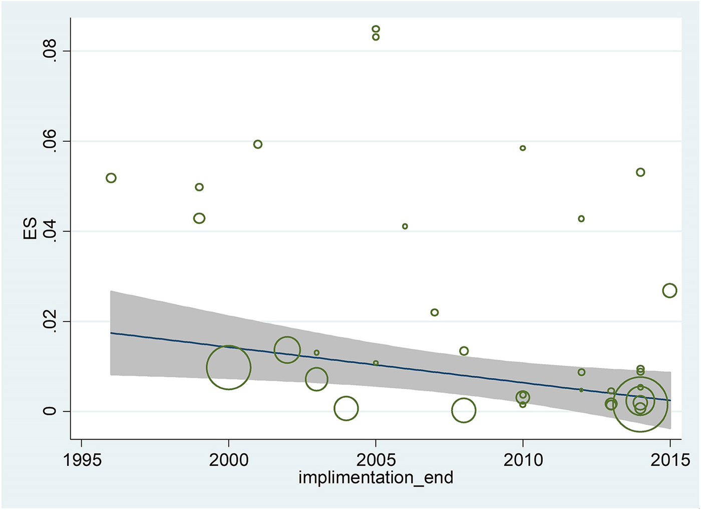
Fig. 3. Meta-regression regarding the effects of during time on the prevalence of Entamoeba infection in Iranian population.

Results of meta-analysis on molecular epidemiology

Seventeen studies involving 44 272 human subjects and 703 Entamoeba complex isolates have performed molecular analysis for inter-species differentiation. Out of 703 Entamoeba complex isolates, 462 isolates were successfully amplified and sequenced. Among these, 396, 55 and 11 isolates were identified as E. dispar E. histolytica and E. moshkovskii, respectively (Table 2). Meta-analysis demonstrated that 83% (95% CI 69–94%; I 2 = 87.8%) and 12% (95% CI 3–24%; I 2 = 85.9%) of isolates were E. dispar and E. histolytica, respectively (Fig. 4a, b). In subgroup analysis based on molecular results, in general population prevalence of E. dispar and E. histolytica were 91% (95% CI 80–99%) and 7% (95% CI 0–19%), while prevalence of these species in patients with gastrointestinal disorders were 75% (95% CI 45–96%) and 18% (95% CI 1–43%), respectively (Fig. 4a, b and Table 2). Related heterogeneities are shown in Figure 4a and b. Due to a low number of E. moshkovskii (11 positive isolates out of only six published articles; two (0.7%) in general population and nine (6%) in patients with gastrointestinal disorders) the prevalence of this Entamoeba based on meta-analysis was not calculable (Table 2).
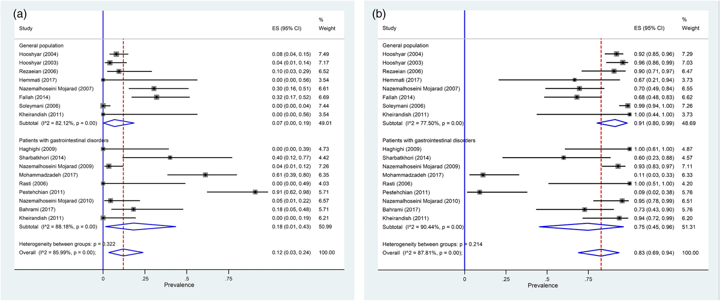
Fig. 4. Forest plot for random-effects meta-analysis on molecular prevalence Entamoeba histolytica (a) and Entamoeba dispar (b) among Iranian general population and patients with gastrointestinal disorders.

Sequence and phylogenetic analyses

The different clades of identified Entamoeba spp. is given in Figure 5 based on the 18S rRNA gene. Cladistic phylogenetic tree indicated the E. dispar clade has a sister relationship with E. histolytica clade in comparison with E. moshkowskii.
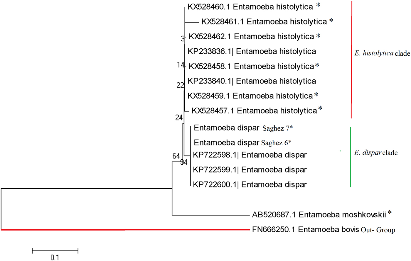
Fig. 5. Phylogenetic analysis of 5S nucleotide sequences of Entamoeba complex isolates recovered from different part of Iran.

Discussion

It is critically important to understand the prevalence of amoebiasis and distributing and molecular epidemiology of Entamoeba complex in developing countries located at tropical and sub-tropical countries. This systematic review and meta-analysis study, based on approximately 331 000 human subjects, 462 PCR-positive Entamoeba complex isolates, resulting from 71 studies, covering 25 provinces in Iran, enables us to judge reliable the prevalence and molecular epidemiology of Entamoeba complex at the national level. Results of our study showed that, nationally, one percent of Iranian people were infected by Entamoeba complex. Prevalence reported here (1%) is much lower than those reported from Ghana (39.8%), South Africa (27%), Mexico (21%), Brazil (21%), India (19%), Malaysia (18.6%) and also lower than reports from Middle East countries, including Yemen (59%), United Arab Emirates (30%), Turkey (2.5%) and Lebanon (2.3%) [Reference Al-Areeqi10, Reference Anuar91Reference Verweij99]. The lower prevalence of Entamoeba complex in Iran could be explained by prevailing of dry climate in most parts of Iran and also higher sanitary status, which had a significant improvement in the three last decades. Our results showed a decrease in the prevalence of Entamoeba complex during the time, indicating that improvement of sanitary status and implementation of health educational programs in the three last decades in Iran was effective to reduce the intestinal parasites including Entamoeba complex. In this present study, we observed the similar prevalence of Entamoeba complex in different population groups. Although in contrast with our results, some previous studies demonstrated that Entamoeba infection is more prevalent in immunocompromised or mentally retarded patients and one of the major health problems in this individuals [Reference Hung100Reference Tachibana104]. This could be explained by the fact that such individuals are suppressed in their immune responses and unable to provide adequate personal hygiene, poor environmental sanitations existing in mentally retarded institutions that is due to lack of toilet training and also direct person-to-person transmission of Entamoeba complex [Reference Sharif88, Reference Moran101]. Another result from the present study was a higher prevalence of Entamoeba infection in undeveloped provinces. In agreement with our results, it is well known that poverty, overcrowding, poor socioeconomic conditions, impoverished sanitation and hygiene conditions, as well as illiteracy and malnutrition, are main factors contributing to the high prevalence of Entamoeba infection [Reference Al-Areeqi10, Reference Lim105, Reference Ngui106].

Regarding the molecular epidemiology of Entamoeba complex in Iran, our result indicated that 396 (83%), 55 (12%) and 11 (2.4%) of the 480 PCR-positive isolates were E. dispar, E. histolytica and E. moshkovskii, respectively. In Yemen and United Arab Emirates, two southern neighbours of Iran, E. histolytica, E. dispar and E. moshkovskii were identified in 44.2%, 34.4% and 39.9% of the 276 PCR-positive products [Reference Al-Areeqi10] and 13.3%, 6.7% and 3.3% of the 120 samples, respectively [Reference ElBakri92]. Results from these studies suggest that in southern neighbour countries of Iran, where hot-humid climate prevails, E. histolytica is more prevalent than E. dispar. While in Turkey, a northwestern neighbour of Iran, study by Kurt et al., 2008 reported that E. histolytica and E. dispar were identified in 23.7% and 52.5% of the 59 PCR-positive products and in a study by Dagci et al., 2007 all obtained isolates were E. dispar, suggesting that E. dispar is the predominant species in Turkey, where hot- or cold-dry climate prevails [Reference Kurt93, Reference Dagci107]. In our study, E. dispar and E. moshkovskii have highest and lowest prevalence in Iranian people. Similar to our results, Anuar et al. [Reference Anuar91] in Malaysia reported that E. dispar was the most prevalent species (13.4%), followed by E. histolytica (3.2%) and E. moshkovskii (1.0%). While in Australia and Colombia E. moshkovskii had a similar prevalence to E. dispar [Reference Fotedar108, Reference López109]. The prevalence E. histolytica, E. dispar and E. moshkovskii were 5.6%, 70.8% and 61.8% in Australia and 0.55%, 23.2% and 25.4% in Colombia, respectively [Reference Fotedar108, Reference López109]. Molecular results observed in the present study highlight higher prevalence of E. histolytica in patients with gastrointestinal disorders (18%) compared with general population (7%) and contrariwise pattern for E. dispar that had a higher prevalence in general population (91%) than patients with gastrointestinal disorders (75%). This result is corroborated by previous studies in different part of world, indicating that E. dispar is responsible for asymptomatic amoebic infection and higher E. histolytica burden is associated with diarrhoeal or gastrointestinal symptoms [Reference Anuar91, Reference Ngobeni95, Reference Gilchrist110Reference Taniuchi112]. An interesting result in our study is a higher prevalence of E. moshkovskii in patients with gastrointestinal disorders (6%) than the general population (0.7%). This result is in agreement with some recent studies suggesting that E. moshkovskii could have potential pathogenic effect in humans [Reference Fotedar5, Reference Ali113, Reference Yakoob114]. In line with these studies, Shimokawa et al. [Reference Shimokawa6], demonstrated that E. moshkovskii is associated with diarrhoea in infants and causes diarrhea and colitis in mice.

The Cladistic phylogenetic tree disclosed that the E. moshkowskii has a greater genetic variability (a distinct branch with distance scale 20%) than E. histolytica/E. dispar clades (Fig. 5). Mohammadzadeh et al.[Reference Mohammadzadeh69] have recently identified a new mutant of E. moshkovskii in a dysentery fecal sample from Saghez city, Kurdistan province, Northwest Iran. This indicates that occurrence of single nucleotide polymorphism can potentially play a pivotal role on pathogenicity rate of E. moshkovskii in clinical isolates. However, more studies with a higher case number are required on ethnic population from different geographic regions of Iran, in order to verify this assumption.

The strengths of this study included the large number of included studies, very large and diverse baseline population, covering different provinces and geographical areas of Iran, rigorous methodology, presentation of pooled data to highlight differences within and between different population groups, determination of molecular epidemiology regarding to high number of Entamoeba isolates and genetic characterisation of Entamoeba species. Moreover, this study is likely limited by significant heterogeneity existing between studies, lacking data for some few provinces and also underestimation the true prevalence, due to the different proficiency of the experimenter in included studies.

In conclusion, despite these limitations, this systematic review and meta-analysis study provides a comprehensive overview of the prevalence and molecular epidemiology of amoebiasis in Iran. We have found that there is the low burden of Entamoeba complex infection (1%) in Iran, although lower developed areas were more influenced. Moreover, our results have shown that less- or non-phatogenic Entamoeba isolate (E. dispar) is the predominant specie in Iran. The decline in prevalence of Entamoeba complex observed across time is likely due to growing standards of living and improved hygiene status in Iran in last years. These data should be taken into consideration by the health authorities of the country and, given the very low incidence of amoebiasis, proper diagnosis and treatment of patients should be done correctly. We suggest additional investigations to further clarify the prevalence of amoebiasis in Iran based on both epidemiological and molecular studies, to guide the development of appropriate public health interventions.

Supplementary material

The supplementary material for this article can be found at https://doi.org/10.1017/S0950268818001863

Acknowledgements

The authors would like to thank Dr Vahid Fallah Omrani, for his assistance during the preparation of this manuscript.

Author contributions

A.R., S.M.R., A.T, A.S, M.E.D, M.M and A.H. conceived the study; A.R., A.T. and A.H initially searched the literature; A.R. A.T, A.S and M.M. collected all data; A.R. S.M.R, A.T and A.H., assessed the included articles; A.R., A.H., M.E.D and S.M.R. analysed and interpreted the data; A.R, A.S and A.H drafted the manuscript; and all authors commented on the drafts of the manuscript and approved the final draft of the paper.

Conflict interest

The authors declare that there is no conflict of interests regarding the publication of this paper.

References

1.Fotedar, R et al. (2007) Laboratory diagnostic techniques for Entamoeba species. Clinical Microbiology Reviews 20, 511532.Google Scholar
2.Royer, TL et al. (2012) Entamoeba bangladeshi nov. sp., Bangladesh. Emerging Infectious Diseases 18, 1543.Google Scholar
3.Haque, R et al. (2003) Amebiasis. New England Journal of Medicine 348, 15651573.Google Scholar
4.Ximénez, C et al. (2009) Reassessment of the epidemiology of amebiasis: state of the art. Infection, Genetics and Evolution 9, 10231032.Google Scholar
5.Fotedar, R et al. (2008) Entamoeba moshkovskii infections in Sydney, Australia. European Journal of Clinical Microbiology & Infectious Diseases 27, 133137.Google Scholar
6.Shimokawa, C et al. (2012) Entamoeba moshkovskii is associated with diarrhea in infants and causes diarrhea and colitis in mice. The Journal of Infectious Diseases 206, 744751.Google Scholar
7.Ali, IKM, Clark, CG and Petri, WA (2008) Molecular epidemiology of amebiasis. Infection, Genetics and Evolution 8, 698707.Google Scholar
8.Nowak, P, Mastalska, K and Loster, J (2015) Entamoeba histolytica-pathogenic protozoan of the large intestine in humans. Journal of Clinical Microbiology and Biochemical Technology 1, 010017.Google Scholar
9.Murray, CJ et al. (2013) Disability-adjusted life years (DALYs) for 291 diseases and injuries in 21 regions, 1990–2010: a systematic analysis for the Global Burden of Disease Study 2010. The Lancet 380, 21972223.Google Scholar
10.Al-Areeqi, MA et al. (2017) First molecular epidemiology of Entamoeba histolytica, E. dispar and E. moshkovskii infections in Yemen: different species-specific associated risk factors. Tropical Medicine & International Health 22, 493504.Google Scholar
11.Wolfe, MS (1973) Nondysenteric intestinal amebiasis: treatment with diloxanide furoate. JAMA 224, 16011604.Google Scholar
12.SCI. Statistical Centre of Iran (2012) Summary and Statistical Report of the 2012 Population and Housing Census. Tehran: Population Census Commission Mazandaran Province: Population Census Commission Amol County.Google Scholar
13.Moher, D et al. (2015) Preferred reporting items for systematic review and meta-analysis protocols (PRISMA-P) 2015 statement. Systematic Reviews 4, 1.Google Scholar
14.Munn, Z et al. (2014) The development of a critical appraisal tool for use in systematic reviews addressing questions of prevalence. International Journal of Health Policy and Management 3, 123.Google Scholar
15.Sabermahani, A et al. (2013) Provincial human development index, a guide for efficiency level analysis: the case of Iran. Iranian Journal of Public Health 42, 149.Google Scholar
16.Freeman, MF and Tukey, JW (1950) Transformations related to the angular and the square root. The Annals of Mathematical Statistics 21, 607611.Google Scholar
17.Harris, R et al. (2008) Metan: fixed-and random-effects meta-analysis. Stata Journal 8, 3.Google Scholar
18.Nyaga, VN, Arbyn, M and Aerts, M (2014) Metaprop: a Stata command to perform meta-analysis of binomial data. Archives of Public Health 72, 39.Google Scholar
19.Riahi, SM and Mokhayeri, Y (2017) Methodological issues in a meta-analysis. Current Medical Research and Opinion 33, 18131813.Google Scholar
20.Vali, GR et al. (1997) Prevalence of parasitic infection among food distributors in Kashan, 1996 . Feyz 1, 4552.Google Scholar
21.Rouhani, S, Kianian, H and Athari, A (2001) Prevalence of intestinal parasites in rural area of Sari, city 1999. Journal of Zanjan University of Medical Sciences 9, 3340.Google Scholar
22.Razavyoon, T and Massoud, J (2002) Intestinal parasitic infections in Fraydoon Kenar, Mazandaran. Journal of School of Public Health and Institute of Public Health Research 1, 3949.Google Scholar
23.Hooshyar, H, Rezaian, M and Kazemi, B (2003) Distribution and differential diagnosis of Entamoeba histolytica from Entamoeba dispar by the PCR-RFLP method in Central Iran. Annals of Saudi Medicine 23, 363366.Google Scholar
24.Asgari, G, Nateghpour, M and Rezaian, M (2003) Prevalence of intestinal parasites in the inhabitants of islam – shahr district. Journal of School of Public Health and Institute of Public Health Research 1, 6774.Google Scholar
25.Hooshyar, H et al. (2004) The distribution of Entamoeba histolytica and Entamoeba dispar in northern, central, and southern Iran. Parasitology Research 94, 96100.Google Scholar
26.Sayyari, A et al. (2005) Prevalence of intestinal parasitic infections in the Islamic Republic of Iran. Eastern Mediterranean Health Journal 11, 377383.Google Scholar
27.Rezaian, M and Hooshyar, H (2006) Differential diagnosis of Entamoeba histolytica from Entamoeba dispar and a study on the intestinal parasites in rural areas of Ahwaz and Hamidieh. Journal of School of Public Health and Institute of Public Health Research 4, 3338.Google Scholar
28.Davami, MH et al. (2006) An investigation on the prevalence of intestinal Parasitic infections in food handlers in Arak. Pars of Jahrom University of Medical Sciences 3, 815.Google Scholar
29.Solaymani-Mohammadi, S et al. (2006) Comparison of a stool antigen detection kit and PCR for diagnosis of Entamoeba histolytica and Entamoeba dispar infections in asymptomatic cyst passers in Iran. Journal of Clinical Microbiology 44, 22582261.Google Scholar
30.Nazemalhoseini Mojarad, E et al. (2007) Prevalence of Entamoeba histolytica and Entamoeba dispar in Gonbad City, 2006, Iran. Iranian Journal of Parasitology 2, 4852.Google Scholar
31.Ebadi, M et al. (2008) Parasitic infections (Helminth and Protozoa) in cases referring to Yazd Central Laboratory, 2002–2004. The Journal of Shahid Sadoughi University of Medical Sciences 15, 5358.Google Scholar
32.Shojaei Arani, A et al. (2008) Prevalence of intestinal parasites in a population in south of Tehran, Iran. Revista do Instituto de Medicina Tropical de São Paulo 50, 145149.Google Scholar
33.Ghorbani, GhA, Izadi, M and Esfahani, AA (2008) Association of drinking water and prevalence of intestinal parasites in military persons. Journal of Military Medicine 10, 159166.Google Scholar
34.Mowlavi, G et al. (2008) Prevalence of intestinal parasites in tribal parts of Khuzestan province during 2005–07. Govaresh 12, 219228.Google Scholar
35.Nasiri, V et al. (2009) Intestinal parasitic infections among inhabitants of Karaj City, Tehran province, Iran in 2006–2008. The Korean Journal of Parasitology 47, 265.Google Scholar
36.Kuzehkanani, AB et al. (2011) Enteric protozoan parasites in rural areas of Bandar-Abbas, southern Iran: comparison of past and present situation. Iranian Journal of Public Health 40, 80.Google Scholar
37.Kheirandish, F et al. (2011) Differential diagnosis of Entamoeba spp. in gastrointestinal disorder patients in Khorramabad, Iran. African Journal of Microbiology Research 5, 28632866.Google Scholar
38.Rahimi Esboei, B et al. (2013) Laboratories performance after outsourcing in the hospitals of Shahid Beheshti University of Medical Sciences. Medical Laboratory Journal 7, 3741.Google Scholar
39.Abedi, M et al. (2013) Prevalence study of intestinal parasitic infections among Health Card applicants Zabol city in 2012. Journal of Zabol University of Medical Sciences and Health Services 5, 5359.Google Scholar
40.Asmar, M et al. (2014) Prevalence of intestinal Parasitic infections in the Urban Areas of Bandar Anzali, Northern Iran. Journal of Guilan University of Medical Sciences 22, 1825.Google Scholar
41.Fallah, E et al. (2014) Differential detection of Entamoeba histolytica from Entamoeba dispar by parasitological and nested multiplex polymerase chain reaction methods. Journal of Analytical Research in Clinical Medicine 2, 2732.Google Scholar
42.Talebimeymand, F, Abasian, L and Rashnavadi, M (2016) Investigating the prevalence of intestinal parasites in Ilam City in 2014. Scientific Journal of Ilam University of Medical Sciences 24, 17.Google Scholar
43.Sharif, M et al. (2015) Prevalence of intestinal parasites among food handlers of Sari, Northern Iran. Revista do Instituto de Medicina Tropical de São Paulo 57, 139144.Google Scholar
44.Balarak, D, Jaffari Modrek, M and Ansari, H (2014) Prevalence of intestinal parasites among the food handlers in the City of Qom, 2014. Community Health Journal 8, 2028.Google Scholar
45.Hemmati, A et al. (2017) Molecular epidemiology and drug resistance study of Entamoeba histolytica in clinical isolates from Tehran, Iran. Journal of Applied Biotechnology Reports 3, 513517.Google Scholar
46.Rahimi, M et al. (2016) The prevalence of intestinal parasites in the patients referred to the laboratories of Baqiyatallah hospital during 2010–2014. Journal of Ardabil University of Medical Sciences 15, 414422.Google Scholar
47.Tork, M et al. (2016) Prevalence of intestinal parasitic infections and associated risk factors in West of Mazandaran province, Iran. Journal of Mazandaran University of Medical Sciences 25, 8188.Google Scholar
48.Norouzi, R and Manochehri, A (2016) Prevalence of intestinal parasites in refereed patients to Shahid Ghazi hospital of Sanandaj province in the year of 2014. Journal of Jiroft University of Medical Sciences 2, 126131.Google Scholar
49.Balarak, D et al. (2016) Prevalence of intestinal parasitic infection among food handlers in Northwest Iran. Journal of Parasitology Research 2016, 8461965.Google Scholar
50.Sarkari, B et al. (2016) Prevalence and risk factors of intestinal protozoan infections: a population-based study in rural areas of Boyer-Ahmad district, Southwestern Iran. BMC Infectious Diseases 16, 703.Google Scholar
51.Halakou, A et al. (2016) Investigating the intestinal parasitic infections in Gonbad e Kavous in 2013. Experimental Animal Biology 4, 7581.Google Scholar
52.Mahni, MB et al. (2016) Prevalence of intestinal parasitic infections in Jiroft, Kerman Province, Iran. Iranian Journal of Parasitology 11, 232.Google Scholar
53.Hemmati, N et al. (2017) Prevalence and risk factors of human intestinal parasites in Roudehen, Tehran province, Iran. Iranian Journal of Parasitology 12, 364.Google Scholar
54.Jafarian, F and Gorgani-Firouzjaee, T (2017) Frequency of parasitic infections among patients referred to Ayatollah Rohani Hospital in Babol, Northern of Iran (2015). Novin Health Journal 1, 3440.Google Scholar
55.Hazrati tappeh, K et al. (2004) A study on frequency the intestinal parasite infections in patients referring to Ghods Clinic of Urmia Medical Sciences University during 78–81. Journal of Nursing and Midwifery Urmia University of Medical Sciences 2, 1120.Google Scholar
56.Rasti, S, Haghighi, A and Hatami, M (2006) Differential diagnosis of Entamoeba histolytica from Entamoeba dispar by PCR. Feyz Journal of Kashan University of Medical Sciences 10, 3034.Google Scholar
57.Nazemalhosseini Mojarrad, E et al. (2008) Genetic diversity among Entamoeba histolytica and Entamoeba dispar strains in gastrointestinal disorder patients in Tehran, Iran. Research in Medicine 32, 213220.Google Scholar
58.Haghighi, A et al. (2009) Frequency of enteric protozoan parasites among patients with gastrointestinal complaints in medical centers of Zahedan, Iran. Transactions of the Royal Society of Tropical Medicine and Hygiene 103, 452454.Google Scholar
59.Nazemalhosseini Mojarad, E et al. (2010) Discrimination of Entamoeba moshkovskii in patients with gastrointestinal disorders by single-round PCR. Japanese Journal of Infectious Diseases 63, 136138.Google Scholar
60.Rostami Nejad, M et al. (2010) Prevalence of intestinal parasites in patients with gastrointestinal symptoms by focus on soil-transmitted helminthes infection. Gastroenterology and Hepatology from Bed to Bench 3, 190194.Google Scholar
61.Kheirandish, F et al. (2011) Prevalence of intestinal parasites in bakery workers in Khorramabad, Lorestan Iran. Iranian Journal of Parasitology 6, 76.Google Scholar
62.Pestehchian, N et al. (2011) Frequency of Entamoeba histolytica and Entamoeba dispar prevalence among patients with gastrointestinal complaints in Chelgerd city, southwest of Iran. Journal of Research in Medical Sciences 16, 1436.Google Scholar
63.Vahedi, M et al. (2012) Prevalence of parasites in patients with gastroenteritis at East of Mazandaran province, Northern Iran. Tropical Biomedicine 29, 568574.Google Scholar
64.Kooshar, F et al. (2013) Frequency of intestinal parasites in diarrhea patients in Gorgan, 2005–2011. Medical Laboratory Journal 4, 5460.Google Scholar
65.Sharbatkhori, M et al. (2014) Discrimination of Entamoeba spp. in children with dysentery. Gastroenterology and Hepatology from Bed to Bench 7, 164.Google Scholar
66.Ayatollahi, J et al. (2014) Frequency of intestinal parasites in the patients referred to the central and Shahid Sadoughi Laboratories of Yazd (2011–2012). Tolooebehdasht 13, 4857.Google Scholar
67.Zebardast, N et al. (2015) Frequency of intestinal parasites in patients with gastrointestinal disorders, in different parts of Iran during 2012–2013. International Journal of Enteric Pathogens 3, e22682.Google Scholar
68.Kiani, H et al. (2016) Prevalence, risk factors and symptoms associated to intestinal parasite infections among patients with gastrointestinal disorders in nahavand, western Iran. Revista do Instituto de Medicina Tropical de São Paulo 58, 42.Google Scholar
69.Mohammadzadeh, A et al. (2017) Gene migration for re-emerging amebiasis in Iran's northwest–Iraq borders: a microevolutionary scale for reflecting epidemiological drift of Entamoeba histolytica metapopulations. Parasitology Research 116, 217224.Google Scholar
70.Saki, J et al. (2017) Prevalence of intestinal parasitic infections in Haftkel County, Southwest of Iran. International Journal of Infection 4, e15593.Google Scholar
71.Salehi, R et al. (2017) Prevalence and subtype identification of Blastocystis isolated from humans in Ahvaz, Southwestern Iran. Gastroenterology and Hepatology from Bed to Bench 10, 235.Google Scholar
72.Bahrami, F et al. (2017) Differential detection of Entamoeba histolytica, Entamoeba dispar and Entamoeba moshkovskii in fecal samples by a nested multiplex PCR in the west of Iran. Shahid Beheshti University of Medical Science 2017, 2533.Google Scholar
73.Ghorbani, R, Pazooki, R and Ahmadian, AR (1999) The prevalence of intestinal parasites and relative factors in children below 2 years old in the urban areas of Semnan in 1996–97. Journal of Gorgan University of Medical Sciences 1, 3945.Google Scholar
74.Ghahramanloo, M, Hassanjani Roshan, M and Haji Ahmadi, M (1999) Prevalence of intestinal parasites in primary school children, Eastern Bandpay, Babol ,1999. Journal of Babol University of Medical Science 3, 4751.Google Scholar
75.Heidari, A and Rokni, M (2003) Prevalence of intestinal parasites among children in day-care centers in Damghan-Iran. Iranian Journal of Public Health 32, 3134.Google Scholar
76.Nematian, J et al. (2004) Prevalence of intestinal parasitic infections and their relation with socio-economic factors and hygienic habits in Tehran primary school students. Acta Tropica 92, 179186.Google Scholar
77.Rostami, M et al. (2012) The prevalence of intestinal parasitic infections in primary school students in Gorgan, Iran. Medical Laboratory Journal 6, 4246.Google Scholar
78.Ghafari, R, Rafiei, A and Tavalla, M (2015) Prevalence of intestinal parasites among children referred to Abozar Hospital in Ahvaz. Jundishapur Scientific Medical Journal 13, 627633.Google Scholar
79.Irankhah, A, Aghaali, M and Damanpak Moghaddam, V (2017) Prevalence of intestinal parasitic diseases in the students of Qom City and hygiene status of their schools, Iran. QOM University of Medical Sciences Journal 10: 6170.Google Scholar
80.Momen Heravi, M et al. (2013) Prevalence of intestinal parasites infections among Afghan children of primary and junior high schools residing Kashan city, Iran, 2009–2010. Iranian Journal of Medical Microbiology 7, 4652.Google Scholar
81.Athari, A, Sadafi, H and Touke, G (2000) Prevalence of intestinal parasites among patients consumed of immunosuppressive drugs in Tehran, Iran. Journal of Zanjan University of Medical Sciences 8, 6168.Google Scholar
82.Togeh, GR et al. (2000) Parasitic infestation in cancer patients chemotherapy. Tehran University Medical Journal 58, 5258.Google Scholar
83.Taherkhani, H et al. (2007) The frequency of intestinal parasites in HIV positive patients admitted to the disease consultation center in Kermanshah province. Medical Laboratory Journal 1, 4144.Google Scholar
84.Daryani, A et al. (2009) Prevalence of intestinal parasites and profile of CD4+ counts in HIV+/AIDS people in north of Iran, 2007–2008. Pakistan Journal of Biological Sciences: PJBS 12, 12771281.Google Scholar
85.Yosefi, F et al. (2012) A study on prevalence of gastrointestinal parasitic infections in HIV (+) patients referred to Ahvaz Razi Hospital in 2008–2009. Jundishapur Journal of Microbiology 5, 424426.Google Scholar
86.Fallah Omrani, V et al. (2015) Prevalence of intestinal parasite infections and associated clinical symptoms among patients with end-stage renal disease undergoing hemodialysis. Infection 43, 537544.Google Scholar
87.Mahyar, A et al. (2000) Intestinal parasitic in mentally retarded children of Qazvin. The Journal of Qazvin University of Medical Sciences 4, 6470.Google Scholar
88.Sharif, M et al. (2010) Intestinal parasitic infections Among intellectual disability children in rehabilitation centers of northern Iran. Research in Developmental Disabilities 31, 924928.Google Scholar
89.Hazrati Tappeh, K et al. (2010) Prevalence of intestinal parasitic infections among mentally disabled children and adults of Urmia, Iran. Iranian Journal of Parasitology 5, 60.Google Scholar
90.Soosaraie, M, Pagheh, A and Gholami, S (2014) Prevalence of intestinal parasitic infections in rehabilitation centers in Golestan province, Iran. Medical Laboratory Journal 8, 4247.Google Scholar
91.Anuar, TS et al. (2012) Molecular epidemiology of amoebiasis in Malaysia: highlighting the different risk factors of Entamoeba histolytica and Entamoeba dispar infections among Orang Asli communities. International Journal for Parasitology 42, 11651175.Google Scholar
92.ElBakri, A et al. (2013) Differential detection of Entamoeba histolytica, Entamoeba dispar and Entamoeba moshkovskii in fecal samples by nested PCR in the United Arab Emirates (UAE). Acta Parasitologica 58, 185190.Google Scholar
93.Kurt, O et al. (2008) Investigation of the prevalence of amoebiasis in Izmir province and determination of Entamoeba spp. using PCR and enzyme immunoassay. The New Microbiologica 31, 393.Google Scholar
94.Nath, J et al. (2015) Molecular epidemiology of amoebiasis: a cross-sectional study among North East Indian population. PLoS Neglected Tropical Diseases 9, e0004225.Google Scholar
95.Ngobeni, R et al. (2017) Entamoeba in South Africa: correlations with the host microbiome, parasite burden and first description of E. bangladeshi outside of Asia. The Journal of Infectious Diseases 19, 15921600.Google Scholar
96.Ramos, F et al. (2005) Entamoeba histolytica and Entamoeba dispar: prevalence infection in a rural Mexican community. Experimental Parasitology 110, 327330.Google Scholar
97.Saab, BR et al. (2004) Intestinal parasites among presumably healthy individuals in Lebanon. Saudi Medical Journal 25, 3437.Google Scholar
98.Santos, HLC et al. (2007) Comparison of multiplex-PCR and antigen detection for differential diagnosis of Entamoeba histolytica. Brazilian Journal of Infectious Diseases 11, 365370.Google Scholar
99.Verweij, JJ et al. (2003) Prevalence of Entamoeba histolytica and Entamoeba dispar in northern Ghana. Tropical Medicine & International Health 8, 11531156.Google Scholar
100.Hung, C-C et al. (2008) Increased risk for Entamoeba histolytica infection and invasive amebiasis in HIV seropositive men who have sex with men in Taiwan. PLoS Neglected Tropical Diseases 2, e175.Google Scholar
101.Moran, P et al. (2005) Entamoeba histolytica and/or Entamoeba dispar: infection frequency in HIV+/AIDS patients in Mexico City. Experimental Parasitology 110, 331334.Google Scholar
102.Ramakrishnan, K et al. (2007) Prevalence of intestinal parasitic infestation in HIV/AIDS patients with diarrhea in Madurai City, South India. Japanese Journal of Infectious Disease 60, 209210.Google Scholar
103.Rivera, WL, Santos, SR and Kanbara, H (2006) Prevalence and genetic diversity of Entamoeba histolytica in an institution for the mentally retarded in the Philippines. Parasitology Research 98, 106110.Google Scholar
104.Tachibana, H et al. (2000) Asymptomatic cyst passers of Entamoeba histolytica but not Entamoeba dispar in institutions for the mentally retarded in Japan. Parasitology International 49, 3135.Google Scholar
105.Lim, Y et al. (2009) Intestinal parasitic infections amongst Orang Asli (indigenous) in Malaysia: has socioeconomic development alleviated the problem? Tropical Biomedicine 26, 110122.Google Scholar
106.Ngui, R et al. (2011) Prevalence and risk factors of intestinal parasitism in rural and remote West Malaysia. PLoS Neglected Tropical Diseases 5, e974.Google Scholar
107.Dagci, H et al. (2007) Differentiation of Entamoeba histolytica and Entamoeba dispar by PCR: a preliminary study in Izmir, Turkey. The New Microbiologica 30, 45.Google Scholar
108.Fotedar, R et al. (2007) PCR detection of Entamoeba histolytica, Entamoeba dispar, and Entamoeba moshkovskii in stool samples from Sydney, Australia. Journal of Clinical Microbiology 45, 10351037.Google Scholar
109.López, MC et al. (2015) Molecular epidemiology of Entamoeba: first description of Entamoeba moshkovskii in a rural area from Central Colombia. PLoS ONE 10, e0140302.Google Scholar
110.Gilchrist, CA et al. (2016) Role of the gut microbiota of children in diarrhea due to the protozoan parasite Entamoeba histolytica. The Journal of Infectious Diseases 213, 15791585.Google Scholar
111.Haghighi, A et al. (2003) Geographic diversity among genotypes of Entamoeba histolytica field isolates. Journal of Clinical Microbiology 41, 37483756.Google Scholar
112.Taniuchi, M et al. (2013) Etiology of diarrhea in Bangladeshi infants in the first year of life analyzed using molecular methods. The Journal of Infectious Diseases 208, 17941802.Google Scholar
113.Ali, IKM et al. (2003) Entamoeba moshkovskii infections in children in Bangladesh. Emerging Infectious Diseases 9, 580.Google Scholar
114.Yakoob, J et al. (2012) Entamoeba species associated with chronic diarrhoea in Pakistan. Epidemiology & Infection 140, 323328.Google Scholar
Figure 0

Fig. 1. Flow chart of the study selection process showing inclusion and exclusion of studies identified.

Figure 1

Table 1. Main characteristics of selected studies reporting the prevalence of Entamoeba complex in Iran

Figure 2

Table 2. Main characteristics of studies reporting molecular distinguish of Entamoeba complex

Figure 3

Fig. 2. Forest plot for random-effects meta-analysis on prevalence Entamoeba infection in different groups of Iranian population (using microscopic results).

Figure 4

Fig. 3. Meta-regression regarding the effects of during time on the prevalence of Entamoeba infection in Iranian population.

Figure 5

Fig. 4. Forest plot for random-effects meta-analysis on molecular prevalence Entamoeba histolytica (a) and Entamoeba dispar (b) among Iranian general population and patients with gastrointestinal disorders.

Figure 6

Fig. 5. Phylogenetic analysis of 5S nucleotide sequences of Entamoeba complex isolates recovered from different part of Iran.

Supplementary material: File

Haghighi et al. supplementary material

Haghighi et al. supplementary material 1

Download Haghighi et al. supplementary material(File)
File 12.2 KB
Supplementary material: Image

Haghighi et al. supplementary material

Haghighi et al. supplementary material 2

Download Haghighi et al. supplementary material(Image)
Image 358 KB
Supplementary material: Image

Haghighi et al. supplementary material

Haghighi et al. supplementary material 3

Download Haghighi et al. supplementary material(Image)
Image 467.6 KB
Supplementary material: Image

Haghighi et al. supplementary material

Haghighi et al. supplementary material 4

Download Haghighi et al. supplementary material(Image)
Image 366.3 KB