EDITOR:
The standard entry point for abdominal insufflation and primary port site placement in gynaecological laparoscopy is the umbilicus. However, where there has been previous abdominal surgery, which increases the risk of bowel adhesions beneath the umbilicus, the use of Palmer’s point (the left subcostal area in the mid-clavicular line) has been advocated [Reference Childers, Brzechffa and Surwit1]. The principal risks associated with this entry technique are damage to the stomach or spleen. Facemask ventilation with positive pressure prior to intubation is a necessary part of anaesthesia, but it is known to insufflate the stomach, and even more so than other forms of ventilation [Reference Ho-Tai, Devitt, Noel and O’Donnell2]. The purpose of this study was to determine whether the insertion of a naso-gastric tube would reduce the degree of gastric distension and, hence, reduce the potential risk of gastric injury during subcostal insertion of a Veress needle and the primary port.
This was a single blinded randomized controlled trial, approved by the local Ethics Committee at the University College London Hospitals’ Foundation Trust. Between June 2005 and March 2006, 42 patients were recruited. Patients were allocated randomly into those who were to receive a naso-gastric tube (NGT Group) and those who were not to have one (no NGT Group). The chosen population consisted of females, ASA Grades I and II, aged 18 and above, undergoing elective gynaecological surgery with planned umbilical port incision. Patients having planned naso-gastric tube or subcostal port insertion were excluded.
After pre-assessment and consenting, anaesthetic induction was performed using intravenous fentanyl 1.5 mcg kg−1, propofol 3 mg kg−1 and vecuronium 0.15 mg kg−1. The same anaesthetist, in all cases, ventilated the patient’s lungs for a period of 2 min using a facemask with 50% oxygen and nitrous oxide mixture and 1.5% isoflurane. After tracheal intubation, an assistant opened the randomization envelope. Only those patients assigned to the NGT Group had a naso-gastric tube passed with a laryngoscope and Magill’s forceps. Placement of the naso-gastric tube was confirmed by air insufflation and auscultation over the stomach. The naso-gastric tube was subsequently aspirated. Next, to blind the surgeon, the head of the patient was covered with a sheet before being transported to the operating theatre. Anaesthesia was maintained with the gas mixture as described above.
At laparoscopy, the scope was pointed upwards in the abdomen, and the degree of stomach distension was assessed. The same surgeon (AC) made all the assessments. Gastric distension was graded according to a visual assessment scale, with a scale of ‘0’ indicating minimum distension and ‘3’ indicating maximum. Surgery proceeded as planned, and the naso-gastric tube was removed prior to emergence.
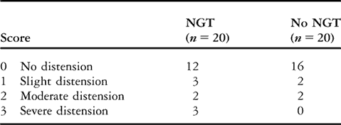
Out of the 42 patients recruited in the study, two patients were excluded, one due to accidental extra-peritoneal insufflation and the other due to inadvertent opening of the randomization envelope. Patients in both groups were matched for age, weight, ASA Grades and airway. The results are summarized in Table 1. An empty stomach (score ‘0’: no distension) was seen in 16 out of 20 patients in the NGT Group compared to 12 out of 20 in the no NGT Group (not significant). In addition, three patients were assessed as having a high risk of gastric injury (score ‘3’: severe distension) and all were in the no NGT Group. The prevalence of severe distension in both the groups was not significantly different. No adverse effects occurred as a result of naso-gastric tube placement in the NGT Group. Although the results were not statistically significant, they convey clinically important information.
Table 1 Visual assessment scale for intraoperative grading of gastric distension.

NGT: naso-gastric tube.
Discussion
Gastric injury during laparoscopy is a serious, but very rare event. Those cases reported in the literature were promptly managed, and no deaths resulted [Reference Reynolds and Pauca3–Reference Erian5]. In a review article by Van der Voort and colleagues [Reference Van der Voort, Heijnsdijk and Gouma6], the incidence of laparoscopy induced gastrointestinal injury was 0.13% and that of bowel perforation 0.22%. Recently, Nezhat and colleagues [Reference Nezhat, de Fazio and Nezhat7] reported a case of gastric perforation following umbilical port insertion for laparoscopic ovarian cyst resection. Most of the cases were reported in the 1970s and are likely to have involved umbilical entry techniques.
Subcostal entry is frequently used to reduce risk of bowel injury in patients who are at an increased risk. However, while avoiding lower abdominal bowel injury, gastric injury may result if the stomach is distended. Donnez and colleagues [Reference Donnez, Bassil and Nisolle8] have reported stomach perforation with a 5-mm port during subcostal insertion. Several factors may influence the degree of gastric distension. These include the use of facemask ventilation, operator experience and patient anxiety. The latter may predispose to gastric distension prior to the induction of anaesthesia, and this has been the cause to attribute gastric perforation in two cases of pelvic laparoscopy [Reference Endler and Moghissi9].
The decision to insert a naso-gastric tube on a prophylactic basis for reducing gastric distension should be weighed against the risk of complications. These include minor risks of naso-pharyngeal trauma and inadvertent insertion into the pulmonary tree. More serious risks and rare sequelae include the tip being lodged within the nasal cavity during removal, perforation of the naso-pharynx and formation of aorto-oesophageal fistulae [Reference Minyard and Smith10].
In our study, gastric distension was significantly more common in patients without a naso-gastric tube and, furthermore, there was substantial potential for gastric perforation in three subjects (all of whom did not have naso-gastric tubes). This gives a risk incidence of 15% (three out of 20) if no naso-gastric tube is sited. There were no associated covariates (age, weight, ASA Grade or airway manageability) relating to these three patients that may have predisposed them to develop distended stomachs. With this method of assessment, the pilot study raises the issue of gastric injury during subcostal port insertion when a naso-gastric tube is not sited. In addition, it would be interesting to observe the effect of varying the size of the naso-gastric tube, but that was not covered in our study. In conclusion, this study supports naso-gastric tube placement when the subcostal approach is used for insertion of the primary port during laparoscopy. A larger study would, however, be required to confirm this.


