Those with childhood-onset schizophrenia, age at onset less than 12 years, have smaller brains (9% on average), enlarged lateral ventricles and smaller mid-thalamic areas (Reference Frazier, Giedd and HamburgerFrazier et al, 1996). Follow-up studies indicate a progressive reduction in temporal lobe volumes and medial temporal lobe structures, including the hippocampus (Reference Jacobsen and RapoportJacobsen & Rapoport, 1998). Progressive ventricular enlargement (P<0.0001) and a reduction in cerebral volumes (P<0.0001) from childhood to adolescence reach an asymptote in late adolescence (Reference Giedd, Jeffries and BlumenthalGiedd et al, 1999). Patients with childhood-onset schizophrenia have up to a fourfold greater rate of loss of grey matter affecting the frontal, parietal and temporal lobes (Reference Rapoport, Giedd and BlumenthalRapoport et al, 1999). Both early and later progressive changes imply that a static neurodevelopmental lesion is not an adequate explanation. Later neurodevelopmental abnormalities of synaptic pruning (Reference FeinbergFeinberg, 1983), apoptosis (Reference WoodsWoods, 1998), altered cortical plasticity (Reference De LisiDe Lisi, 1999) or subtle neurodegenerative processes, particularly in those with poor outcome (Reference Lieberman, Chakos and WuLieberman et al, 2001), have been implicated.
METHOD
Subjects
A total of 16 subjects diagnosed with adolescent-onset schizophrenia following a semi-structured interview (K-SADS; Reference Kaufman, Birmaher and BrentKaufman et al, 1997) according to DSM-III-R criteria for schizophrenia (296) (American Psychiatric Association, 1987) and 16 matched controls were included in a follow-up study. The subjects and controls were part of an initial study of 29 subjects and 20 controls (Reference James, Crow and RenowdenJames et al, 1999). The same protocol was used throughout the study and there were no systematic differences between those who participated and those who defaulted from follow-up in terms of demography and brain structure volumes measured on magnetic resonance imaging (MRI) scan at time 1. Patients and controls with mental impairment (IQ<70) and those with histories of head injuries or neurological disorder such as cerebral palsy, encephalitis, epilepsy, etc. were excluded. Normal controls were recruited from the community via their general practitioner, with screening for any history of emotional, behavioural or medical problems. All subjects and controls attended normal schools. Unfortunately, owing to initial difficulties in recruiting controls, the follow-up period differed between the groups. Comparison of those who agreed to complete the study with those who declined showed no differences with respect to demographics and initial brain structure volumes. The study was carried out under the auspices of the Oxford Psychiatric Research Ethics Committee (OPREC no. 95/43).
The patients were a severely ill group but they were compliant with medication throughout the study period. The majority received typical neuroleptics at the onset of the study; although most were transferred to atypical neuroleptics, three were treatment resistant and on clozapine.
Magnetic resonance imaging
Subjects were scanned on a General Electric Signa 1.5 Tesla MRI machine, which remained the same throughout the study with regular quality control checks. The chin was elevated so that the volumetric gradient echo sequence (which cannot be angled) was perpendicular to the temporal lobe, to minimise partial volume effects. The initial two scans were to ensure correct patient orientation — the anterior genu of the corpus callosum and the clivus should follow a vertical line. These sequences were repeated if necessary to ensure a horizontal anterior commissure—posterior commissure (AC-PC) line. Image sequences were as follows:
-
(a) Sagittal T1-weighted spin echo (time echo, TE=300 ms; field of view, FOV=24 cm × 24 cm; slice thickness=5 mm; slice gap=0 mm; matrix=256 × 128; number of excitations, NEX=-0.5; slices=9).
-
(b) A coronal volumetric T1-weighted radio-frequency-spoiled gradient echo, SPRG (TE=5 ms; repetition time, TR=35 ms; flip=35°; FOV=20 cm × 20 cm; thickness=3 mm; matrix=256 × 256; NEX=1.0; slices=64).
Anatomical markers
The temporal lobe was defined posteriorly at the level where all four colliculi were visualised. Temporal lobe and medial temporal lobe structures were measured manually on sequential coronal slices. The temporal stem was demarcated by a line connecting the most inferior point of the insular cisterns to the most lateral point of the basal cisterns above the hippocampus. Medially, the boundary between the temporal lobe and cerebrum was determined by drawing a perpendicular line from the most inferior aspect of the Sylvian fissure across the narrowest portion of the temporal lobe. The hippocampus and amygdala were outlined using a manual tracing method. The hippocampus was defined posteriorly by the separation of the crus of the fornix from the hippocampus, and anteriorly from the head of the amygdala by the uncal recess of the inferior horn of the lateral ventricle. The anterior amygdala was measured only in those slices where the grey matter was 2.5 times the thickness of the adjacent cortical grey matter. Images were displayed in three-dimensional orthogonal views using the RESCUE program (Reference Griffin, Colchester, Röll and HancockGriffin et al, 1994). A hierarchical semi-automated method of segmentation of grey—white matter of the temporal lobes was undertaken using RESCUE. The third ventricle was defined posteriorly at the level of the suprapineal recess and anteriorly at the level of the anterior commissure. Lateral ventricular volumes included measurement of the temporal horn. Total brain volumes were measured with the cerebellar tonsils as the inferior marker. The cerebral hemispheres were separated from the brain-stem at the superior limit of the pons. All measurements, including assessments of interrater reliability (A. C. D. J., A. J.), were made blind to diagnosis. The hippocampal and amygdala measurements were undertaken by one rater (A. C. D. J.).
Reliability studies
Inter-/intrarater reliability studies were undertaken with three raters (A. C. D. J., S. J., A. J.). Intraclass correlation coefficients (ICCs) (Reference BartkoBartko, 1966) were 0.95 for total brain volume, 0.94 for ventricular volume, 0.87 for amygdala volume, and 0.90 for hippocampal volume.
Statistical methods
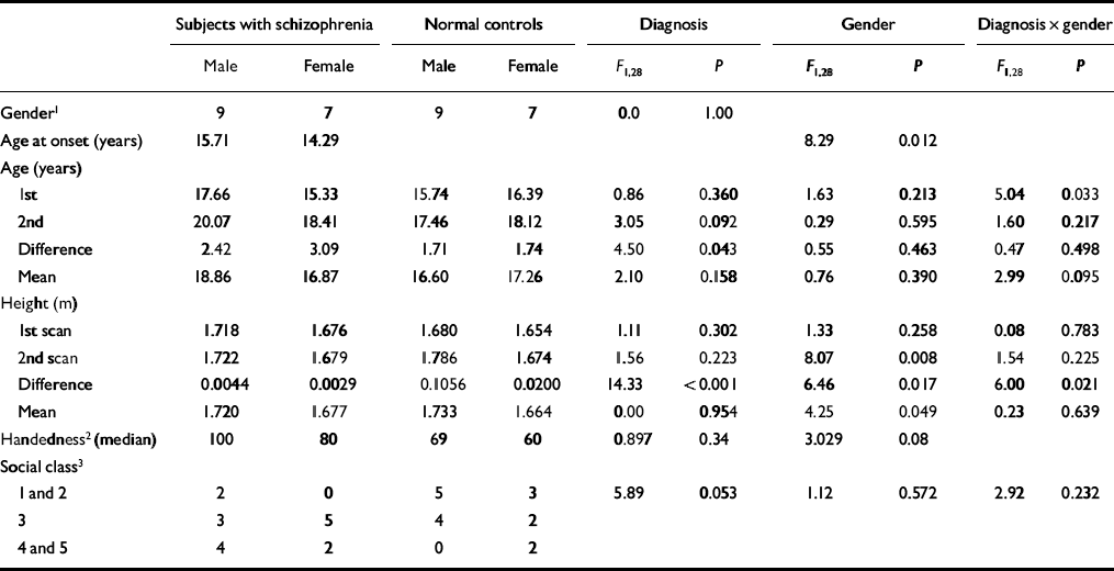
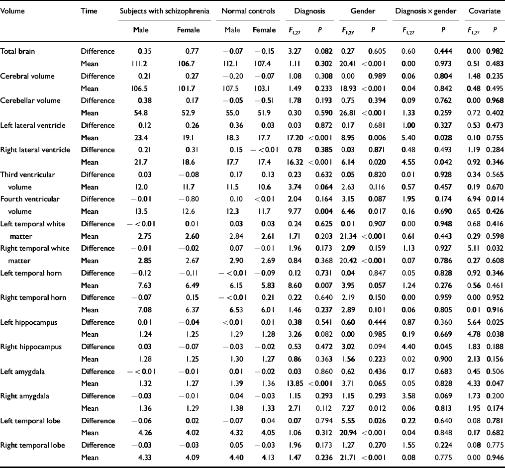
Categorical variables such as gender were analysed by χ2 tests (Table 1). Handedness was analysed using the Kruskal—Wallis test. The majority of the variables, in particular the volumes, were analysed by analysis of variance (ANOVA), where the model concerned involved diagnosis (schizophrenia, normal), gender (male, female) and their interaction. It was decided to cube-root-transform the volumes prior to performing the ANOVA. This was done because generally they showed a strong mean variance relationship (ANOVA assumes equal variances) and because volume is (distance)3. An initial analysis of the transformed volume results at the first and second measurement times strongly suggested that there were no differences between the two diagnosis groups with regard to the change between the two times but that there were differences between the averages. To confirm this, the volumes were re-expressed as difference and mean between and over the two measurement times. The results of the analyses of differences and means are given in Table 2. An adjustment for age differences was made by introducing age difference into the ANOVA as a covariate for the difference in cube-rooted volumes and mean age for the mean cube-rooted volumes. For some of the volumes these covariates were significant, so the results are reported here. The means given in Table 2 are those for cube-rooted volumes after adjustment for differences in the covariate value.
Table 1 Demographic details

| Subjects with schizophrenia | Normal controls | Diagnosis | Gender | Diagnosis × gender | ||||||
|---|---|---|---|---|---|---|---|---|---|---|
| Male | Female | Male | Female | F 1,28 | P | F 1,28 | P | F 1,28 | P | |
| Gender1 | 9 | 7 | 9 | 7 | 0.0 | 1.00 | ||||
| Age at onset (years) | 15.71 | 14.29 | 8.29 | 0.012 | ||||||
| Age (years) | ||||||||||
| 1st | 17.66 | 15.33 | 15.74 | 16.39 | 0.86 | 0.360 | 1.63 | 0.213 | 5.04 | 0.033 |
| 2nd | 20.07 | 18.41 | 17.46 | 18.12 | 3.05 | 0.092 | 0.29 | 0.595 | 1.60 | 0.217 |
| Difference | 2.42 | 3.09 | 1.71 | 1.74 | 4.50 | 0.043 | 0.55 | 0.463 | 0.47 | 0.498 |
| Mean | 18.86 | 16.87 | 16.60 | 17.26 | 2.10 | 0.158 | 0.76 | 0.390 | 2.99 | 0.095 |
| Height (m) | ||||||||||
| 1st scan | 1.718 | 1.676 | 1.680 | 1.654 | 1.11 | 0.302 | 1.33 | 0.258 | 0.08 | 0.783 |
| 2nd scan | 1.722 | 1.679 | 1.786 | 1.674 | 1.56 | 0.223 | 8.07 | 0.008 | 1.54 | 0.225 |
| Difference | 0.0044 | 0.0029 | 0.1056 | 0.0200 | 14.33 | <0.001 | 6.46 | 0.017 | 6.00 | 0.021 |
| Mean | 1.720 | 1.677 | 1.733 | 1.664 | 0.00 | 0.954 | 4.25 | 0.049 | 0.23 | 0.639 |
| Handedness2(median) | 100 | 80 | 69 | 60 | 0.897 | 0.34 | 3.029 | 0.08 | ||
| Social class3 | ||||||||||
| 1 and 2 | 2 | 0 | 5 | 3 | 5.89 | 0.053 | 1.12 | 0.572 | 2.92 | 0.232 |
| 3 | 3 | 5 | 4 | 2 | ||||||
| 4 and 5 | 4 | 2 | 0 | 2 | ||||||
Table 2 Cube-rooted volume measurements, as differences and means

| Volume | Time | Subjects with schizophrenia | Normal controls | Diagnosis | Gender | Diagnosis × gender | Covariate | ||||||
|---|---|---|---|---|---|---|---|---|---|---|---|---|---|
| Male | Female | Male | Female | F 1,27 | P | F 1,27 | P | F 1,27 | P | F 1,27 | P | ||
| Total brain | Difference | 0.35 | 0.77 | -0.07 | -0.15 | 3.27 | 0.082 | 0.27 | 0.605 | 0.60 | 0.444 | 0.00 | 0.982 |
| Mean | 111.2 | 106.7 | 112.1 | 107.4 | 1.11 | 0.302 | 20.41 | <0.001 | 0.00 | 0.973 | 0.51 | 0.483 | |
| Cerebral volume | Difference | 0.21 | 0.27 | -0.20 | -0.07 | 1.08 | 0.308 | 0.00 | 0.989 | 0.06 | 0.804 | 1.48 | 0.235 |
| Mean | 106.5 | 101.7 | 107.5 | 103.1 | 1.49 | 0.233 | 18.93 | <0.001 | 0.04 | 0.842 | 0.48 | 0.495 | |
| Cerebellar volume | Difference | 0.38 | 0.17 | -0.05 | -0.51 | 1.78 | 0.193 | 0.75 | 0.394 | 0.09 | 0.762 | 0.00 | 0.968 |
| Mean | 54.8 | 52.9 | 55.0 | 51.9 | 0.30 | 0.590 | 26.81 | <0.001 | 1.33 | 0.259 | 0.72 | 0.402 | |
| Left lateral ventricle | Difference | 0.12 | 0.26 | 0.36 | 0.03 | 0.03 | 0.872 | 0.17 | 0.681 | 1.00 | 0.327 | 0.53 | 0.473 |
| Mean | 23.4 | 19.1 | 18.3 | 17.7 | 17.20 | <0.001 | 8.95 | 0.006 | 5.40 | 0.028 | 0.10 | 0.755 | |
| Right lateral ventricle | Difference | 0.21 | 0.31 | 0.15 | -<0.01 | 0.78 | 0.385 | 0.03 | 0.871 | 0.48 | 0.493 | 1.19 | 0.284 |
| Mean | 21.7 | 18.6 | 17.7 | 17.4 | 16.32 | <0.001 | 6.14 | 0.020 | 4.55 | 0.042 | 0.92 | 0.346 | |
| Third ventricular | Difference | 0.03 | -0.08 | 0.17 | 0.13 | 0.23 | 0.632 | 0.05 | 0.820 | 0.01 | 0.928 | 0.34 | 0.565 |
| volume | Mean | 12.0 | 11.7 | 11.5 | 10.6 | 3.74 | 0.064 | 2.63 | 0.116 | 0.57 | 0.457 | 0.19 | 0.670 |
| Fourth ventricular | Difference | -0.01 | -0.80 | 0.10 | <0.01 | 2.04 | 0.164 | 3.15 | 0.087 | 1.95 | 0.174 | 6.94 | 0.014 |
| volume | Mean | 13.5 | 12.6 | 12.3 | 11.7 | 9.77 | 0.004 | 6.46 | 0.017 | 0.16 | 0.690 | 0.65 | 0.426 |
| Left temporal white | Difference | — <0.01 | 0.01 | 0.03 | 0.03 | 0.24 | 0.625 | 0.01 | 0.907 | 0.00 | 0.948 | 0.68 | 0.416 |
| matter | Mean | 2.75 | 2.60 | 2.84 | 2.61 | 1.71 | 0.203 | 21.34 | <0.001 | 0.61 | 0.443 | 0.29 | 0.598 |
| Right temporal white | Difference | -0.01 | -0.02 | 0.07 | -0.01 | 1.96 | 0.173 | 2.09 | 0.159 | 1.13 | 0.927 | 5.11 | 0.032 |
| matter | Mean | 2.85 | 2.67 | 2.90 | 2.69 | 0.84 | 0.368 | 20.42 | <0.001 | 0.07 | 0.786 | 0.27 | 0.608 |
| Left temporal horn | Difference | -0.12 | -0.11 | — <0.01 | -0.09 | 0.12 | 0.731 | 0.04 | 0.847 | 0.05 | 0.828 | 0.92 | 0.346 |
| Mean | 7.63 | 6.49 | 6.15 | 5.83 | 8.60 | 0.007 | 3.95 | 0.057 | 1.24 | 0.276 | 0.56 | 0.461 | |
| Right temporal horn | Difference | -0.07 | 0.15 | — <0.01 | 0.21 | 0.22 | 0.640 | 2.19 | 0.150 | 0.00 | 0.959 | 0.00 | 0.952 |
| Mean | 7.08 | 6.37 | 6.53 | 6.01 | 1.46 | 0.237 | 2.89 | 0.101 | 0.06 | 0.805 | 0.01 | 0.916 | |
| Left hippocampus | Difference | 0.01 | -0.04 | <0.01 | 0.01 | 0.38 | 0.541 | 0.60 | 0.444 | 0.87 | 0.360 | 5.64 | 0.025 |
| Mean | 1.24 | 1.25 | 1.29 | 1.28 | 3.26 | 0.082 | 0.00 | 0.985 | 0.19 | 0.669 | 4.78 | 0.038 | |
| Right hippocampus | Difference | 0.03 | -0.07 | -0.03 | -0.02 | 0.53 | 0.472 | 3.02 | 0.094 | 4.40 | 0.045 | 1.83 | 0.188 |
| Mean | 1.28 | 1.25 | 1.30 | 1.27 | 0.86 | 0.363 | 1.56 | 0.223 | 0.02 | 0.900 | 2.13 | 0.156 | |
| Left amygdala | Difference | — <0.01 | -0.01 | 0.01 | -0.02 | 0.03 | 0.860 | 0.62 | 0.436 | 0.17 | 0.683 | 0.45 | 0.506 |
| Mean | 1.32 | 1.27 | 1.39 | 1.36 | 13.85 | <0.001 | 3.71 | 0.065 | 0.05 | 0.828 | 4.33 | 0.047 | |
| Right amygdala | Difference | -0.03 | -0.01 | 0.04 | -0.03 | 1.15 | 0.293 | 1.15 | 0.293 | 3.58 | 0.069 | 1.73 | 0.200 |
| Mean | 1.36 | 1.29 | 1.38 | 1.33 | 2.71 | 0.112 | 7.27 | 0.012 | 0.06 | 0.813 | 1.95 | 0.174 | |
| Left temporal lobe | Difference | -0.06 | 0.02 | -0.07 | 0.04 | 0.07 | 0.794 | 5.55 | 0.026 | 0.22 | 0.640 | 0.08 | 0.781 |
| Mean | 4.26 | 4.02 | 4.32 | 4.05 | 1.06 | 0.312 | 20.94 | <0.001 | 0.04 | 0.848 | 0.17 | 0.682 | |
| Right temporal lobe | Difference | -0.03 | -0.03 | 0.05 | -0.03 | 1.96 | 0.173 | 1.27 | 0.270 | 1.55 | 0.224 | 0.08 | 0.775 |
| Mean | 4.33 | 4.09 | 4.40 | 4.13 | 1.47 | 0.236 | 21.71 | <0.001 | 0.08 | 0.775 | 0.00 | 0.946 | |
For bilateral volumes the asymmetry, calculated as:
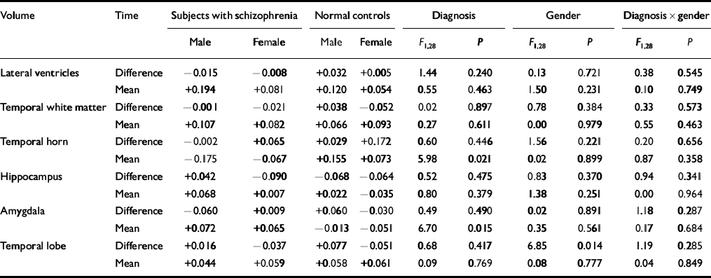
was re-expressed as differences and means and analysed in the same way as the volumes, except that the volumes were not transformed before the asymmetry was calculated. The results of these analyses are given in Table 3. Analyses of the asymmetries were conducted with handedness introduced into the ANOVA as a covariate. However, for all asymmetries the covariate was not significant and the conclusions did not change, so the results of these analyses are not reported.
Table 3 Asymmetry, as differences and means

| Volume | Time | Subjects with schizophrenia | Normal controls | Diagnosis | Gender | Diagnosis × gender | |||||
|---|---|---|---|---|---|---|---|---|---|---|---|
| Male | Female | Male | Female | F 1,28 | P | F 1,28 | P | F 1,28 | P | ||
| Lateral ventricles | Difference | -0.015 | -0.008 | +0.032 | +0.005 | 1.44 | 0.240 | 0.13 | 0.721 | 0.38 | 0.545 |
| Mean | +0.194 | +0.081 | +0.120 | +0.054 | 0.55 | 0.463 | 1.50 | 0.231 | 0.10 | 0.749 | |
| Temporal white matter | Difference | -0.001 | -0.021 | +0.038 | -0.052 | 0.02 | 0.897 | 0.78 | 0.384 | 0.33 | 0.573 |
| Mean | +0.107 | +0.082 | +0.066 | +0.093 | 0.27 | 0.611 | 0.00 | 0.979 | 0.55 | 0.463 | |
| Temporal horn | Difference | -0.002 | +0.065 | +0.029 | +0.172 | 0.60 | 0.446 | 1.56 | 0.221 | 0.20 | 0.656 |
| Mean | -0.175 | -0.067 | +0.155 | +0.073 | 5.98 | 0.021 | 0.02 | 0.899 | 0.87 | 0.358 | |
| Hippocampus | Difference | +0.042 | -0.090 | -0.068 | -0.064 | 0.52 | 0.475 | 0.83 | 0.370 | 0.94 | 0.341 |
| Mean | +0.068 | +0.007 | +0.022 | -0.035 | 0.80 | 0.379 | 1.38 | 0.251 | 0.00 | 0.964 | |
| Amygdala | Difference | -0.060 | +0.009 | +0.060 | -0.030 | 0.49 | 0.490 | 0.02 | 0.891 | 1.18 | 0.287 |
| Mean | +0.072 | +0.065 | -0.013 | -0.051 | 6.70 | 0.015 | 0.35 | 0.561 | 0.17 | 0.684 | |
| Temporal lobe | Difference | +0.016 | -0.037 | +0.077 | -0.051 | 0.68 | 0.417 | 6.85 | 0.014 | 1.19 | 0.285 |
| Mean | +0.044 | +0.059 | +0.058 | +0.061 | 0.09 | 0.769 | 0.08 | 0.777 | 0.04 | 0.849 | |
RESULTS
A pattern of generalised ventricular (lateral, 3rd and 4th ventricle) enlargement that was roughly constant over time was found. The differences in the volumes of the ventricles between times 1 and 2 were not significant for those with schizophrenia and the normal controls, whereas the mean values over time were. Within both diagnosis groups, for all ventricle volumes the male brain structures were bigger than those in females. A significant gender by diagnosis interaction was evident for the left and right lateral and total ventricular volumes. The total ventricular volumes of those with schizophrenia were approximately 87% and 24% greater than those of the normal controls for males and females, respectively. The outstanding feature was the comparatively large size for the males with schizophrenia. Apart from the ventricles, the only volumes displaying a significant difference between diagnosis groups were the left temporal horn mean and the left amygdala mean. For left temporal horn volumes the general pattern of the mean volumes was the same as for total ventricular volumes, where those with schizophrenia were approximately 79% and 39% greater than the controls for males and females, respectively. For the amygdala volumes, the general pattern of means was different from that for total ventricular and left temporal horn volumes. The left amygdala volumes of the patients with schizophrenia were approximately 12% smaller and 7% smaller than the normal controls for males and females, respectively.
The ‘normal’ pattern (Reference Giedd, Valtuzis and HamburgerGieddet al, 1996) of right greater than left asymmetry for the temporal lobe and hippocampus was evident, if not significant, for the patients with schizophrenia and the normal controls. No differences in asymmetry between times 1 and 2 were significant. Two mean (over times 1 and 2) asymmetries displayed evidence of a difference between diagnostic groups, these being temporal horn and amygdala. The temporal horn showed a left greater than right asymmetry for patients with schizophrenia but a right greater than left asymmetry for the normal controls. For the amygdala the patients with schizophrenia showed a right greater than left asymmetry, whereas the normal controls showed a left greater than right asymmetry.
DISCUSSION
Brain and ventricular changes
There was no progressive decline in total brain volumes in the subjects with schizophrenia. The most striking finding in this sample with adolescent-onset schizophrenia is of a non-progressive, generalised ventricular enlargement. The initial degree of ventricular enlargement is substantial, particularly for males with schizophrenia. The pattern for the lateral ventricular volumes suggests that male patients with schizophrenia have substantially enlarged lateral ventricles, whereas the lateral ventricles of female patients are only marginally bigger than those of female normal controls. This is the same pattern displayed in the total ventricular volumes. This pattern is consistent with that of the meta-analysis of Wright et al (Reference Wright, Rabe-Hesketh and Woodruff2000) and with the gender dimorphic picture seen in schizophrenia research, with males having an earlier onset, poorer outcome, greater neuropsychological deficits and structural brain abnormalities (Reference Leung and ChueLeung & Chue, 2000). The disease process, although generalised in both males and females, is perhaps initially more active and severe in males. The findings of a static total ventricular enlargement in adolescence would imply a generalised brain disorder, with the initial ventricular enlargement in childhood, before the onset of schizophrenic symptoms. The initial scans were done at first presentation, on average 18 months (s.d.=13 months) after the appearance of the psychosis. The age of onset of psychosis in this study ranged from 12.75 years to 16.5 years (mean=15.1, s.d.=1.1), suggesting that the initial changes of ventricular enlargement occurred before this date. This contrasts with the conclusions of Giedd et al (Reference Giedd, Jeffries and Blumenthal1999) that the changes are progressive in late adolescence.
Temporal lobe
There were no differences between groups in temporal lobe volumes, or temporal lobe grey or white matter volumes, or over time. Several longitudinal studies of first-episode adult patients have failed to find progressive temporal lobe volume changes (De Lisi et al, Reference De Lisi, Tew and Xie1995, Reference De Lisi, Sakuma and Tew1997; Reference Gur, Cowell and TuretskyGur et al, 1998), although there are reports of loss of left superior temporal gyral volumes (Reference Hirayasu, Shenton and SalisburyHirayasu et al, 1999) and loss of grey matter (Reference Mathalon, Sullivan and LimMathalon et al, 2001) over periods of 1 to 4 years. The findings contrast with the reported loss of 7% of temporal grey matter (Reference Rapoport, Giedd and BlumenthalRapoport et al, 1999) in adolescents with childhood-onset schizophrenia over a 4-year period, and a recent study of 100 non-chronic patients where the loss of temporal lobe grey matter was 7% for men and 8.5% for women (Reference Gur, Cowell and FinkelmanGur et al, 2000). The findings of a left amygdala volume reduction of 15% (95% CI 4-25) is slightly larger than the 9% (95% CI 6-13) in meta-analytical studies (Reference Wright, Rabe-Hesketh and WoodruffWright et al, 2000) and greater than any other temporal lobe volume reduction. A time-yoked study of 42 adolescents with childhood-onset schizophrenia and 74 matched controls over three time periods showed relative stability of the amygdala and a non-linear reduction in hippocampal volumes (Reference Giedd, Jeffries and BlumenthalGiedd et al, 1999). Although not all studies have reported hippocampal reductions, recent meta-analyses (Reference Nelson, Saykin and FlashmanNelson et al, 1998) indicate bilateral reductions (effect size: 0.37 left, 0.39 right). There was a trend towards a reduction in left hippocampal volume at time 2 (F=3.4, P=0.07), which is in line with the findings of loss of hippocampal volume during adolescence after onset of the illness (Reference Matsumoto, Simmons and WilliamsMatsumoto et al, 2001).
Gender dimorphism
Male subjects with schizophrenia have larger lateral ventricles. Despite others' findings of gender dimorphism in the amygdala changing with age (Reference Goldstein, Kennedy and CavinessGoldstein et al, 1999; Reference Gur, Cowell and FinkelmanGur et al, 2000), here there were no gender by diagnosis interactions. Female subjects with schizophrenia consistently had the smallest amygdala. Bryantet al (Reference Bryant, Buchanan and Vladar1999) argue that temporal lobe structures are gender dimorphic, with male subjects with schizophrenia having smaller temporal lobe volumes. A meta-analysis (Reference Wright, Rabe-Hesketh and WoodruffWright et al, 2000) found little supporting evidence for a gender effect. All the structures examined were larger in males, with a gender by diagnosis interaction only for the right hippocampus.
Asymmetry
In this study the pathology in adolescent-onset schizophrenia appears to be predominantly left-sided with left temporal horn enlargement, together with a reduced left amygdala volume. The left-lateralised changes have been noted previously (Reference Crow, Ball and BloomCrow et al, 1989; Reference Bogerts, Ashtari and DegreefBogerts et al, 1990) and have been hypothesised to be of aetiological significance to the aberrant neurodevelopment of schizophrenia (Reference CrowCrow, 1997), particularly in view of the lateralisation to the left temporal lobe of certain language functions.
Clinical Implications and Limitations
CLINICAL IMPLICATIONS
-
• The brain changes, which probably reflect neurodevelopmental abnormalities, are non-progressive during late adolescence.
-
• Significant ventricular enlargement at the outset of the illness suggests that global brain changes occur prior to the development of the psychosis.
-
• Males appear to be affected more severely.
LIMITATIONS
-
• The small numbers involved limit the power of the study.
-
• The imaging protocol with 3-mm slices is limited.
-
• The differing period of follow-up for the subjects and controls makes comparisons more problematic.
Acknowledgements
The authors are grateful for support and help from Professor T. Crow, Dr P. Anslow and Rebecca Craven. The authors are particularly grateful for the help and cooperation of the patients, controls and families involved and the Donnington Health Centre, Oxford.


eLetters
No eLetters have been published for this article.