To the Editor
It was with great pleasure that we recently read two papers in your journal, one regarding the history of neuropsychiatry and the other about the comorbidity between neurological illness and psychiatric disorders. We totally agree that “neuropsychiatry is a discipline in its own right and not just a wing of psychiatry or a bridge between neurology and psychiatry,”Reference Trimble 1 while on the other hand we truly grieve the “lack of partnerships between psychiatrists and neurologists and the paucity of neuropsychiatrists.”Reference Hesdorffer 2
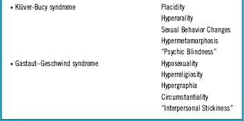
Both articles reminded us of a patient of ours who came to us presenting with an incredible collection of neuropsychiatric symptoms that many neurologists would easily recognize as classic syndromes related to temporal lobe dysfunction, mimicking both Klüver–Bucy syndrome (KBS) and Gastaut–Geschwind syndrome (GGS)Reference Trimble, Mendez and Cummings 3 (Table 1).
Table 1 Neuropsychiatric symptoms from the temporal lobes (adapted from Trimble, 1997)

A 33-year-old man was admitted for aggressive behavior secondary to auditory hallucinations and messianic delusions: “The voice of God was calling me! I am the new Jesus, the son of Joseph and Mary.”
In his extensive clinical file, we found 13 psychiatric admissions in the previous 10 years. We easily found enough information compatible with various affective and nonaffective psychotic episodes to suggest a diagnosis of schizoaffective disorder, going back at least to when he was 18 years of age.
The patient had been on many different combinations of benzodiazepines, antidepressants, mood stabilizers, and antipsychotics. Various attempts with psychotherapy, group therapy, occupational therapy, and electroconvulsive therapy were also made. Full recovery was never achieved due to complete absence of insight, and severe burnout was quite evident among his family and caregivers. The patient’s mother suffered from Hashimoto’s thyroiditis and his younger brother from postpartum hypoxic–ischemic encephalopathy with secondary epilepsy and cerebral palsy. Descriptions of his father’s and grandfather’s behavior were highly suggestive of what we could describe as undisclosed personality disorders.
Both family interview and a full-records review pointed to fluctuating bizarre behavior throughout his life that included: hypergraphia (writing on hundreds of pages of paper); hyperreligiosity (wearing a rosary and praying for overlong periods of time); interpersonal stickiness (viscosity in social contact, even with strangers); circumstantiality (being very obsessive with irrelevant details during conversation); changes in sexual behavior (alternating practice of voyeurism, exibicionism, onanism, mechanophilia, gerontophilia, and incestual and bisexual satyriasis); hypermetamorphosis (uncontrolled urge to touch other people’s genitalia); hyperorality (pica, coprophagia, indiscriminate fellatio practice with other patients); and placidity (tameness, docility, never showing any embarrassment about his socially disturbing behavior). No other changes were found on anamnesis and neurologic or psychiatric exams.
Although the Gastaut–Geschwind and the Klüver–Bucy syndromes were fluctuating and alternating, they were both undoubtedly present, at many points during his clinical evolution, and that called for a better assessment of the patient. We thus started a diagnostic workup in order to discard a possible psychotic disorder due to another medical condition.
Initial routine electroencephalography presented diffuse slowing, but these changes were not found on 24-hour EEG. Brain magnetic resonance imaging showed mild frontal lobe atrophy with corpus callosum thinning, while brain technetium exametazime (99mTc-HMPAO) single-photon emission computed tomography (SPECT) showed bilateral discrete decrease of temporal lobes perfusion (Figure 1). Repeated lumbar puncture showed no changes at all, even when we looked for exquisite causes of encephalitis such as N-methyl-d-aspartate receptor antibodies. Neuropsychological evaluation pointed to moderate impairment of attention and associative memory. No other findings were relevant, including karyotyping, blood work, and urine drug tests.

Figure 1 Brain 99-mTc HMPAO single-photon emission computed tomography showing bilateral discrete decrease of temporal lobes perfusion.
Surprisingly, the initial diagnosis of schizoaffective disorder was confirmed, and we stabilized the patient with oral daily doses of lorazepam (7.5 mg), aripiprazol (30 mg), and clozapine (800 mg). He was then transferred to a state prison hospital ward, following a judicial decision by the court, accused of sexual assault.
Blood flow decrease in both temporal areas has been recently described in brain SPECT of schizophrenic patients.Reference Wake, Miyaoka and Kawakami 4 We believe that these kinds of neurophysiological changes might be present in patients with schizoaffective or schizophrenia disorder, resulting in Klüver–Bucy and/or Gastaut–Geschwind syndrome, as some authors have suggested,Reference Tracy, de Leon, Qureshi, McCann, McGrory and Josiassen 5 – Reference Gama Marques, Teixeira and Carnot 7 depending on the temporal lobe structures affected by progressive physiopathology. These kinds of patients are clear examples on how psychiatry and neurology still need clinical neuropsychiatry to grow as an independent and strong field in the area of the neurosciences and mental health.
Disclosures
Dr. Gama Marques has nothing to disclose.


