Hostname: page-component-78c5997874-s2hrs Total loading time: 0 Render date: 2024-11-05T16:19:59.286Z Has data issue: false hasContentIssue false

‘Scientific Strabismus’ or two related pandemics: coronavirus disease and vitamin D deficiency

Published online by Cambridge University Press:  12 May 2020

Murat Kara
Affiliation:
Department of Physical Medicine and Rehabilitation, Hacettepe University Medical School, Ankara, Turkey
Timur Ekiz*
Affiliation:
Department of Physical Medicine and Rehabilitation, Türkmenbaşı Medical Center, Adana, Turkey
Vincenzo Ricci
Affiliation:
Department of Biomedical and Neuromotor Science, Physical and Rehabilitation Medicine Unit, IRCCS Rizzoli Orthopaedic Institute, Bologna, Italy
Özgür Kara
Affiliation:
Geriatrics Unit, Yenimahalle Training and Research Hospital, Yıldırım Beyazıt University, Ankara, Turkey
Ke-Vin Chang
Affiliation:
Department of Physical Medicine and Rehabilitation, National Taiwan University Hospital, Bei-Hu Branch, Taipei, Taiwan
Levent Özçakar
Affiliation:
Department of Physical Medicine and Rehabilitation, Hacettepe University Medical School, Ankara, Turkey
*
* Corresponding author: Timur Ekiz, email [email protected]
Rights & Permissions [Opens in a new window]

Abstract

The WHO has announced the novel coronavirus disease (COVID-19) outbreak to be a global pandemic. The distribution of community outbreaks shows seasonal patterns along certain latitude, temperature and humidity, that is, similar to the behaviour of seasonal viral respiratory tract infections. COVID-19 displays significant spread in northern mid-latitude countries with an average temperature of 5–11°C and low humidity. Vitamin D deficiency has also been described as pandemic, especially in Europe. Regardless of age, ethnicity and latitude, recent data showed that 40 % of Europeans are vitamin D deficient (25-hydroxyvitamin D (25(OH)D) levels <50 nmol/l), and 13 % are severely deficient (25(OH)D < 30 nmol/l). A quadratic relationship was found between the prevalences of vitamin D deficiency in most commonly affected countries by COVID-19 and the latitudes. Vitamin D deficiency is more common in the subtropical and mid-latitude countries than the tropical and high-latitude countries. The most commonly affected countries with severe vitamin D deficiency are from the subtropical (Saudi Arabia 46 %; Qatar 46 %; Iran 33·4 %; Chile 26·4 %) and mid-latitude (France 27·3 %; Portugal 21·2 %; Austria 19·3 %) regions. Severe vitamin D deficiency was found to be nearly 0 % in some high-latitude countries (e.g. Norway, Finland, Sweden, Denmark and Netherlands). Accordingly, we would like to call attention to the possible association between severe vitamin D deficiency and mortality pertaining to COVID-19. Given its rare side effects and relatively wide safety, prophylactic vitamin D supplementation and/or food fortification might reasonably serve as a very convenient adjuvant therapy for these two worldwide public health problems alike.

Type
Full Papers
Creative Commons
Creative Common License - CCCreative Common License - BY
This is an Open Access article, distributed under the terms of the Creative Commons Attribution licence (http://creativecommons.org/licenses/by/4.0/), which permits unrestricted re-use, distribution, and reproduction in any medium, provided the original work is properly cited.
Copyright
© The Authors 2020. Published by Cambridge University Press

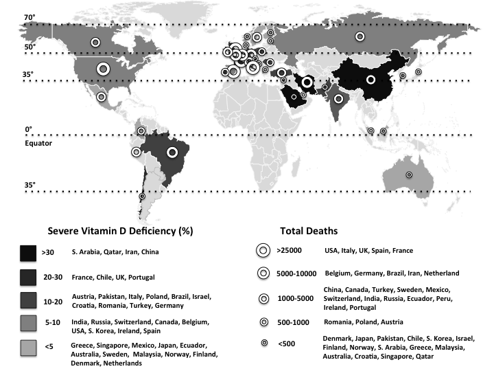
On 11 March 2020, the WHO announced the novel coronavirus disease (COVID-19) outbreak to be a global pandemic(1). The spread of COVID-19 is becoming unstoppable, and as of 15 May, more than 4 500 000 people have been infected and more than 300 000 people have died (Fig. 1)(1). The severe acute respiratory syndrome coronavirus 2 (SARS-Cov-2) is the pathogen of COVID-19. SARS-CoV-2, classified into two β coronaviruses, is an enveloped, positive-sense and single-stranded RNA virus of about 30 kb. The life cycle of the virus with the host comprises mainly five steps as follows: attachment, penetration, biosynthesis, maturation and release. Once SARS-CoV-2 attaches to the host receptors, it penetrates the cells via endocytosis/membrane fusion. Herein, angiotensin-converting enzyme 2 is the entry and functional receptor of SARS-CoV-2. It has been shown that the spike for SARS-CoV-2, structural membrane proteins formed by the trans-membrane trimetric glycoprotein protruding from the viral surface, also binds to angiotensin-converting enzyme 2. After the viral contents are released inside the host cells, viral RNA enters the nucleus to replicate. As for the biosynthesis, viral mRNA is used to make viral proteins. The new viral particles are formed in the maturation step and then released(Reference Yuki, Fujiogi and Koutsogiannaki2).

Fig. 1. The world map illustrates the total deaths and percentage of severe vitamin D deficiency in countries most commonly affected by COVID-19(1,Reference Eymundsdottir, Chang and Geirsdottir5Reference Mechenro, Venugopal and Kumar43) . Severe vitamin D deficiency (%): (), >30 (South Arabia, Qatar, Iran, China); (), 20–30 (France, Chile, UK, Portugal); (), 10–20 (Austria, Pakistan, Italy, Poland, Brazil, Israel, Croatia, Romania, Turkey, Germany); (), 5–10 (India, Russia, Switzerland, Canada, Belgium, USA, South Korea, Ireland, Spain); (), <5 (Greece, Singapore, Mexico, Japan, Ecuador, Australia, Sweden, Malaysia, Norway, Finland, Denmark, Netherlands). Total deaths: (), >25 000 (USA, UK, Italy, France, Spain); (), 5000–10 000 (Brazil, Belgium, Germany, Iran, The Netherlands, Canada); (), 1000–5000 (China, Mexico, Turkey, Sweden, India, Ecuador, Russia, Peru, Switzerland, Ireland, Portugal, Romania); (), 500–1000 (Poland, Pakistan, Japan, Austria, Denmark); (), <500 (Chile, Finland, Saudi Arabia, Israel, South Korea, Norway, Greece, Malaysia, Australia, Croatia, Singapore, Qatar).

Angiotensin-converting enzyme 2 plays an important role for the interaction between the classical and non-classical pathway of the renin angiotensin system. The former acts through the angiotensin II type 1 receptors, and its increased activity leads to fibrosis, inflammation and angiogenesis. The latter acts through the Mas receptors and has opposing effects to the angiotensin II type 1 receptors(Reference Vaduganathan, Vardeny and Michel3). Angiotensin-converting enzyme 2 is expressed by the epithelial cells of lungs, intestines, kidneys and blood vessels; therefore, the aforementioned tissues/organs are vulnerable to SARS-CoV-2 infection(Reference Zou, Chen and Zou4). Additionally, activation of the renin angiotensin system is significantly associated with increased morbidity and mortality as in hypertension(Reference Vaduganathan, Vardeny and Michel3).

On the other hand, vitamin D deficiency has also been described as pandemic and a global public health problem, especially in Europe (Table 1)(Reference Eymundsdottir, Chang and Geirsdottir5Reference Mechenro, Venugopal and Kumar43). Regardless of age, ethnicity and latitude, recent data showed that 40 % of Europeans are vitamin D deficient (25-hydroxyvitamin D (25(OH)D) levels <50 nmol/l), and 13 % are severely deficient (25(OH)D < 30 nmol/l)(Reference Lips, Cashman and Lamberg-Allardt44). According to regression analyses, a quadratic relationship was found between the prevalences of vitamin D deficiency in most commonly affected countries by COVID-19 and the latitudes (Fig. 2). Interestingly, vitamin D deficiency is more common in the subtropical and mid-latitude countries than the tropical and high-latitude countries. Contrary to the expectation, the most commonly affected countries with severe vitamin D deficiency are from the subtropical (Saudi Arabia 46 %; Qatar 46 %; Iran 33·4 %; Chile 26·4 %) and mid-latitude (France 27·3 %; Portugal 21·2 %; Austria 19·3 %) regions. On the other hand, severe vitamin D deficiency was found to be nearly 0 % in some high-latitude countries (e.g. Norway, Finland, Sweden, Denmark and Netherlands). The low prevalences of severe vitamin D deficiencies in high-latitude countries (except for the UK; 23·7 %) can possibly be attributed to the high awareness of vitamin D deficiency, high amount of vitamin D supplementation, food fortification and health policies as well(Reference Lips, Cashman and Lamberg-Allardt44). Indeed, as the main source of vitamin D is exposure of the skin to sun (UV-B), it has long been supposed that living in a sunny country guarantees sufficient vitamin D levels. However, there is increasing evidence that vitamin D deficiency may have been underestimated/ignored in low latitude, even in tropical countries(Reference Mendes, Hart and Botelho45).

Table 1. Available data for vitamin D deficiency among older adults in countries most commonly affected by COVID-19

n, Number; 25(OH)D, 25-hydroxyvitamin D; N/A, not applicable; W, women; M, men.

* The most commonly infected countries and regions with COVID-19 in descending order.

Percentages of severe vitamin D deficiency.

Fig. 2. The histogram shows the prevalence of vitamin D deficiency (<50 nmol/l) and severe deficiency (<25 nmol/l) among the forty countries most commonly affected by COVID-19. The number above each column represents the country’s position in the world ranking concerning the number of total cases of infections. The colour band is a graphical representation of the four main climatic areas in the world. Regression lines show the prevalence of overall (solid black line) and severe (dotted line) vitamin D deficiencies. (), Vitamin D deficiency; (), severe vitamin D deficiency.

The risks for vitamin D deficiency encompass obesity, elderly, lack of proper sun (UV-B) exposure, dark skin, smoking, living with air pollution and the presence of co-morbid diseases such as infection, cancer, CVD, chronic respiratory disease, osteoporosis, sarcopenia and diabetes mellitus(Reference Schleicher, Sternberg and Looker46,Reference Grant, Lahore and McDonnell47) . Further, it is known that severe vitamin D deficiency dramatically increases the risk of mortality, infections and many other diseases. As such, it should indisputably be prevented whenever detected/possible(Reference Schleicher, Sternberg and Looker46).

Vitamin D hormone has important functions – including immunomodulant, anti-inflammatory and anti-infective roles(Reference Grant, Lahore and McDonnell47). It acts via monocyte and cell-mediated immunity stimulation, suppression of lymphocyte proliferation, antibody production and cytokine synthesis(Reference Beard, Bearden and Striker48). Human lung cells are able to intracellularly convert the inactive 25(OH)D to its active form 1,25(OH)D which reduces proinflammatory cytokines and increases peptides (e.g. the innate antimicrobial peptide cathelicidin)(Reference Beard, Bearden and Striker48). Cathelicidin has direct antiviral activity against enveloped respiratory viruses such as hepatitis B, influenza, respiratory syncytial virus and possibly the COVID-19 as well(Reference Beard, Bearden and Striker48). Other than the above-mentioned functions, vitamin D has also anti-fibrotic effects. The renin-inhibiting activity and down-regulation of the renin angiotensin system activity seem to be the beneficial effects of vitamin D. Moreover, vitamin D has been shown to suppress angiotensinogen and regulate its expression(Reference Feldman, Pike and Bouillon49).

The distribution of community outbreaks shows seasonal patterns along certain latitude, temperature and humidity, that is, similar to the behaviour of seasonal viral respiratory tract infections. It has been reported that COVID-19 displays significant spread in mid-latitude (35–50° N′) regions and/or in those with an average temperature of 5–11°C and low humidity (Fig. 1)(1,Reference Sajadi, Habibzadeh and Vintzileos50) . Coronaviruses are very stable at 4°C (viable for up to 3 d) and can survive at −20°C (for up to 2 years)(1). Depending on some parameters (e.g. temperature, humidity and sunlight), they can live on different surfaces for a few days. They are thermolabile; decreased sunlight, low temperatures and less humidity seem to be favourable for COVID-19(1). Although natural UV (UV-C) from the sunlight may not be strong enough to kill COVID-19, its antimicrobial efficacy has long been shown to inactivate, thus preventing the transmission of airborne-mediated infections such as influenza and tuberculosis(Reference Welch, Buonanno and Grilj51). Further, UV-B from the sun can induce endogenous synthesis of vitamin D in the skin – being the main source of vitamin D other than the dietary intake or supplementation. These factors might possibly be explanatory as regards the low prevalence of COVID-19 in subtropical and southern countries.

Patients infected with COVID-19 have higher mortality rates if they are older, that is, 8·0 % (70–79 years) and 14·8 % (>80 years). The similar rates for co-morbid conditions are as 10·5 % (CVD), 7·3 % (diabetes mellitus), 6·3 % (chronic respiratory disease), 6·0 % (hypertension) and 5·6 % (cancer)(52). Older adults with any of these co-morbid diseases are at high risk for COVID-19 infection – especially in the presence of severe vitamin D deficiency(Reference Alipio53). To this end, since there is positive/strong evidence concerning the effects of vitamin D against viral respiratory infections, it would not be unsound to say that vitamin D supplementation may decrease viral induction and inflammatory genes, and incidence/severity of respiratory tract infections(Reference Beard, Bearden and Striker48). In this sense, a meta-analysis of twenty-five randomised controlled trials showed that vitamin D supplementation has a preventive effect against acute respiratory tract infections and that the benefit is higher in those subjects receiving daily or weekly vitamin D without additional bolus doses, and in those having severe vitamin D deficiency at baseline(Reference Martineau, Jolliffe and Hooper54).

Although vitamin D was primarily recognised for bone metabolism, increasing evidence indicates its proper function for nearly every tissue in the body including brain, heart, lung, muscle, immune system and skin(Reference Mostafa and Hegazy55). Therefore, the treatment of vitamin D deficiency would be vital for several diseases including cardiovascular and neurological disorders, cancers, autoimmune diseases and infections as well(Reference Mostafa and Hegazy55). Likewise, a recent review recommended that in people at risk of influenza/COVID-19 infection, 250 μg/d of vitamin D3 for a few weeks (or a month),that is, to rapidly increase the 25(OH)D concentrations and then 125 μg/d in the follow-up can be considered(Reference Grant, Lahore and McDonnell47). The target should be to raise its value above 40–60 ng/ml. Additionally, the authors also suggested higher vitamin D3 doses for infected patients with COVID-19. For sure, attention should be paid not to take high calcium supplementation for potential risk of hypercalcaemia while taking high doses of vitamin D3. Needless to say, as vitamin D is synthesised mainly in the skin, sun (UV-B) exposure (15–20 min daily) inducing the light pink colour of minimal erythema would be the natural way of production and activation of vitamin D by keratinocytes(Reference Mostafa and Hegazy55).

Accordingly, presenting this paper, we would like to call attention to the possible association between severe vitamin D deficiency and mortality pertaining to COVID-19. Given its rare side effects and relatively wide safety, prophylactic vitamin D supplementation and/or food fortification might reasonably serve as a very convenient and incomparable/invaluable adjuvant therapy for these two worldwide public health problems alike.

Acknowledgements

This research received no specific grant from any funding agency, commercial or not-for-profit sectors.

M. K. performed study concept and design, acquisition of the data, writing and drafting the manuscript, and final approval and is the guarantor of the manuscript. T. E. performed study concept and design, acquisition of the data, writing and drafting the manuscript and final approval. V. R. performed study concept and design, writing and drafting the manuscript and final approval. Ö. K. performed study concept and design, writing and drafting the manuscript and final approval. K.-V. C. performed study concept and design, writing and drafting the manuscript and final approval. L. Ö. performed study concept and design, writing and drafting the manuscript, and final approval and is the supervisor.

The authors report no conflicts of interest.

References

World Health Organization (2020) Coronavirus disease (COVID-2019) situation reports. Geneva: World Health Organization. https://www.who.int/emergencies/diseases/novel-coronavirus-2019/situation-reports/ (accessed May 2020).Google Scholar
Yuki, K, Fujiogi, M & Koutsogiannaki, S (2020) COVID-19 pathophysiology: a review. Clin Immunol 2020, 108427.CrossRefGoogle Scholar
Vaduganathan, M, Vardeny, O, Michel, T, et al. (2020) Renin-angiotensin-aldosterone system inhibitors in patients with Covid-19. N Engl J Med 382, 16531659.CrossRefGoogle ScholarPubMed
Zou, X, Chen, K, Zou, J, et al. (2020) Single-cell RNA-seq data analysis on the receptor ACE2 expression reveals the potential risk of different human organs vulnerable to 2019-nCoV infection. Front Med (epublication ahead of print version 12 March 2020).CrossRefGoogle ScholarPubMed
Eymundsdottir, H, Chang, M, Geirsdottir, OG, et al. (2020) Lifestyle and 25-hydroxy-vitamin D among community-dwelling old adults with dementia, mild cognitive impairment, or normal cognitive function. Aging Clin Exp Res (epublication ahead of print version 4 April 2020).CrossRefGoogle ScholarPubMed
Lopes, JB, Fernandes, GH, Takayama, L, et al. (2014) A predictive model of vitamin D insufficiency in older community people: from the Sao Paulo Aging & Health study (SPAH). Maturitas 78, 335340.CrossRefGoogle Scholar
Sarafin, K, Durazo-Arvizu, R, Tian, L, et al. (2015) Standardizing 25-hydroxyvitamin D values from the Canadian Health Measures Survey. Am J Clin Nutr 102, 10441050.CrossRefGoogle ScholarPubMed
Urrunaga-Pastor, D, Guarnizo-Poma, M, Macollunco-Flores, P, et al. (2019) Association between vitamin D deficiency and insulin resistance markers in euthyroid non-diabetic individuals. Diabetes Metab Syndr 13, 258263.CrossRefGoogle ScholarPubMed
Orces, CH (2015) Vitamin D status among older adults residing in the littoral and Andes mountains in Ecuador. ScientificWorldJournal 2015, 545297.CrossRefGoogle ScholarPubMed
Clark, P, Vivanco-Muñoz, N, Piña, JT, et al. (2015) High prevalence of hypovitaminosis D in Mexicans aged 14 years and older and its correlation with parathyroid hormone. Arch Osteoporos 10, 225.CrossRefGoogle ScholarPubMed
Solis-Urra, P, Cristi-Montero, C, Romero-Parra, J, et al. (2019) Passive commuting and higher sedentary time is associated with vitamin D deficiency in adult and older women: results from Chilean National Health Survey 2016–2017. Nutrients 11, 300.CrossRefGoogle ScholarPubMed
Formiga, F, Ferrer, A, Megido, MJ, et al. (2014) Low serum vitamin D is not associated with an increase in mortality in oldest old subjects: the Octabaix three-year follow-up study. Gerontology 60, 1015.CrossRefGoogle Scholar
Veronese, N, Sergi, G, De Rui, M, et al. (2014) Serum 25-hydroxyvitamin D and incidence of diabetes in elderly people: the PRO.V.A. Study. J Clin Endocrinol Metab 99, 23512358.CrossRefGoogle ScholarPubMed
Aspell, N, Laird, E, Healy, M, et al. (2019) The prevalence and determinants of vitamin D status in community-dwelling older adults: results from the English Longitudinal Study of Ageing (ELSA). Nutrients 11, 1253.CrossRefGoogle ScholarPubMed
Cougnard-Grégoire, A, Merle, BM, Korobelnik, JF, et al. (2015) Vitamin D deficiency in community-dwelling elderly is not associated with age-related macular degeneration. J Nutr 145, 18651872.CrossRefGoogle Scholar
Vetter, VM, Spira, D, Banszerus, VL, et al. (2020) Epigenetic clock and leukocyte telomere length are associated with vitamin D status, but not with functional assessments and frailty in the Berlin Aging Study II. J Gerontol A Biol Sci Med Sci (epublication ahead of print version 23 April 2020).CrossRefGoogle Scholar
Karonova, T, Andreeva, A, Nikitina, I, et al. (2016) Prevalence of vitamin D deficiency in the North-West region of Russia: a cross-sectional study. J Steroid Biochem Mol Biol 164, 230234.CrossRefGoogle ScholarPubMed
Öztürk, ZA, Gol, M & Türkbeyler, İH (2017) Prevalence of vitamin D deficiency in otherwise healthy individuals between the ages of 18 and 90 years in southeast Turkey. Wien Klin Wochenschr 129, 854855.CrossRefGoogle ScholarPubMed
Hoge, A, Donneau, AF, Streel, S, et al. (2015) Vitamin D deficiency is common among adults in Wallonia (Belgium, 51°30’ North): findings from the Nutrition, Environment and Cardio-Vascular Health study. Nutr Res 35, 716725.CrossRefGoogle ScholarPubMed
Ten Haaf, DSM, Balvers, MGJ, Timmers, S, et al. (2019) Determinants of vitamin D status in physically active elderly in the Netherlands. Eur J Nutr 58, 31213128.CrossRefGoogle ScholarPubMed
Sakem, B, Nock, C, Stanga, Z, et al. (2013) Serum concentrations of 25-hydroxyvitamin D and immunoglobulins in an older Swiss cohort: results of the Senior Labor Study. BMC Med 11, 176.CrossRefGoogle Scholar
Duarte, C, Carvalheiro, H, Rodrigues, AM, et al. (2020) Prevalence of vitamin D deficiency and its predictors in the Portuguese population: a nationwide population-based study. Arch Osteoporos 15, 36.CrossRefGoogle ScholarPubMed
Buchebner, D, McGuigan, F, Gerdhem, P, et al. (2014) Vitamin D insufficiency over 5 years is associated with increased fracture risk-an observational cohort study of elderly women. Osteoporos Int 25, 27672775.CrossRefGoogle ScholarPubMed
Cashman, KD, Kiely, M, Kinsella, M, et al. (2013) Evaluation of vitamin D standardization program protocols for standardizing serum 25-hydroxyvitamin D data: a case study of the program’s potential for national nutrition and health surveys. Am J Clin Nutr 97, 12351242.CrossRefGoogle Scholar
Elmadfa, I, Meyer, AL, Wottawa, D, et al. (2017) Vitamin D intake and status in Austria and its effects on some health indicators. Austin J Nutr Metab 4, 1050.Google Scholar
Płudowski, P, Ducki, C, Konstantynowicz, J, et al. (2016) Vitamin D status in Poland. Pol Arch Med Wewn 126, 530539.Google ScholarPubMed
Niculescu, DA, Capatina, CAM, Dusceac, R, et al. (2017) Seasonal variation of serum vitamin D levels in Romania. Arch Osteoporos 12, 113.CrossRefGoogle ScholarPubMed
Cashman, KD, Dowling, KG, Škrabáková, Z, et al. (2015) Standardizing serum 25-hydroxyvitamin D data from four Nordic population samples using the vitamin D standardization program protocols: shedding new light on vitamin D status in Nordic individuals. Scand J Clin Lab Invest 75, 549561.CrossRefGoogle ScholarPubMed
Cashman, KD, Dowling, KG, Škrabáková, Z, et al. (2016) Vitamin D deficiency in Europe: pandemic? Am J Clin Nutr 103, 10331044.CrossRefGoogle ScholarPubMed
Kassi, EN, Stavropoulos, S, Kokkoris, P, et al. (2015) Smoking is a significant determinant of low serum vitamin D in young and middle-aged healthy males. Hormones (Athens) 14, 245250.Google Scholar
Laktasic-Zerjavic, N, Korsic, M, Crncevic-Orlic, Z, et al. (2010) Vitamin D status, dependence on age, and seasonal variations in the concentration of vitamin D in Croatian postmenopausal women initially screened for osteoporosis. Clin Rheumatol 29, 861867.CrossRefGoogle ScholarPubMed
Wei, J, Zhu, A & Ji, JS (2019) A comparison study of vitamin D deficiency among older adults in China and the United States. Sci Rep 9, 19713.CrossRefGoogle ScholarPubMed
Robien, K, Butler, LM, Wang, R, et al. (2013) Genetic and environmental predictors of serum 25-hydroxyvitamin D concentrations among middle-aged and elderly Chinese in Singapore. Br J Nutr 109, 493502.CrossRefGoogle ScholarPubMed
Nakamura, K, Kitamura, K, Takachi, R, et al. (2015) Impact of demographic, environmental, and lifestyle factors on vitamin D sufficiency in 9084 Japanese adults. Bone 74, 1017.CrossRefGoogle ScholarPubMed
Shin, JH, Lee, HT, Lim, YH, et al. (2015) Defining vitamin D deficiency and its relationship to hypertension in postmenopausal Korean women. J Women’s Health (Larchmt) 24, 10211029.CrossRefGoogle ScholarPubMed
Gill, TK, Hill, CL, Shanahan, EM, et al. (2014) Vitamin D levels in an Australian population. BMC Public Health 14, 1001.CrossRefGoogle Scholar
Chin, KY, Ima-Nirwana, S, Ibrahim, S, et al. (2014) Vitamin D status in Malaysian men and its associated factors. Nutrients 6, 54195433.CrossRefGoogle ScholarPubMed
Khosravi-Boroujeni, H, Sarrafzadegan, N, Sadeghi, M, et al. (2017) Prevalence and trends of vitamin D deficiency among Iranian adults: a longitudinal study from 2001–2013. J Nutr Sci Vitaminol (Tokyo) 63, 284290.CrossRefGoogle ScholarPubMed
Alfawaz, H, Tamim, H, Alharbi, S, et al. (2014) Vitamin D status among patients visiting a tertiary care center in Riyadh, Saudi Arabia: a retrospective review of 3475 cases. BMC Public Health 14, 159.CrossRefGoogle ScholarPubMed
Mehboobali, N, Iqbal, SP & Iqbal, MP (2015) High prevalence of vitamin D deficiency and insufficiency in a low income peri-urban community in Karachi. J Pak Med Assoc 65, 946949.Google Scholar
Saliba, W, Rennert, HS, Kershenbaum, A, et al. (2012) Serum 25(OH)D concentrations in sunny Israel. Osteoporos Int 23, 687694.CrossRefGoogle ScholarPubMed
El-Menyar, A, Rahil, A, Dousa, K, et al. (2012) Low vitamin D and cardiovascular risk factors in males and females from a sunny, rich country. Open Cardiovasc Med J 6, 7680.CrossRefGoogle ScholarPubMed
Mechenro, J, Venugopal, G, Kumar, B, et al. (2018) Vitamin D status in Kancheepuram District, Tamil Nadu, India. BMC Public Health 18, 1345.CrossRefGoogle ScholarPubMed
Lips, P, Cashman, KD, Lamberg-Allardt, C, et al. (2019) Current vitamin D status in European and Middle East countries and strategies to prevent vitamin D deficiency: a position statement of the European Calcified Tissue Society. Eur J Endocrinol 180, P23P54.CrossRefGoogle ScholarPubMed
Mendes, MM, Hart, KH, Botelho, PB, et al. (2018) Vitamin D status in the tropics: is sunlight exposure the main determinant? Nutr Bull 43, 428434.CrossRefGoogle Scholar
Schleicher, RL, Sternberg, MR, Looker, AC, et al. (2016) National estimates of serum total 25-hydroxyvitamin D and metabolite concentrations measured by liquid chromatography-tandem mass spectrometry in the US population during 2007–2010. J Nutr 146, 10511061.CrossRefGoogle ScholarPubMed
Grant, WB, Lahore, H, McDonnell, SL, et al. (2020) Evidence that vitamin D supplementation could reduce risk of influenza and COVID-19 infections and deaths. Nutrients 12, E988.CrossRefGoogle ScholarPubMed
Beard, JA, Bearden, A & Striker, R (2011) Vitamin D and the anti-viral state. J Clin Virol 50, 194200.CrossRefGoogle ScholarPubMed
Feldman, D, Pike, JW, Bouillon, R, et al. (editors) (2017) Vitamin D: Volume 1: Biochemistry, Physiology And Diagnostics. Cambridge: Academic Press.Google Scholar
Sajadi, MM, Habibzadeh, P, Vintzileos, A, et al. (2020) Temperature, humidity and latitude analysis to predict potential spread and seasonality for COVID-19. https://ssrn.com/abstract=3550308 (accessed March 2020).CrossRefGoogle Scholar
Welch, D, Buonanno, M, Grilj, V, et al. (2018) Far-UVC light: a new tool to control the spread of airborne-mediated microbial diseases. Sci Rep 8, 2752.CrossRefGoogle Scholar
Novel Coronavirus Pneumonia Emergency Response Epidemiology Team (2020) The epidemiological characteristics of an outbreak of 2019 novel coronavirus diseases (COVID-19) in China. Zhonghua Liu Xing Bing Xue Za Zhi 41, 145151.Google Scholar
Alipio, M (2020) Vitamin D supplementation could possibly improve clinical outcomes of patients infected with Coronavirus-2019 (COVID-2019). https://ssrn.com/abstract=3571484 (accessed April 2020).CrossRefGoogle Scholar
Martineau, AR, Jolliffe, DA, Hooper, RL, et al. (2017) Vitamin D supplementation to prevent acute respiratory tract infections: systematic review and meta-analysis of individual participant data. BMJ 356, i6583.CrossRefGoogle ScholarPubMed
Mostafa, WZ & Hegazy, RA (2015) Vitamin D and the skin: focus on a complex relationship: a review. J Adv Res 6, 793804.CrossRefGoogle ScholarPubMed
Figure 0

Fig. 1. The world map illustrates the total deaths and percentage of severe vitamin D deficiency in countries most commonly affected by COVID-19(1,5–43). Severe vitamin D deficiency (%): (), >30 (South Arabia, Qatar, Iran, China); (), 20–30 (France, Chile, UK, Portugal); (), 10–20 (Austria, Pakistan, Italy, Poland, Brazil, Israel, Croatia, Romania, Turkey, Germany); (), 5–10 (India, Russia, Switzerland, Canada, Belgium, USA, South Korea, Ireland, Spain); (), <5 (Greece, Singapore, Mexico, Japan, Ecuador, Australia, Sweden, Malaysia, Norway, Finland, Denmark, Netherlands). Total deaths: (), >25 000 (USA, UK, Italy, France, Spain); (), 5000–10 000 (Brazil, Belgium, Germany, Iran, The Netherlands, Canada); (), 1000–5000 (China, Mexico, Turkey, Sweden, India, Ecuador, Russia, Peru, Switzerland, Ireland, Portugal, Romania); (), 500–1000 (Poland, Pakistan, Japan, Austria, Denmark); (), <500 (Chile, Finland, Saudi Arabia, Israel, South Korea, Norway, Greece, Malaysia, Australia, Croatia, Singapore, Qatar).

Figure 1

Table 1. Available data for vitamin D deficiency among older adults in countries most commonly affected by COVID-19

Figure 2

Fig. 2. The histogram shows the prevalence of vitamin D deficiency (<50 nmol/l) and severe deficiency (<25 nmol/l) among the forty countries most commonly affected by COVID-19. The number above each column represents the country’s position in the world ranking concerning the number of total cases of infections. The colour band is a graphical representation of the four main climatic areas in the world. Regression lines show the prevalence of overall (solid black line) and severe (dotted line) vitamin D deficiencies. (), Vitamin D deficiency; (), severe vitamin D deficiency.