I. INTRODUCTION
Selected area electron diffraction (SAED) in the transmission electron microscope (TEM) is a standard technique for identifying unknown phases in materials research when small sample volumes or ultrafine particles must be characterized (Phillips, Reference Phillips1960; Carr et al., Reference Carr, Chambers, Melgaard, Himes, Stalick and Mighell1989; Lyman and Carr, Reference Lyman, Carr and Cowley1993). While electron diffraction spot patterns from aligned single crystals and Debye-Scherrer-like electron diffraction ring patterns from nanocrystalline samples are in principle equally suitable for this task, the assignment of the diffraction spots to a particular structure is often quite challenging when the materials are in the size range between the two or when not enough crystallites contribute to the pattern. In such cases, a large number of reflections are arranged on incomplete concentric rings due to random crystal orientation, making it difficult to assign the spots to a particular phase (Ferrell Jr and Paulson, Reference Ferrell and Paulson1977). Previously developed software for analyzing electron diffraction ring patterns focuses strongly on analyzing the extracted radially integrated intensity profile as a whole (e.g. Lábár, Reference Lábár2002; Li, Reference Li2012; Shi et al., Reference Shi, Luo and Wang2019) and is therefore insensitive to a small number of (weak) additional spots, which are then likely to be overlooked in the analysis. To solve this issue, an ImageJ script was written that allows the user to first superimpose ring masks on the diffraction patterns to clearly identify diffraction spots or rings that belong to known phases. This approach then allows easy identification of the remaining reflection positions and determination of their d-values for subsequent phase identification by database search. The developed script has been thoroughly tested in the author's laboratory and has been demonstrated to be an effective tool for aiding the phase identification process in multiphase samples.
II. MATERIALS AND METHODS
A. Program description
The macro code of the program FINDS (FINDS Identifies Non-matrix Diffraction Spots) was developed using the current freely available FIJI distribution of ImageJ (Schindelin et al., Reference Schindelin, Arganda-Carreras and Frise2012; Schneider et al., Reference Schneider, Rasband and Eliceiri2012). The macro code is fully accessible and thus can be easily adjusted and extended to meet the specific needs of the user (see for an introduction Ferreira and Ehrenfeuchter, Reference Ferreira and Ehrenfeuchter2022). Since the ImageJ/FIJI software offers a wide range of file readers for conventional bitmap images, including a plugin (see https://imagej.net/ij/plugins/DM3_Reader.html) that can read the native DM format (Gatan Inc., Pleasanton, California, USA), the users of FINDS are able to process diffraction patterns from a broad spectrum of sources. The program code can be executed by loading the code-file either from the program editor or via the ImageJ plugins menu. In addition, the macro can be permanently added to the list of available tools, making it easily accessible for frequent use.
In the first step, the program requests a user-defined project file that contains, line by line, the name of the image file with the SAED pattern, the camera constant (CC), the X and Y position of the center of the diffraction pattern, and the name of the text file with the d-values for plotting the diffraction rings of the known phase(s). The last argument can be left empty if no information about known phases is available, which is often the case in the early stages of a phase identification. A log file is displayed on the monitor, keeping track of the input data and giving warnings to the user, e.g. that a subroutine to define the center of the diffraction pattern will be called if the values for X and Y of the pattern center both are zero in the project file. After loading and displaying the diffraction pattern on the screen, the user is requested to define the diffraction pattern center using the ImageJ Oval tool if the center is not defined (Figure 3) or the user is immediately taken to the main menu (Figure 4). In the main menu, it is possible to switch from the default greyscale image to different false color modes, which can improve the visibility of weak diffraction spots in the pattern. The main menu also allows the user to invert the contrast of the diffraction pattern, to toggle the display of the superimposed diffraction rings, and select the color and line width of the overlay diffraction rings. In addition, it is also possible to auto-trim the diffraction pattern by setting a limiting d-value for the display. After confirming the options in the main menu, the diffraction rings are plotted according to the user-defined d-value list and the user is prompted to select the mode for determining the diffraction peak positions. This can be done by manually setting the peak positions with a mouse click or by using the automatic peak detection feature of ImageJ's built-in Find Maxima tool. In the latter case, each of the diffraction peaks has to be encircled by using the ImageJ Oval tool to define the region of interest. Once all diffraction peak positions have been evaluated, their d-values are calculated via ![]() $d_{hkl}{\rm} = {\rm CC/}R_{hkl}$ using the determined distances R hkl from the center of the pattern and the user-provided CC. For information on the TEM CC calibration procedure, the reader is referred to McCaffrey (Reference McCaffrey2005) and Walck (Reference Walck2020), as this matter is beyond the scope of the present paper. Once the d-values of the indicated diffraction spots have been determined, FINDS ends by writing the project log file and the processed diffraction pattern with superimposed diffraction rings and marked peak positions with a time stamp in the filenames to the local directory.
$d_{hkl}{\rm} = {\rm CC/}R_{hkl}$ using the determined distances R hkl from the center of the pattern and the user-provided CC. For information on the TEM CC calibration procedure, the reader is referred to McCaffrey (Reference McCaffrey2005) and Walck (Reference Walck2020), as this matter is beyond the scope of the present paper. Once the d-values of the indicated diffraction spots have been determined, FINDS ends by writing the project log file and the processed diffraction pattern with superimposed diffraction rings and marked peak positions with a time stamp in the filenames to the local directory.
B. Use case: phase identification of metallic spray coatings
Focused ion beam (FIB) prepared lamellae of metallic spray coatings produced under two different conditions (here referred to as sample A and sample B) were examined in a FEI Tecnai F20 TEM operated at 200 kV. The energy-dispersive X-ray (EDX) spectra and SAED patterns obtained during the investigation are used for phase identification in this case study. According to standardless EDX quantifications of the K lines, both samples are composed of approximately 24 at-% Cr and 76 at-% Fe with some traces of nickel and oxygen. SAED patterns were recorded on Kodak SO-163 film at a nominal camera length of 970 mm and then digitized using a flatbed scanner (Figure 1). As can be clearly seen from the comparison of the SAED patterns, sample A shows only a few, but clearly distinct, spotty rings with some diffuse background. The SAED pattern of sample B shows a less diffuse background than sample A, but many additional diffraction spots located on rings, indicating that this sample may contain less disorder, but at least two different phases. Furthermore, visual inspection of the pattern from sample A indicates that this phase is also likely to be present in sample B. Therefore, the first step in the analysis is to address and identify the phase present in sample A. As the X and Y positions of the pattern center are typically unknown at the beginning of an analysis, these values are set to zero in lines 3 and 4 of the FINDS project file. In response to the latter, the program will launch a subroutine to assist in determining the center coordinates (Figure 3). In the absence of lists of d-spacings for drawing the superimposed diffraction rings at this stage, line 5 of the project file should remain empty. So, the minimum information of the user has to provide in the project file is the name of the file containing the diffraction pattern in line 1 and the CC in Å⋅pixel units in line 2. However, each time the program is started, a help message is displayed to ensure that the project file has been set up correctly (Figure 2). The diffraction pattern named in the project file is then loaded and displayed on the screen, together with a predefined circle created by the ImageJ Oval tool, which the user is required to fit to one of the diffraction rings in the pattern (Figure 3). If the ring pattern has a slight elliptical distortion, the diffraction ring can also be fitted using the same tool by dragging the shown anchor points onto the diffraction ring. After user confirmation, the X and Y coordinates of the diffraction pattern center are copied from the center of the user-defined circle or ellipse and displayed in the on-screen project report. Once the pattern center has been defined, the main menu allows the user to invert the image contrast and/or change the color mode from gray to the desired false color display (Figure 4). It is important to note that the current version of the program is designed as a straight-through protocol, which does not allow for the user to try different color schemes within a loop. Thus, if an inappropriate color scheme has been selected, it cannot be modified within the same run of the program. In such an instance, it is advisable to exit the program at any time via “Cancel”. To avoid the necessity of recalculating the coordinates of the diffraction pattern center after a new start of FINDS, it is recommended that the X and Y coordinates are copied from the screen log into the project file prior to the new run. In the absence of a file containing the d-values for drawing the diffraction rings, the program will skip this subroutine and all related settings in the main menu will have no effect. This mode can be used, for example, to obtain a centered and cropped copy of the diffraction pattern by defining a minimum d-spacing for display (Figure 7). In the final stage, the positions of the reflection spots of interest are determined, and the corresponding d-values are calculated. As mentioned in section A, the user has the option of selecting either the manual mode of defining the spot positions by the crosshair cursor and mouse click or to use the ImageJ inbuilt peak maximum finder function. It should be noted that this menu also offers the option to skip the definition of the spot positions entirely via “none”. This is particularly useful when only the SAED pattern with the overlayed diffraction ring positions is of primary interest, e.g. for some documentary. In the present case, the determination of the d-spacings can be used within the following scenarios:
• A list of d-values can be prepared for subsequent superimposition of the diffraction rings in the same or another SAED pattern. It should be acknowledged that this approach does not require any of the phases to be known, nor does it require the d-spacings to be indexed. This allows for the different phases to be separated from one another at the early stages of phase identification as demonstrated in the present case.
• The list of obtained d-values can be used for indexing and refining the lattice parameters, e.g. with the program UNITCELL (Holland and Redfern, Reference Holland and Redfern1997).
• The d-values can be utilized in the conventional approach for phase identification within the ICDD Powder Diffraction File (PDF) database (Kabekkodu et al., Reference Kabekkodu, Dosen and Blanton2024).
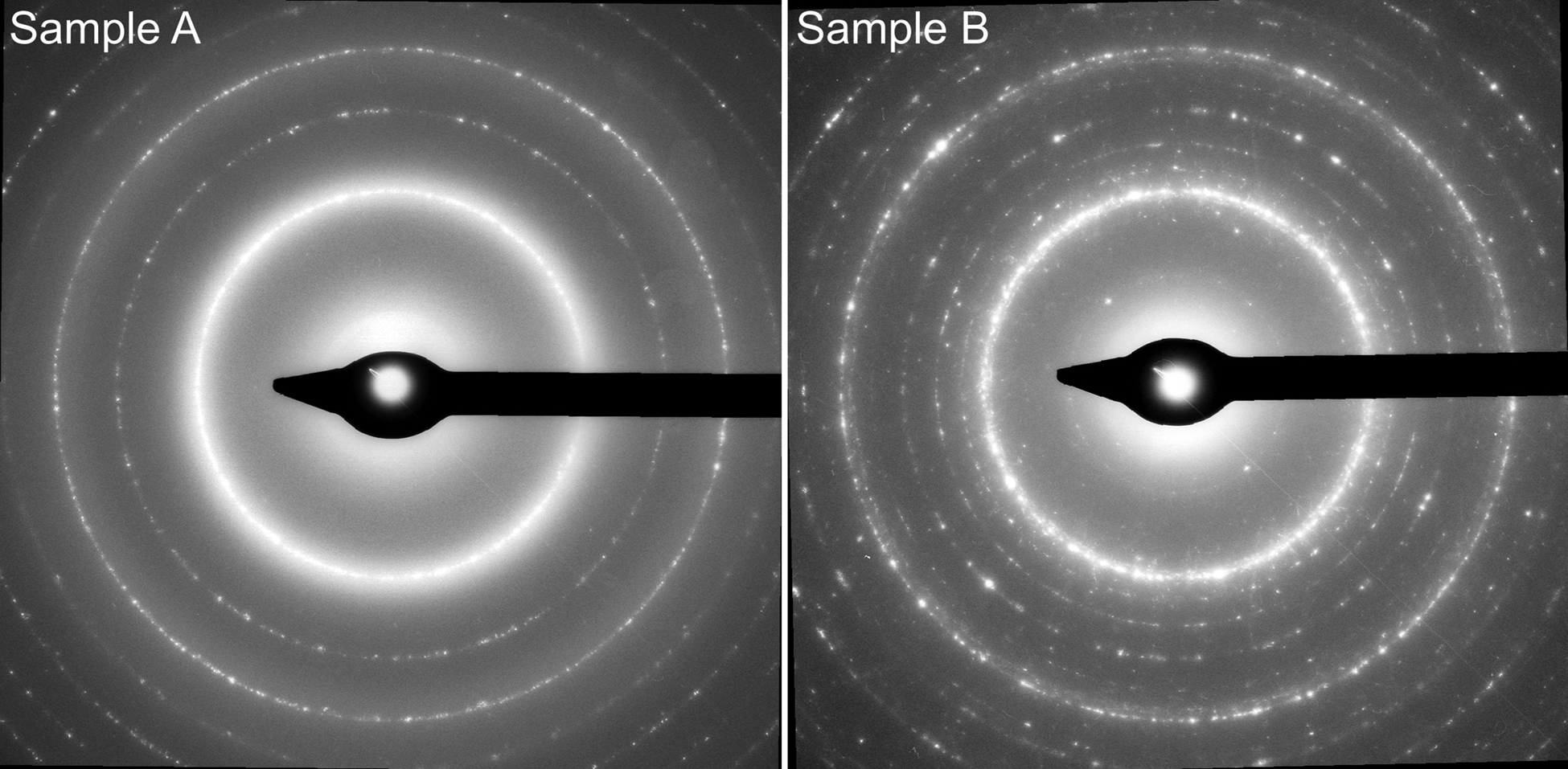
Figure 1. SAED patterns of the Cr–Fe spray coatings of samples A and B, as obtained from cross-sections prepared with a focused ion beam (FIB).
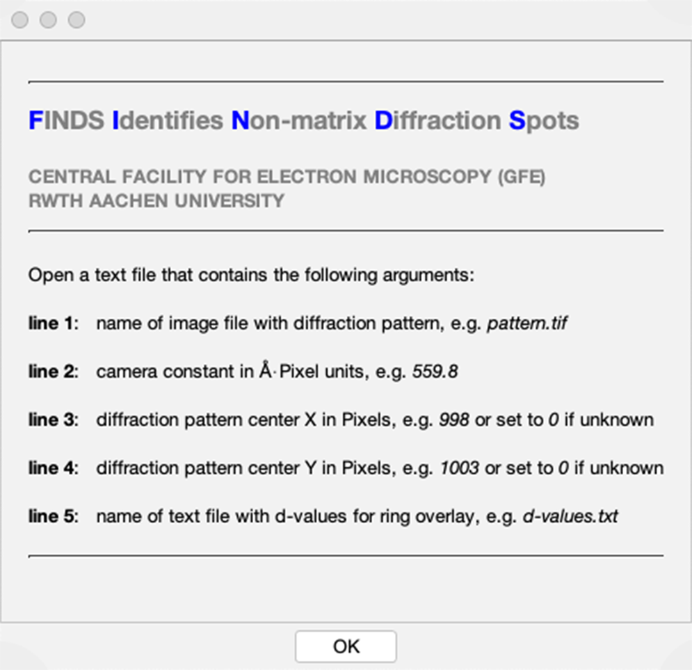
Figure 2. The FINDS start screen, where the format of the project file to be loaded in the next step is specified.
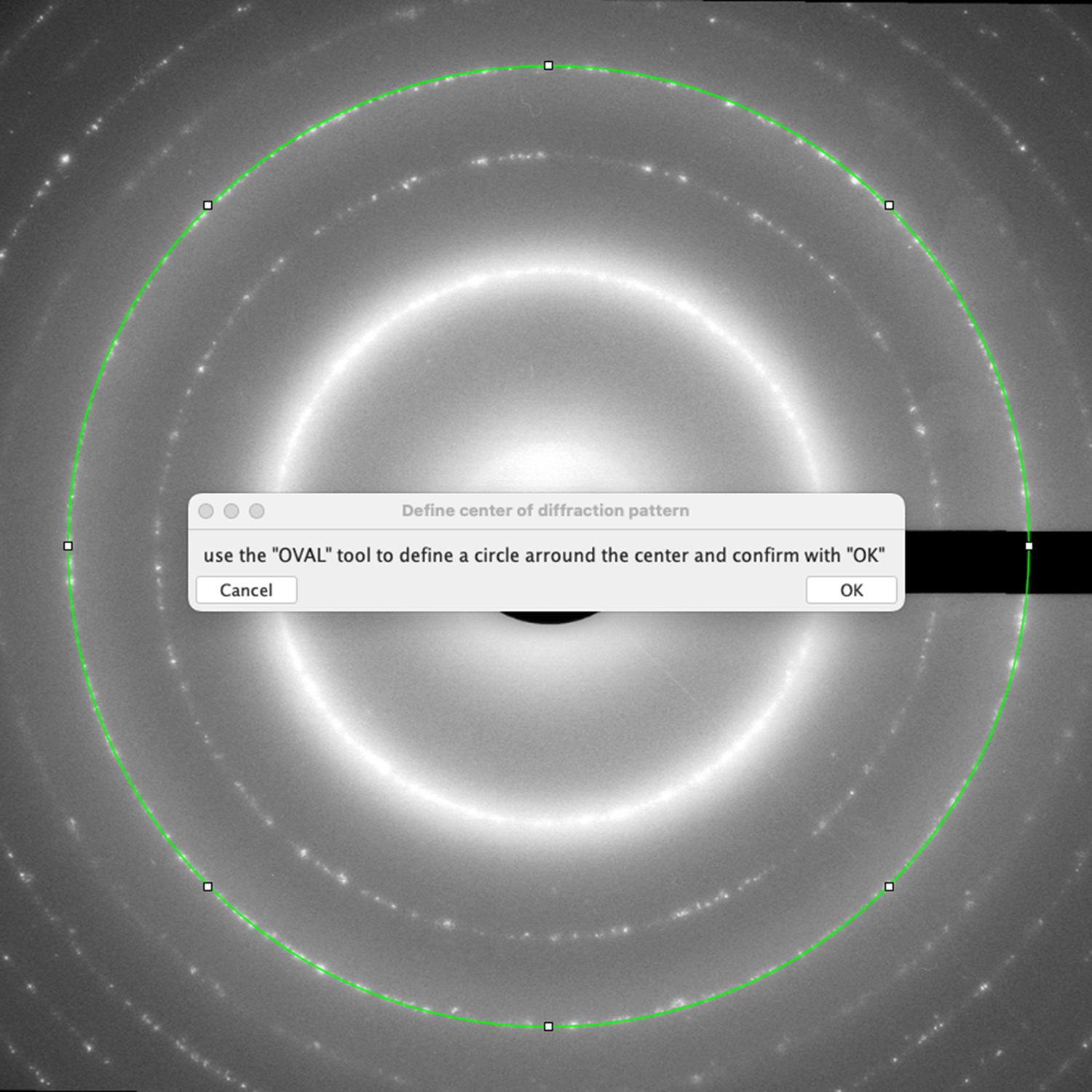
Figure 3. If both the X and Y centers of the SAED pattern have been set to zero in lines 3 and 4 of the project file, the diffraction pattern is displayed with a predefined circle which the user is asked to fit to one of the diffraction rings in the pattern. Note that a slight elliptical distortion of the diffraction ring can also be fitted by dragging the indicated anchor points of the ImageJ oval tool onto the reflection ring. The X and Y coordinates of the center of the diffraction pattern are obtained by the program from the center of the user-defined circle or ellipse.

Figure 4. FINDS main menu, which allows the user to make adjustments to the display of the processed SAED pattern. The “color mode” option can be used to change the default greyscale display to three different false colors and the “display mode” option allows the user to toggle between normal and inverted display of the diffraction pattern. The menu additionally allows the display of the simulated ring pattern to be toggled and to set different colors and line widths for drawing the superimposed rings. As low-angle diffraction spots are often most important for the identification of the phase, it is possible to focus on this part and to crop the SAED pattern by using the “d-value limit” option (see Figure 7 for an example).
To demonstrate the first approach, 58 diffraction spots from 5 different rings were determined by manual selection (Figure 5), giving the following averaged d-values (number of averaged reflections in brackets): 2.022 Å (9), 1.431 Å (9), 1.167 Å (13), 1.011 Å (15), and 0.902 Å (12). Furthermore, the averaged d-values could be indexed in agreement with a body-centered cubic (bcc) cell of lattice parameter a = 2.858(2) Å. A subsequently performed ICDD PDF database search with the averaged d-values and composition from EDX yielded the best match for the bcc phase Fe3Cr (PDF card no. 04-022-2502) with a = 2.870(2) Å. The diffraction pattern of sample A with the superimposed diffraction rings from the averaged d-values is shown on the left in Figure 6. The same ring pattern was superimposed on the SAED pattern of sample B to verify the presence of bcc Fe3Cr in this material (right image in Figure 6). The latter hypothesis is supported by the good match of the bcc phase rings with the diffraction rings. However, as visual inspection readily reveals, the diffraction pattern of sample B contains a considerable number of additional diffraction spots that belong to at least another structure with a larger unit cell. This situation is a typical scenario for which FINDS was designed, as it now allows the clear identification of the reflections of the unknown phases and the determination of the spot positions using the peak locator functionality described earlier (see green cross markers in the diffraction pattern on the right in Figure 6). A search of the ICDD database with the determined d-values of the additional reflections yielded a reasonable match with the tetragonal phase Fe1.13Cr0.87 (PDF card no. 01-071-7531). Calculated d-spacings of this phase were used to generate and overly the ideal ring pattern onto the SAED pattern of sample B. With the exception of the second Laue ring with index 200 of the bcc Fe3Cr phase, all diffraction spots are now covered by the superimposed diffraction rings (Figure 7). Hence, it can be concluded that the material of sample B consists only of cubic Fe3Cr and tetragonal Fe1.13Cr0.87.
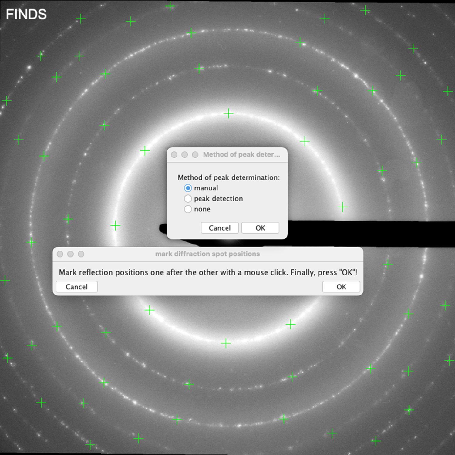
Figure 5. For the SAED pattern of sample A, the manual mode was used to locate the positions of the diffraction spots by means of a crosshair cursor. For statistical reasons, several reflections were addressed for each diffraction ring (green crosses) and, after averaging of the d-values, were used to generate the superimposed rings on the SAED patterns in Figure 6.
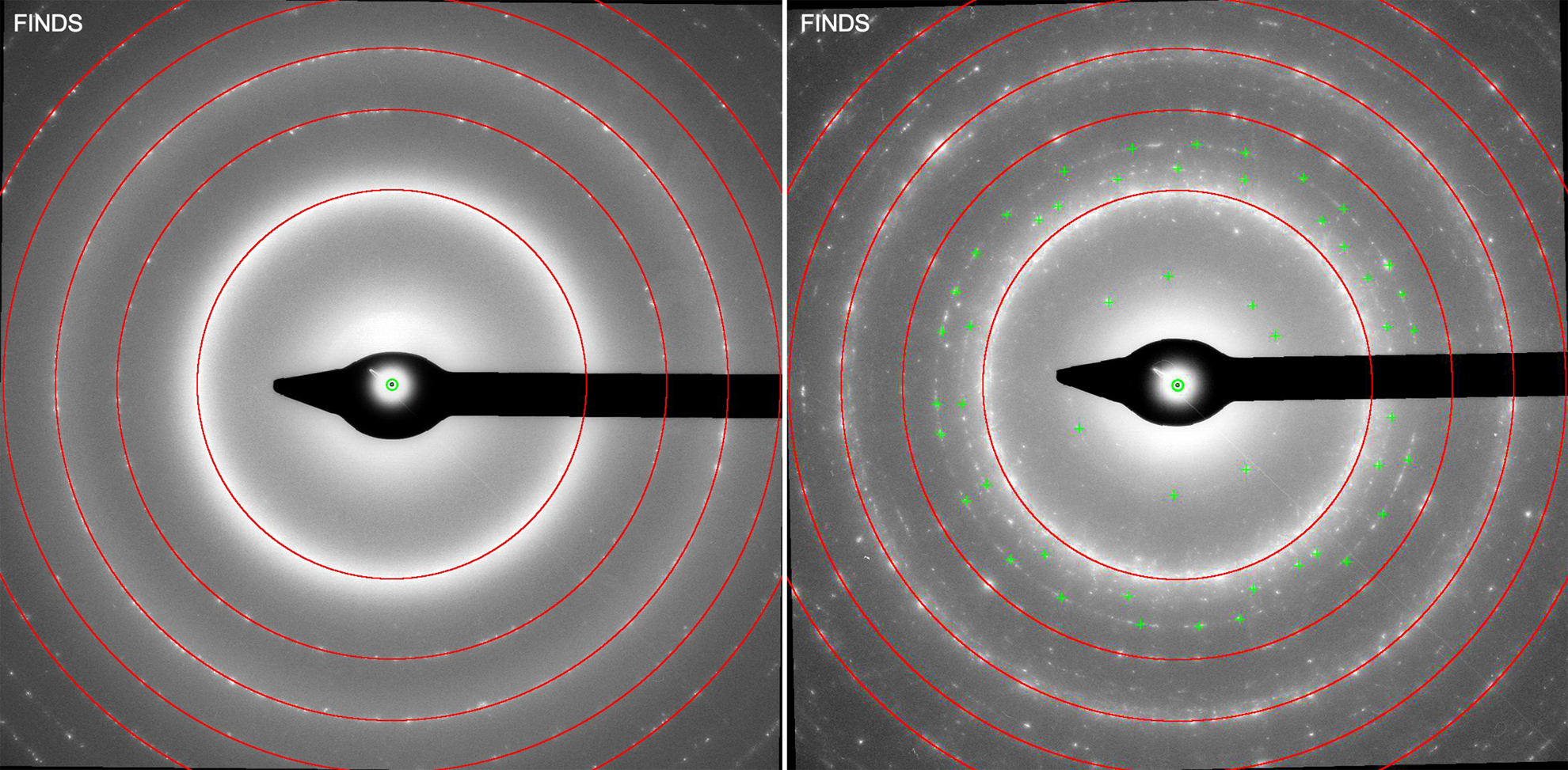
Figure 6. SAED patterns of sample A (left) and sample B (right) with the superimposed ring pattern calculated from the experimental d-values obtained for sample A. The simulated ring positions are compatible with the bcc phase Fe3Cr (PDF card no. 04-022-2502) with refined lattice parameter a = 2.870(2) Å.
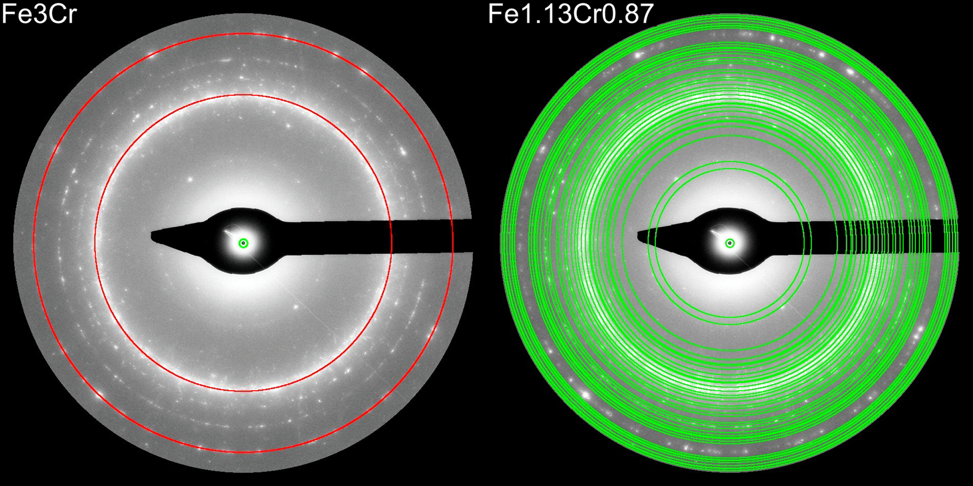
Figure 7. SAED pattern of sample B with the superimposed ring patterns of the bcc phase Fe3Cr (PDF card no. 04-022-2502) and the tetragonal phase Fe1.13Cr0.87 (PDF card no. 01-071-7531), respectively. The SAED patterns have been cropped to a resolution of 1.3 Å using the “d-value limit” option in the main menu (see Figure 4).
C. Use case: quality control of magnetic iron oxide nanoparticles for biomedical applications
Magnetic iron oxide nanoparticles for biomedical applications were synthesized and subsequently investigated in a FEI Tecnai F20 TEM operated at 200 kV for a quality check (Göpfert et al., Reference Göpfert, Schoenen, Buhl, Weirich, Schmitz-Rode and Slabu2024). Within the TEM characterization, a Veleta S04F side-entry TEM camera (Emsis GmbH, Münster, Germany) was used to record SAED patterns at a nominal camera length of 970 mm for subsequent processing with FINDS. The target structure of the synthesis is that of spinel-type Fe3O4, which was confirmed within an earlier investigation of the present author to be compatible with ICDD PDF card no. 01-080-7683 (a = 8.405 Å). The calculated d-spacings of the latter phase were used to superimpose the ring positions on the SAED pattern (right image in Figure 8), allowing easy detection of additional reflections that may arise from an unwanted by-product. As illustrated in the magnified center region of the SAED pattern in Figure 9, three additional weak diffraction spots are visible between the first and second diffraction inner rings of Fe3O4 that require further investigation. In order to achieve optimal precision, the ImageJ inbuilt peak detection function “Find Maxima” was employed to ascertain the d-values of the diffraction spots in question. The mean value of these d-values was determined to be 4.23(4) Å, which was subsequently used for a search in the ICDD PDF database (Kabekkodu et al., Reference Kabekkodu, Dosen and Blanton2024). The matches found for this d-value suggest that the sample contains some traces of monoclinic Fe2O3 (I2/a, a = 7.405 Å, b = 5.035 Å, c = 5.427 Å, β = 95.87°) or orthorhombic Fe2O3 (Amam, a = 6.379 Å, b = 8.609 Å, c = 2.665 Å) in addition to the target structure. The corresponding ICDD PDF files are nos. 00-071-0073 and 04-019-9517, respectively.
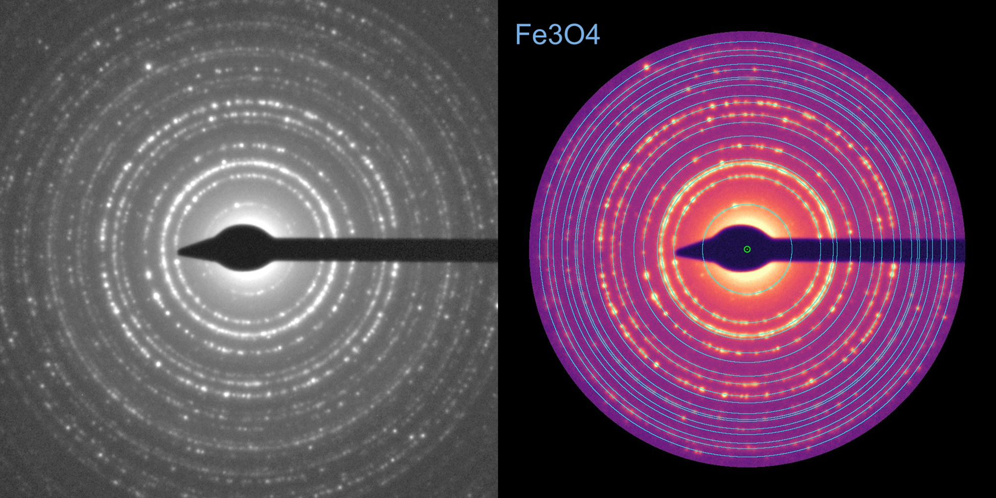
Figure 8. SAED pattern from a sample of magnetic iron oxide nanoparticles for biomedical applications. The diffraction pattern on the right is displayed in false color “mpl-magma” and has been cropped to a resolution of 1.0 Å. The superimposed rings correspond to the d-values of spinel-type Fe3O4 (PDF card no. 01-080-7683, a = 8.405 Å).
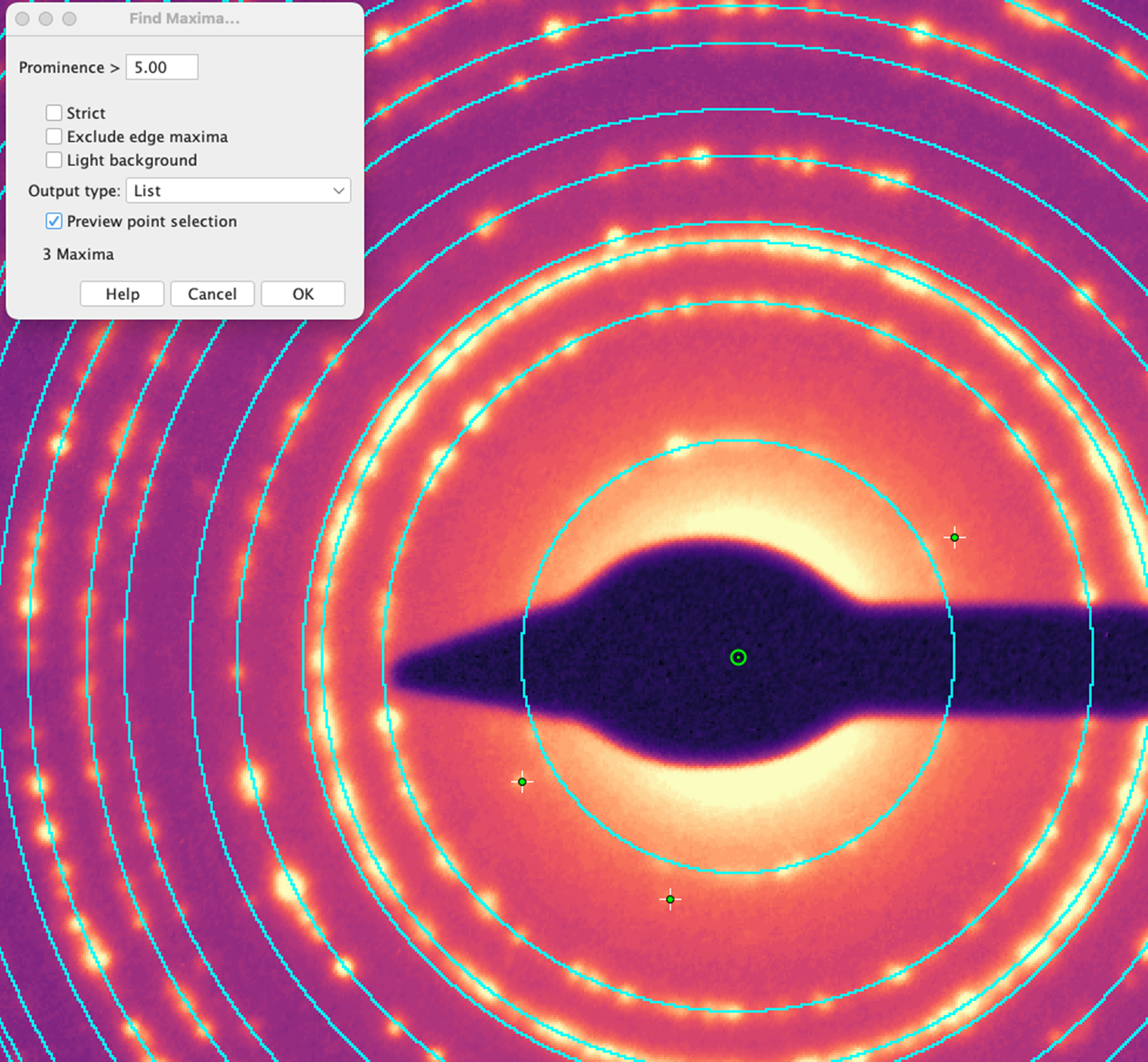
Figure 9. Magnified center section of the SAED pattern in Figure 8. The three weak diffraction spots that are indicated by the crosshair markers are not consistent with cubic spinel-type Fe3O4 and must therefore belong to a second phase. In order to obtain the most accurate d-values of these reflections, the ImageJ's built-in peak detection function “Find Maxima” was applied.
III. RESULTS AND DISCUSSION
The examples shown here illustrate that a particularly challenging situation for SAED phase characterization arises in cases where the pattern consists of a large number of diffraction spots arranged in concentric but incomplete rings due to random crystal orientation of crystallites that are neither large enough to form a usable single crystal pattern nor small enough to form continuous rings. This usually results in a confusing situation that makes it extremely difficult to sort out the reflections that belong to a particular phase or to identify additional reflections that come from other phases. For these use cases, the ImageJ macro script FINDS was developed, which allows to superimpose user-defined reflection rings on the experimental SAED pattern. Once the reflections of the known phase(s) are evident, additional reflections are easily identified and addressed as they lie outside the superimposed ring pattern. FINDS not only provides the full functionality to draw the superimposed ring patterns in different styles, but also provides tools to locate the positions of the additional diffraction spots and determine their d-values. Two examples, characterized by the challenges outlined above, demonstrated the full workflow and usefulness of the developed script to aid the phase identification process in multiphase samples.
AVAILABILITY OF THE PROGRAM CODE
The macro code of FINDS is published under GNU General Public License v3.0 or later at https://doi.org/10.5281/zenodo.13748483 (Weirich, Reference Weirich2024) and complies with the FAIR Principles for research software (Barker et al., Reference Barker, Chue Hong, Katz, Lamprecht, Martinez-Ortiz, Psomopoulos, Harrow, Castro, Gruenpeter, Martinez and Honeyman2022).
ACKNOWLEDGMENTS
The development of the macro code has been carried out within Collaborative Research Centre Transregio 188: Damage Controlled Forming Processes (DFG – German Research Foundation, Project-ID 278868966). The author would like to express his gratitude to Mr. Sebastian Zischke (GFE, RWTH Aachen University) for recording the SAED images of the iron oxide nanoparticles and to Prof. Dr. Ioana Slabu (Institute of Applied Medical Engineering, RWTH Aachen University) for providing the sample.
CONFLICTS OF INTEREST
The author declares no conflicts of interest.


