Introduction
Chemical modification of proteins has become a valuable tool for developing modified proteins. Playing complementary roles to genetic techniques, we have a broad toolkit that allows us to create an almost unlimited number of protein constructs with natural or synthetically modified residues using chemical modifications (Stephanopoulos and Francis, Reference Stephanopoulos and Francis2011). The ideal requirements for such reactions include functional group tolerance/compatibility, water as a reaction medium, selectivity, high reaction rates, neutral pH and room temperature (or up to 40 °C), nontoxic reagents, and low reactant concentrations. Reactions must be designed and implemented to achieve high modification efficiencies without the need for tedious and inefficient purification/characterization protocols. For in vivo studies, chemical modification methods involving those listed above are appropriate since they do not interfere with normal cell function (Boutureira and Bernardes, Reference Boutureira and Bernardes2015). Protein modifications have a significant impact on signaling, migration, differentiation, and trafficking as important cellular processes through sulfation, phosphorylation, methylation, acylation, ubiquitination, glycosylation, farnesylation, and so on (Walsh et al., Reference Walsh, Garneau-Tsodikova and Gatto2005).
Posttranslational protein modifications (PTMs) are commonly thought to be responsible for the vast biodiversity found in nature today (Boutureira and Bernardes, Reference Boutureira and Bernardes2015). These modifications usually occur after protein translation. In this regard, considering the characteristics of the natural modification of proteins, it can be concluded that the efficient and controlled reproducing of PTM provides a valuable tool in the study and precise function of proteins. In addition to the facilities provided by (bio)orthogonal methods; it allows precise and site-selective modification of proteins, making it a valuable tool for in vivo and in vitro studies (Bernardes et al., Reference Bernardes, Chalker, Davis and Pignataro2010; Kee and Muir, Reference Kee and Muir2012). Considering the various methods used for chemical modification, it will now be feasible to choose the target residue and modification type to provide the required property/function such as chemical affinity probes, fluorophores, reactive tags, and so forth. Despite the enormous progress made in the bioconjugation of proteins, there are still many serious challenges, not only from a synthetic point of view but also in manufacturing, processing, stability, and safety. Some types of proteins can be modified using methods that are not appropriate for all kinds of proteins (Boutureira and Bernardes, Reference Boutureira and Bernardes2015). Therefore, there is still a need for the development of complementary reactions for site-selective modifications of proteins that are efficient, robust, and mild. The various aspects of protein synthesis have been discussed in detail (Bernardes et al., Reference Bernardes, Chalker, Davis and Pignataro2010), from general chemical ligation strategies (Hackenberger and Schwarzer, Reference Hackenberger and Schwarzer2008; Kent, Reference Kent2009; Siman and Brik, Reference Siman and Brik2012), endogenous amino acid modification (Baslé et al., Reference Baslé, Joubert and Pucheault2010), to click modification protocols (Lallana et al., Reference Lallana, Riguera and Fernandez-MEGIA2011; Van Berkel et al., Reference Van Berkel, Van Eldijk and Van Hest2011; Palomo, Reference Palomo2012; Tasdelen and Yagci, Reference Tasdelen and Yagci2013), which are more specialized on specific PTMs, including glycosylation (Gamblin et al., Reference Gamblin, Scanlan and Davis2009; Schmaltz et al., Reference Schmaltz, Hanson and Wong2011; Villalonga et al., Reference Villalonga, Diez, Sanchez, Gamella, Pingarron and Villalonga2014; Wang and Amin, Reference Wang and Amin2014), PEGylation (Nischan and Hackenberger, Reference Nischan and Hackenberger2014), and polymerization of protein-based initiator (Matyjaszewski and Tsarevsky, Reference Matyjaszewski and Tsarevsky2014; Wallat et al., Reference Wallat, Rose and Pokorski2014). However, most coupling methods are inspired by naturally occurring reactions or engineered protein-catalyzed reactions (Lange and Polizzi, Reference Lange and Polizzi2021). Based on biomimetic research studies, there is a general attraction toward carbon–heteroatom bond formation over carbon–carbon bonds; for instance, proteins, polysaccharides, and nucleic acids are condensed polymeric assemblies of subunits bound by carbon-heteroatom (Kolb and Sharpless, Reference Kolb and Sharpless2003).
With the emergence of click chemistry, a new promising line of chemical protein modification has been developed. Click reactions were first introduced by Kolb et al. (Reference Kolb, Finn and Sharpless2001). The basis of this method is the simple assembly of molecules. These reactions are based on covalent bonds that can be used in various applications (Kolb et al., Reference Kolb, Finn and Sharpless2001). Caroline Bertozzi published a new click reaction in 2004, called strain-promoted azide-alkyne cycloaddition (SPAAC). This achievement opened a new field in click chemistry (Agard et al., Reference Agard, Prescher and Bertozzi2004). This method has simplified the use of the click method in biological studies by eliminating the need for copper. In 2022, the Nobel Prize in Chemistry was awarded to three influential scientists, Barry Sharpless, Morten Meldahl, and Caroline Bertozzi, who were pioneered the field of click reactions. The click strategy is a powerful tool to produce new molecules and can be summarized by the following statement: ˝All searches must be restricted to molecules that are easy to make˝ (Kolb et al., Reference Kolb, Finn and Sharpless2001). Click reactions are modular, applicable reactions that give very high yields, produce no unpleasant by-products, and require simple reaction conditions (Shaabani et al., Reference Shaabani, Maleki and Mofakham2008; Shaabani et al., Reference Shaabani, Afshari and Hooshmand2017a; Shaabani et al., Reference Shaabani, Tavousi Tabatabaei and Shaabani2017b; Shaabani et al., Reference Shaabani, Shadi, Mohammadian, Javanbakht, Nazeri and Bahri2019; Khodkari et al., Reference Khodkari, Nazeri, Javanbakht and Shaabani2023). When there is a need for new molecular properties, small molecular building blocks can be joined together to produce such properties (Suazo et al., Reference Suazo, Park and Distefano2021). As shown in Figure 1, there has been an increase in the number of publications on the topic of click protein chemistry with biological applications in recent years. Click protein chemistry has given rise to a myriad of highly interesting developments in the field of medicine and has significantly impacted the synthesis of structurally diverse molecules through shorter, stereoselective, and efficient synthetic routes. In continuation of our previous review articles on modified/functionalized materials via multicomponent reactions strategy as “click” reactions (Afshari and Shaabani, Reference Afshari and Shaabani2018; Javanbakht and Shaabani, Reference Javanbakht and Shaabani2019; Javanbakht et al., Reference Javanbakht, Nasiriani, Farhid, Nazeri and Shaabani2022), herein, an attempt has been made to review the medicinal applications of protein click chemistry.
Bioorthogonal click chemistry
The widespread use of straightforward click reactions in biological environments led to the advent of new definition “bio-click chemistry” which can be understood toward reactions of functional groups commonly found in biological molecules like proteins or live cells and by exploiting the potential of click reactions (Rodríguez et al., Reference Rodríguez, Moglie, Ramírez-Sarmiento, Singh, Dua and Zacconi2022). There is a close overlap between click and bioorthogonal chemistry. Consequently, click chemistry tools will enable the development of bioorthogonal reactions, while technological advancements in chemical biology will stimulate the possibility of developing novel click chemistry methods (Suazo et al., Reference Suazo, Park and Distefano2021). Bioorthogonal reactions and protein labeling in complex cellular mixtures have also been studied in several types of research over the past decade (Lang and Chin, Reference Lang and Chin2014; Patterson et al., Reference Patterson, Nazarova and prescher2014). In another definition, bioorthogonal chemistry represents high-yielding rapid and selective chemical reactions that proceed in biological environments without any by-products (Scinto et al., Reference Scinto, Bilodeau, Hincapie, Lee, Nguyen, Xu, Am Ende, Finn, Lang and Lin2021). The significant advantage of bioorthogonal approaches is related to the fact that they usually do not affect other normal biochemical processes (Sletten and Bertozzi, Reference Sletten and Bertozzi2009). For example, bioorthogonal click chemistry has enabled selective labeling of enzymatic processes in vitro and in vivo, allowing real-time analysis of enzymatic processes in both environments. In this way, we have better understood about many biomedical challenges and biological questions. These include the causes of Alzheimer’s disease, Coronavirus, and Cancer, among others (Suazo et al., Reference Suazo, Park and Distefano2021).
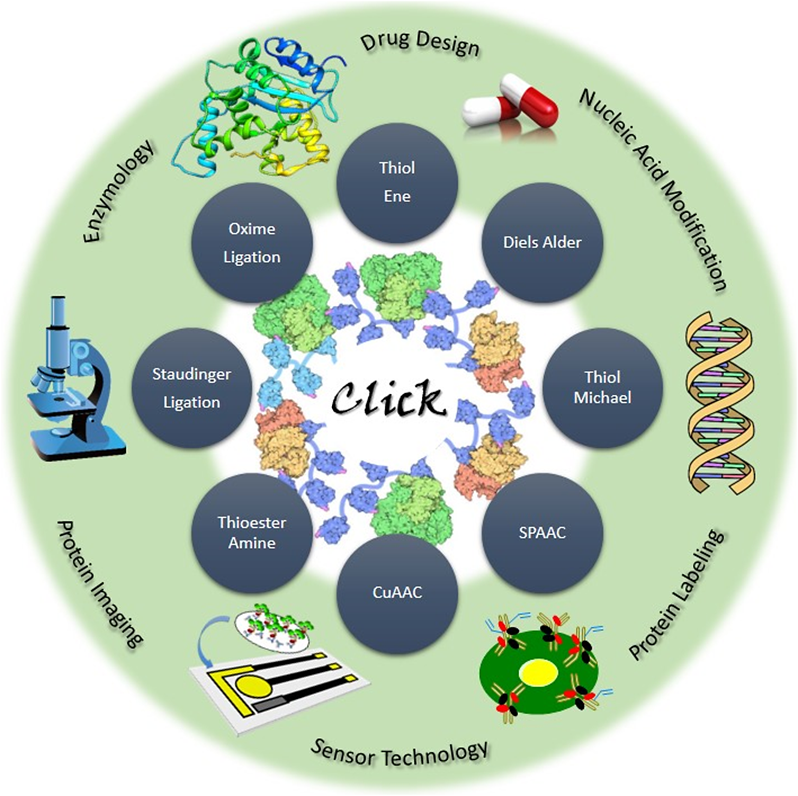
Different amino acids are used in protein click reactions, including histidine, lysine, cysteine, and so forth. Besides the direct use of amino acids in the protein click reactions, many reports suggest that the amino acids can be modified before the click reaction. For instance, the use of iodoacetamide can be used to modify nucleophilic amino acids such as cysteine, lysine, and histidine nonspecifically. It has also been noted that activated carboxylic acid derivatives, such as N-hydroxysuccinimide esters, have been extensively used to modify the ε-amine of lysine residues in peptides and proteins (Fisher et al., Reference Fisher, Baker and Shoichet2017). With the aim of modifying proteins, it should be considered that the bioactivity of the protein may be affected if the modification disrupts the active site. Currently, modification of site-specific proteins with unnatural amino acids while maintaining protein structure and function is very popular (Raliski et al., Reference Raliski, Howard and Young2014; Maza et al., Reference Maza, McKenna, Raliski, Freedman and Young2015). Click chemistry involves a range of important reactions in synthetic organic chemistry, including strained ring opening, conjugate addition, aldehyde capture by α-effect nucleophiles, cycloaddition, and acylation/sulfonylation (Devaraj and Finn, Reference Devaraj and Finn2021). Figure 2 presents various types of click reactions. Therefore, click methods are among the most efficient strategies for protein modification, binding the protein to a new substrate or another protein.
Thiol-alkene (thiol-ene) and thiol-alkyne (thiol-yne) couplings have attracted great attention in recent years because they utilize readily available chemical functions found in the structure of proteins, peptides, polymers, and materials (Campos et al., Reference Campos, Killops, Sakai, Paulusse, Damiron, Drockenmuller, Messmore and Hawker2008; Hensarling et al., Reference Hensarling, Doughty, Chan and Patton2009; Hoogenboom, Reference Hoogenboom2010). An advantage of this method is that it requires only low concentrations of initiators, typically proceeds rapidly, and can be easily isolated from the products (Colak et al., Reference Colak, Da Silva, Soares and Gautrot2016). In general, the kinetics of a reaction is strongly influenced by the structure of the alkene moiety. A strained alkene moiety, as well as an electron-rich alkene moiety, will have the greatest reactivity (Reddy et al., Reference Reddy, Cramer and Bowman2006; Hoyle and Bowman, Reference Hoyle and Bowman2010). The thiol-Michael addition reaction has been widely applied for protein conjugation because it can work under mild conditions in an aqueous environment (Jones et al., Reference Jones, Mantovani, Ryan, Wang, Brayden and Haddleton2009). While UV irradiation or photoinitiators may be harmful to cells or materials, it is often preferable to use thiol-Michael addition instead of thiol-ene reactions (Fisher et al., Reference Fisher, Baker and Shoichet2017). The Cu-catalyzed azide-alkyne cycloaddition (CuAAC) reaction is used in pharmacy and chemical biology as an efficient method for the covalent modification of active and proactive biological molecules (McKay and Finn, Reference McKay and Finn2014). CuAAC offers a unique advantage over other non-Cu click technologies. The small triazole linkage is identical in size and polarity to the peptide linkage, minimizing disruption of the biological function of the conjugate (Valverde et al., Reference Valverde, Lecaille, Lalmanach, Aucagne and Delmas2012; Birts et al., Reference Birts, Sanzone, El-Sagheer, Blaydes, Brown and Tavassoli2014). However, there is a significant drawback in using CuAAC reactions for bioconjugations related to the presence of Cu, which generates reactive oxygen species (Thirumurugan et al., Reference Thirumurugan, Matosiuk and Jozwiak2013). Because of the oxidation of Cu(I) to Cu(II), it is often necessary to carry out the reaction under inert gas or to use reducing agents such as ascorbic acid or sodium ascorbate (Rostovtsev et al., Reference Rostovtsev, Green, Fokin and Sharpless2002). In contrast to CuAAC, SPAAC reaction does not require a metal catalyst. A cycloalkyne’s electronic structure strongly influences the reaction rate and efficiency (Fisher et al., Reference Fisher, Baker and Shoichet2017). Despite SPAAC is effectively applied for conjugating proteins and peptides, strained cyclooctyin exhibits high reactivity, resulting in poor stability and difficult synthetic processes (Jewett et al., Reference Jewett, Sletten and Bertozzi2010). In a Diels–Alder reaction, electron-rich dienophiles react with electron-poor dienophiles to form a [4 + 2] cyclization reaction (Fisher et al., Reference Fisher, Baker and Shoichet2017). It is important to remember that the Diels–Alder reaction is a highly selective transformation and can proceed more rapidly and selectively in water than in organic solvents (Li and Chan, Reference Li and Chan1997). In general, the Diels–Alder reaction is quite slow; however, when heated to higher temperatures, the reaction rate can be increased. However, temperatures above 37 °C should be avoided because of the possibility of protein denaturation, while at higher temperatures the Diels–Alder reaction is reversible (Koehler et al., Reference Koehler, Anseth and Bowman2013). Binding of the C-terminal part of a thioester to the N-terminal part of a cysteine residue provides the formation of amide moiety in the thioester-amine method (native chemical ligation (NCL)) (Dawson et al., Reference Dawson, Muir, Clark-Lewis and Kent1994). NCL allows peptides larger than 50 amino acids to be constructed by synthesizing the peptide fragments and connecting them together. This method overcomes the limitations of the solid-phase peptide synthetic method (Dawson and Kent, Reference Dawson and Kent2000). The NCL procedure is performed in aqueous solutions, at neutral pH levels, in the presence of denaturing agents such as guanidine hydrochloride to prevent protein aggregation. This reaction is very sensitive to the pH value of the system, whereas high pH values can hydrolyze thioesters, and low pH values can reduce the reactivity of cysteine thiol amine, decreasing the rate of the reaction (Fisher et al., Reference Fisher, Baker and Shoichet2017). The Bertozzi group developed Staudinger ligation for cell surface modification to form amide bonds between azides and triarylphosphine derivatives, in which cells with azide moieties reacted with phosphines to generate amide bonds (Saxon and Bertozzi, Reference Saxon and Bertozzi2000). Proteins can be conjugated to polymers or immobilized on the glass or gold surfaces through this reaction (Fisher et al., Reference Fisher, Baker and Shoichet2017). Nucleophilic oxyamines attack the electron-deficient aldehydes or ketones in the oxime ligation reaction, resulting in the formation of an oxime bond, with water molecules being formed as a by-product of the reaction. It is a highly selective bioorthogonal process that has almost quantitative conversions, proceeds under mildly acidic aqueous conditions, and does not require metal catalysts (Fisher et al., Reference Fisher, Baker and Shoichet2017).
In summary, click reactions play notable roles in the design of chemical motifs and help to construct bioorthogonal covalent conjugations under physiological conditions. Considering the extent of click chemistry, new strategies have emerged to improve target specificity and increase efficacy in diagnosing and treating disease. With this background, we highlight a range of medicinal applications that involve the bioorthogonal chemistry toolbox.
Diagnosis
Protein labeling
There are tens of thousands of proteins in the human proteome, many of them occurring in minute concentrations below the limits of detection (LODs) of current technologies such as ELISA, mass spectrometry (MS), and protein microarrays (Wilson, Reference Wilson2013). It is imperative to develop a molecular instrument that can detect disease-related protein biomarkers at low levels in the body without the need for any further manipulation (Senapati et al., Reference Senapati, Manna, Lindsay and Zhang2013).
Imaging is a method of distinguishing the target biomolecule from a living system. This is possible through the use of a spectroscopic probe (Baskin et al., Reference Baskin, Prescher, Laughlin, Agard, Chang, Miller, Lo, Codelli and Bertozzi2007). If less structural disruption is desired, the target protein can be chemically (Carrico et al., Reference Carrico, Carlson and Bertozzi2007) or enzymatically (Chen and Ting, Reference Chen and Ting2005) labeled with small molecules. There seems to be a growing interest in imaging biomolecules that are not easily modified by genetic modification, even though proteins remain the primary targets of cellular imaging (Carrico et al., Reference Carrico, Carlson and Bertozzi2007). For example, because lipids and glycans have independent functions and these molecules are the result of PTM, they cannot be imaging using protein-specific methods. Although these biomolecules have been studied in vitro in static systems, the dynamic behavior of these biomolecules in living cells is poorly understood (Carrico et al., Reference Carrico, Carlson and Bertozzi2007). Therefore, in vivo biomolecular labeling can be achieved without genetic manipulation using a bioorthogonal click strategy (Carrico et al., Reference Carrico, Carlson and Bertozzi2007). In this regard, bioorthogonal functional groups are installed in target biomolecules by a cell’s metabolic machinery. The next step involves the covalent labeling of a probe to the functional group (Prescher and Bertozzi, Reference Prescher and Bertozzi2005).
There are various conventional methods for click chemistry, depending on the type of chemical protein modification. Combining photoaffinity labeling with liquid chromatography (LC)–MS/MS method can effectively identify cognate proteins in biological systems (Preston and Wilson, Reference Preston and Wilson2013). Because these analyses are based on comparing the signal strength of unlabeled peptides, small MS signals at the LOD are still difficult to distinguish from false positives. However, the multifunctional cross-linkers with highly sensitive functions can be applied as alternatives that can effectively distinguish labeled peptides from various contaminants (He et al., Reference He, Xie, Yang, Zhang, Su, Ge, Song and Chen2017; Horne et al., Reference Horne, Walko, Calabrese, Levenstein, Brockwell, Kapur, Wilson and Radford2018). Hence, the sulfo-click reaction can simplify the synthesis of photoprobes of biomolecules with different functional groups. In addition, highly controlled cleavage of the N-acylsulfonamide linkage significantly improves the handling of the labeled proteins. A synthetic advantage makes it possible to determine the specific direction in which ligands bind to the surface of the interacting proteins. Another advantage of this method is that it can be combined with a cross-linking photocyclization reaction to produce a special MS tag whose mass changes under the influence of irradiation, leading to an understandable distinction between labeled peptides and false positive signals. Thanks to these improvements, target proteins and interacting sites of biomolecules can be identified more quickly in practical applications (Hayashi et al., Reference Hayashi, Morimoto and Tomohiro2019).
Through a metal- and oxidant-free synthetic approach, a monosubstituted 3 bromo-1,2,4,5-tetrazine fragment (Tz) was synthesized. Since it has superb reactivity toward different nucleophiles, its application as a biomolecule labeling agent was investigated, especially as a simple and straightforward way to incorporate a bioorthogonal handle into proteins. Based on the results of LC–MS/MS analyses, this compound appears to be chemoselective for lysines and forms monosubstituted amino-Tz on the surface of the protein with preserved folding. The chemical properties of the labeled lysines were investigated for click-to-release (CtR) reactions. They were additionally can be used in cell culture as a therapeutically relevant context (Figure 3) (Ros et al., Reference Ros, Bellido, Verdaguer, Ribas De Pouplana and Riera2020).
It has been reported that nitroso species can be selectively incorporated into a number of proteins, including lysozyme, BSA, KRAS, MiaA, and HRAS, using lysine residues as targets. Under physiological conditions, it was observed that the corresponding azo functionalities were formed in a highly selective manner with excellent yields, displaying rather good stability. It was found that a fluorescent and/or dual fluorescent labeling protocol could be efficiently and selectively applied for the preparation of different labeled proteins, including HRAS, KRAS, and lysozyme. Consequently, the interactions between prenylated proteins and enzymes were evaluated through fluorescence resonance energy transfer (FRET) assays. By incorporating pyrenyl functionality into specific proteins at lysine residues, established via the so-called NEL process, along with the Selectfluor and click reaction approach targeting prenyl functionality, chemically modified proteins with a 1-pyrenyl-fluorophore by 254 nm UV irradiation were interestingly synthesized. The sequential azidation and click reaction of the protein prenyl functionality allow the incorporation of naphthene to increase the transmitted fluorescence energy. Moreover, significantly increased absorbance at 218 nm in lysed HEK293T cells and greatly enhanced greenish fluorescence in live HEK293T cells were observed (Gan et al., Reference Gan, Li, Zhou and Wang2022). This was the first report of using a chemical approach to figure out protein–protein interactions through the FRET assay.
A bioorthogonal reaction for dynamic cellular imaging was developed by combining the biocompatibility of Staudinger ligation with the rapid reaction kinetics of the click strategy. The development of an alternative method to activate alkynes for [3 + 2]-cycloaddition with azides would be one approach to achieve this goal (Carrico et al., Reference Carrico, Carlson and Bertozzi2007). However, cyclooctynes as the smallest of the stable cycloalkynes, have achieved bioorthogonal azide labeling by using ring strain (Agard et al., Reference Agard, Prescher and Bertozzi2004). Even though the SPAAC reaction showed no better sensitivity than that of the Staudinger ligation, the cyclooctyne probes demonstrated no cellular cytotoxicity (Agard et al., Reference Agard, Baskin, Prescher, Lo and Bertozzi2006). By means of ring strain and electron-withdrawing groups as two important rate-accelerating factors, the sensitivity of cyclooctynes for azide detection can be increased in Cu-free click reagent design (Baskin et al., Reference Baskin, Prescher, Laughlin, Agard, Chang, Miller, Lo, Codelli and Bertozzi2007). It is expected that azide labeling and Cu-free click chemistry will find numerous applications in glycobiology. Cu-free clicks using difluorinated cyclooctyne have been demonstrated to proceed selectively inside living mice (Baskin et al., Reference Baskin, Prescher, Laughlin, Agard, Chang, Miller, Lo, Codelli and Bertozzi2007). In addition, Cu-free click chemistry can be used to characterize other metabolites and enzyme activities (Speers and Cravatt, Reference Speers and Cravatt2004), PTMs (Prescher and Bertozzi, Reference Prescher and Bertozzi2005), and site-specifically labeled proteins (Chin et al., Reference Chin, Cropp, Anderson, Mukherji, Zhang and Schultz2003; Chen and Ting, Reference Chen and Ting2005) in living systems.
Protein biomarkers can be detected using affinity molecules attached to atomic force microscopy (AFM) tips using catalyst-free click reactions. Molecular recognition force spectroscopy (MRFS), a technique based on the use of AFM measurements, is being developed to determine and characterize the interactions between antibodies and antigens, ligands and receptors, DNA probes and targets, and so forth, at the single-molecule level (Florin et al., Reference Florin, Moy and Gaub1994; Dammer et al., Reference Dammer, Hegner, Anselmetti, Wagner, Dreier, Huber and Güntherodt1996; Avci et al., Reference Avci, Schweitzer, Boyd, Wittmeyer, Steele, Toporski, Beech, Arce, Spangler and Cole2004; Neuert et al., Reference Neuert, Albrecht, Pamir and Gaub2006; Carvalho et al., Reference Carvalho, Connell, Miltenberger-Miltenyi, Pereira, Tavares, Ariëns and Santos2010; Meng et al., Reference Meng, Paetzell, Bogorad and Soboyejo2010; Zapotoczny et al., Reference Zapotoczny, Biedroń, Marcinkiewicz and Nowakowska2012). AFM, with its single-molecule sensitivity, is a candidate for nanodiagnostics (Archakov and Ivanov, Reference Archakov and Ivanov2007). In combination with irreversible binding, it has been shown that AFM can reach a concentration sensitivity limit of 10−17 M (Archakov et al., Reference Archakov, Ivanov, Lisitsa and Zgoda2007). AFM is being used in DNA, protein, and cell analysis, and its chemical sensibility has also been greatly enhanced. When the tip of the AFM is coated with an affinity molecule, it can see and count the target molecules (Senapati et al., Reference Senapati, Manna, Lindsay and Zhang2013). With an affinity molecule tethered to the tip, AFM can scan individual proteins on a surface, called recognition imaging (RI) (Stroh et al., Reference Stroh, Wang, Bash, Ashcroft, Nelson, Gruber, Lohr, Lindsay and Hinterdorfer2004; Lin et al., Reference Lin, Wang, Liu, Yan and Lindsay2006; Wang et al., Reference Wang, Dalal, Henikoff and Lindsay2008; Chtcheglova and Hinterdorfer, Reference Chtcheglova and Hinterdorfer2011; Wang et al., Reference Wang, Guo, Zhang, Park and Xu2012). Several protein biomarkers can be identified and detected by MRFS and RI in the clinical setting. A new method combining two orthogonal click chemistries, thiol-vinyl sulfone-Michael addition and catalyst-free azide-alkyne cycloaddition, was developed for affinity molecule attachment to AFM tips for force spectroscopy and RI. All reactions were carried out in aqueous solutions. It is noteworthy that this method is used for both AFM-based force measurement and detection imaging. In addition, no specific reaction conditions are required for the attachment process (Senapati et al., Reference Senapati, Manna, Lindsay and Zhang2013).
By using positron emission tomography (PET) and single photon emission computed tomography techniques, scientists have the ability to take imaging of blood pools (Kumar and Boddeti, Reference Kumar, Boddeti, Baum and Rösch2013). Imaging plays an important role in the diagnosis of cardiovascular function evaluation (Millar et al., Reference Millar, Hannan, Sapru and Muir1979; Nishimura et al., Reference Nishimura, Hamada, Hayashida, Uehara, Katabuchi and Hayashi1989), a bleeding gastrointestinal tract (Grady, Reference Grady2016), and testing cancerous tissues for vascular permeability (Niu et al., Reference Niu, Lang, Kiesewetter, Ma, Sun, Guo, Guo, Wu and Chen2014). Radiolabeling of human serum albumin (HSA) with 99mTc using the chelate-then-click SPAAC method was developed and optimized to make an agent that can be used to image blood pools. Under mild reaction conditions, 99mTc and HSA are paired in excellent radiochemical yields. When compared with commercially available conventional 99mTc-HSA, 99mTc-DPA-HSA, 2,2′-dipicolylamine (DPA) as chelator agent, showed a high degree of stability in vivo, indicating higher blood retention and improved visualization of vasculatures in healthy mice for up to 3 hours following injection. Accordingly, the present radiolabeled method for the detection of biomolecules with similar properties can also be applied to other biomolecules with similar sensitivity (Figure 4) (Lodhi et al., Reference Lodhi, Park, Kim, Kim, Shin, Lee, Im, Jeong, Khalid and Cheon2019).
Specific functional group labeling in living cells is one of the most potent applications of bioorthogonal couplings. Tetrazines, for example, irreversibly form dihydropyrazine products and dinitrogen by reacting with a strained dienophile norbornene, which is a rapid, selective, and high-yield reaction in aqueous media. A norbornene-modified monoclonal antibody was used in human breast cancer cells to target Her2/neu receptors. In the presence of serum, tetrazines conjugated to a near-infrared fluorochrome label the pre-targeted antibody selectively and rapidly in the presence of the pre-targeted antibody. Based on these findings, this chemistry may be useful for pre-targeted imaging in vivo under numerous modalities under in vitro labeling experiments (Figure 5) (Devaraj et al., Reference Devaraj, Weissleder and Hilderbrand2008).
A series of novel heterobifunctional linkers have been developed based on substituted dimethylmaleic anhydride. This hybrid linker combines the advantages of click chemistry with pH-sensitive binding between conjugated biomolecules that can be used in various applications. Due to the acidic environment of tumors or early endosomes (Murphy et al., Reference Murphy, Powers and Cantor1984; Engin et al., Reference Engin, Leeper, Cater, Thistlethwaite, Tupchong and McFarlane1995), the pH labiality of linkers to the acidic environment is helpful. A further advantage of this method is that the linker is cleaved off traceless, yielding an unmodified molecule of interest. The versatility of the technique in protein modification is illustrated in examples such as the reversible dye labeling of proteins, pH-sensitive modification with polyethylene glycol (PEG), and the intracellular transduction of proteins in bioactive form (Maier and Wagner, Reference Maier and Wagner2012).
A biaryl-linker probe produced by the click strategy has a unique T-shaped conformation that significantly enhances its labeling performance. A new generation of biaryl-linker probes made by click reactions makes activity-based protein profiling analysis of trace membrane targets a powerful tool for identifying bioactive compounds (Nakamura et al., Reference Nakamura, Inomata, Ebine, Manabe, Iwakura and Ueda2010).
PET is widely acknowledged as a robust and noninvasive molecular imaging technique due to its unique properties. By utilizing PET, valuable functional insights into various physiological, biochemical, and pharmacological processes can be obtained (Phelps, Reference Phelps2000). The expanding array of novel targets utilized for PET imaging has led to the advent of new advanced radiolabeling techniques, particularly for high-weight molecules like peptides, proteins, antibodies, and antibody fragments. The short-lived positron emitter fluorine-18 (18F) has proven to be beneficial in designing and synthesizing PET radiotracers, thanks to its advantageous nuclear and chemical properties. Nevertheless, incorporating 18F into high-molecular-weight compounds, such as proteins, presents a significant challenge (Ramenda et al., Reference Ramenda, Kniess, Bergmann, Steinbach and Wuest2009). In this regard, a Cu(I)-mediated 1,3-dipolar [3 + 2] cycloaddition reaction was employed to label azide-functionalized HSA with the short-lived positron emitter 18F. This groundbreaking achievement expands the potential of click chemistry as a versatile tool for a wide range of radiolabeling reactions in future applications. The study showcases the promising application of click chemistry in the field of molecular imaging (Ramenda et al., Reference Ramenda, Kniess, Bergmann, Steinbach and Wuest2009). It highlights its importance in developing innovative radiotracers for noninvasive PET imaging of high-molecular-weight compounds like proteins.
In another study, a new pyrene-based excimer fluorescence labeling technique was developed. Through a thiol-ene click strategy, biological samples were linked to an excimer fluorescent precursor (EFP). Thiol groups of proteins in reduced cells and tissues could be successfully labeled with radicals generated by UV light or electron beams. Correlative light/electron microscopy (CLEM) was used to observe cells or tissues with atmospheric scanning electron microscope (SEM). The labeling reaction was induced by the electron beam from an inverted SEM, while the subsequent excimer fluorescence of the samples was observed using fluorescence microscopy. Because the labeling process is limited to the specific area scanned by the electron beam, there is potential for space-limited labeling within a narrow electron beam trajectory, which results in high resolution in the imaging process. The presented technique can be helpful in various biological analyses such as oxidative stress sensing and protein folding. EFP-based CLEM techniques together with protein dynamics studies hold immense potential to improve our understanding of diseases and contribute to the development of clinical applications, including X-ray radiodynamic therapy for cancer (Naya and Sato, Reference Naya and Sato2020).
In general, bioorthogonal click chemistry has dramatically affected the field of radiochemistry and molecular imaging via making radiotracers with specific functions more feasible, accessible, and efficient (Zhong et al., Reference Zhong, Yan, Ding, Su, Xu and Yang2023).
Sensor technology
Absolutely, the assembly of proteins at the electrode surfaces presents an exciting opportunity to harness the diverse capabilities of biology and electronics in a synergistic manner. By combining the molecular recognition properties of proteins with the signal transduction capabilities of electronics, bioelectronic devices can be developed with a wide range of potential applications. For example, the successful development of a device to monitor blood glucose levels shows promise for the broader application of multiplex biosensors in wearable devices. This achievement suggests that this technology can be extended to other essential functions, including on-site contamination detection and point-of-care disease diagnosis (Shi et al., Reference Shi, Qiu, Nie, Xiao, Payne and Du2013). The surface-immobilized biological material acts as a connector that interacts with the target analyte to produce a measurable signal. For practical applications, the biosensor design focuses on miniaturization and portability. By optimizing the size of the biosensor, it becomes more practical and can be easily integrated into various systems for different applications.
In recent years, there has been a significant interest in the field of glycobiology, which involves the study of carbohydrates and glycoconjugates (glycans) in biological systems and their intricate interactions with proteins, cells, and other biomolecules (Norberg et al., Reference Norberg, Deng, Aastrup, Yan and Ramstrom2011). The exploration of the complex functions of glycans is restricted as a result of a number of challenges, including the synthesis and/or purification of glycans, as well as analyzing the biomolecular interactions (Gabius, Reference Gabius2011). To achieve robust and efficient high-throughput analysis, several powerful glycan array formats have been developed using more or less specific chemical ligation techniques to selective immobilization of glycans (Norberg et al., Reference Norberg, Deng, Aastrup, Yan and Ramstrom2011). In this regard, the photoclick surface functionalization method has been suggested as a general polymeric system for protein–carbohydrate interactions (Norberg et al., Reference Norberg, Deng, Yan and Ramstrom2009; Norberg et al., Reference Norberg, Deng, Aastrup, Yan and Ramstrom2011). A panel of carbohydrate structures were applied to fabricate lectin-binding evaluating sensors using a quartz crystal microbalances setup that was possible to efficiently demonstrate protein binding (Norberg et al., Reference Norberg, Deng, Aastrup, Yan and Ramstrom2011).
The availability of simple and powerful detection technologies for protein biomarkers is essential for disease diagnosis. ELISAs, Western blots, and antibody-based enzyme-linked immunosorbent assays are commonly used methods to detect proteins. However, these traditional techniques are time-consuming and labor-intensive. In recent years, a variety of new biosensors have been introduced that feature high sensitivity and specificity, inexpensive instruments or devices that do not require washing, and rapid response times. With this background, a fast affinity-induced reaction sensor (FAIRS) technique based on differential kinetics was developed for rapid, one-step antigen detection. The FAIRS detection is based on the rapid affinity of antibodies and antigens and the slow reaction of fluorogenic click chemistry. Complete characterization of the sensor revealed a response time of 6.5 ± 1.0 minutes. The significant increase in the local concentration of click chemicals results from the binding of tagged antibodies to antigens quantitatively matches the difference in the intrinsic second-order rate constants. A kinetic discrepancy provides a guideline for further designing such sensors. Considering the important properties of the FAIRS method, including its high specificity, sensitivity, and simple detection procedure, this method has found wide application in drug and biomarker discovery, inflammatory disease diagnosis, and similar cases (Liu et al., Reference Liu, Abdullah, Yang and Wang2019).
Impedimetric biosensors allow the detection of biological targets with no need to use any prior labeling steps (fluorophores, redox enzymes, etc.) (Daniels and Pourmand, Reference Daniels and Pourmand2007). Impedimetric technique has also been used for monitoring the catalyzed reactions of enzymes or the biomolecular recognition events of lectins, specific binding proteins, nucleic acids, receptors, antibodies, antibody-related substances, and whole cells (Prodromidis, Reference Prodromidis2010). Physical adsorption, intermolecular cross-linking, covalent bonding, and entrapment are techniques commonly used to immobilize biomolecules to develop specific biosensors (Turner et al., Reference Turner, Karube and Wilson1987). However, electrochemical immobilization and especially electro-addressing are new approaches to immobilize active biomolecules on conductive surfaces. Biomolecules can be immobilized on an electrode (chip) by applying an electrochemical potential that must be compatible with biochip microarray fabrication for multiple detections. With this background, an electro-addressing strategy compatible with the fabrication of multidetector microarrays was used to monitor the label-free immobilization of proteins on a gold surface using electrochemical impedance spectroscopy (EIS). Azide-alkyne cycloaddition click strategy is used to achieve the functionalization process. The main advantage of this method lies in the fact that proteins can be spatially addressed on a single gold chip in a microarray while avoiding any unspecific addressing by maintaining the other chips at a positive potential (+300 mV) (Meini et al., Reference Meini, Ripert, Chaix, Farre, De Crozals, Kherrat and Jaffrezic-Renault2014).
Nucleic acid modifications
Indeed, functionalized oligonucleotides hold significant potential for a wide range of applications in various fields, including nucleic acid diagnostics, therapy, and nanobiotechnology (Nåbo et al., Reference Nåbo, Madsen, Jensen, Kongsted and Astakhova2015). There has been a growing interest in the conjugation of proteins and nucleic acids for diverse applications in both scientific research and industrial settings (Baranda Pellejero et al., Reference Baranda Pellejero, Nijenhuis, Ricci and Gothelf2023; Freitag et al., Reference Freitag, Möser, Belay, Altattan, Grasse, Pothineni, Schnauß and Smith2023; Rück et al., Reference Rück, Mishra, Sørensen, Liisberg, Sloth, Cerretani, Mollerup, Kjaer, Lou and Jensen2023). This emerging field offers exciting possibilities as it combines the multiple functionalities of proteins with the precise recognition and encoding properties of nucleic acid (Manderville and Wetmore, Reference Manderville and Wetmore2016; Trads et al., Reference Trads, Tørring and Gothelf2017). Absolutely, proteins offer a wide range of functionalities that include catalyzing chemical reactions (Köhler and Turner, Reference Köhler and Turner2015), generating forces (Derr et al., Reference Derr, Goodman, Jungmann, Leschziner, Shih and Reck-Peterson2012), and participating in receptor–ligand interactions (Li et al., Reference Li, Wong and Liu2014). On the other hand, nucleic acid provides a powerful tool for positioning a molecule of interest with nanometer-level accuracy (Chandrasekaran, Reference Chandrasekaran2016; Ramakrishnan et al., Reference Ramakrishnan, Krainer, Grundmeier, Schlierf and Keller2016; Zhang et al., Reference Zhang, Yang, Jiang, Liu and Yan2016). Although protein-nucleic acid conjugation has been achieved by a variety of chemical (Baranda Pellejero et al., Reference Baranda Pellejero, Nijenhuis, Ricci and Gothelf2023) and genetic methods (Siggers and Gordân, Reference Siggers and Gordân2014; Manderville and Wetmore, Reference Manderville and Wetmore2016; Praetorius and Dietz, Reference Praetorius and Dietz2017; Trads et al., Reference Trads, Tørring and Gothelf2017; Chen et al., Reference Chen, Cheng, Huang and Chen2018), there are still few strategies that have high reaction rates, specificity, and biocompatibility (Manderville and Wetmore, Reference Manderville and Wetmore2016; Trads et al., Reference Trads, Tørring and Gothelf2017). By using click chemistry-based methods, researchers can overcome the challenges of conventional protein-nucleic acid conjugation approaches. These strategies provide a versatile and efficient platform for developing bioconjugates with high reaction speed, specificity, and biocompatibility.
RNA–protein interactions (RPIs) are critical in modulating many aspects of coding and noncoding RNA biology and are potential future drug targets (Gerstberger et al., Reference Gerstberger, Hafner and Tuschl2014). In this regard, the researchers developed a homogeneous complementation assay using click chemistry for studying RPIs. The newly developed assay shares many of the same advantages as catalytic enzyme-linked click chemistry assay. One of these benefits is catalytic signal amplification, which allows for the creation of a robust and highly sensitive assay system. This approach should be adaptable to other RPI systems for high-throughput screening design and development, as chemical RNA synthesis allows the synthesis of a variety of labeled RNAs and HT fusion products are available at both the N-terminal and C-terminal (Sherman et al., Reference Sherman, Lorenz and Garner2019).
In DNA sequencing and diagnostics, electrochemical detection of redox-labeled DNA is an alternative to fluorescence techniques (Palecek et al., Reference Palecek, Scheller and Wang2005; Palecek and Bartosik, Reference Palecek and Bartosik2012). Even though, there has been extensive research and numerous oxidizable or reducible labels available, redox labeling of DNA is often encounters challenges related to stability, sensitivity, and cross-reactivity due to the labels (Brázdilová et al., Reference Brázdilová, Vrábel, Pohl, Pivoňková, Havran, Hocek and Fojta2007; Hocek and Fojta, Reference Hocek and Fojta2011). Therefore, redox labeling and electrochemistry have been applied to study DNA–protein interactions that are relatively scarce. This application is limited to methods based on changes in DNA-mediated charge transfer (CT) upon protein binding. Accordingly, developing a new redox labeling method for DNA involving an azido group represents a significant advancement in the field. This novel approach allows for the chemical transformation of the azido group to either nitrophenyltriazole or silence phenyltriazole, while this technique provides electrochemical detection of DNA–protein interactions. The preparation of 5-(4-azidophenyl)-20-deoxycytidine and 7-(4-azidophenyl)-7-deaza-20-deoxyadenosine nucleosides involved an aqueous-phase Suzuki cross-coupling reaction. These azido-labeled nucleosides were then converted into nucleoside triphosphates to serve as substrates for incorporation into DNA. The incorporation process was facilitated by DNA polymerase. Due to a reduction in the azido function, the azidoophenyl-modified nucleotides and azidophenyl-modified DNA have shown a strong signal in voltammetry studies at 0.9 V. It has been shown that the Cu-catalyzed click reactions of azidophenyl-modified nucleosides or azidophenyl-modified DNA with 4-nitrophenylacetylene lead to nitrophenyl-substituted triazoles that exhibit a reduction peak of −0.4 V under voltammetry; however, the click reaction with phenylacetylene leads to electrochemically silent phenyltriazoles. In this study, converting the azidophenyl label to nitrophenyltriazole was used for the electrochemical detection of DNA–protein interactions, in which the p53 protein is explicitly involved. This conversion served as a selective indicator, as only the azidophenyl groups in the regions of DNA that were not protected by the bound p53 protein were converted to nitrophenyltriazoles (Figure 6) (Balintová et al., Reference Balintová, Špaček, Pohl, Brázdová, Havran, Fojta and Hocek2015).
There has been considerable interest in developing methods for measuring Mn2+ in biological and environmental samples due to the important role and wide distribution of Mn2+ in biological systems, as well as its extensive utility as an excellent contrast agent for MRI scanning, which has attracted a lot of attention (Carter et al., Reference Carter, Young and Palmer2014). For example, overexposure to Mn2+ poses a significant risk to cells and can potentially lead to serious diseases, including neurodegenerative disorders and developmental disorders in children (Guilarte, Reference Guilarte2010; Horning et al., Reference Horning, Caito, Tipps, Bowman and Aschner2015). However, the development of fluorescent Mn2+ sensors has been challenged, due to the chemical similarity between Mn2+ and Mg2+/Ca2+, and quenching of fluorescence, which originates from the paramagnetic properties of Mn2+. Consequently, researchers employed the click-type MnDDC (Mn2+-activated DCV-DNA conjugate) reaction as a fluorescent sensor for Mn2+ detection. In this approach, a molecular beacon was used to facilitate the ligation of DCV-DNA (rep protein of duck circovirus-DNA) and signal readout. The click properties of the MnDDC reaction make this assay extremely versatile for Mn2+ detection and extend its applicability beyond serum and food samples. Importantly, it enables Mn2+ detection directly in live cells without the need for additional washing steps. It must also be noted that using DCV’s protein nature, it could be used on the cell surface as a subcellular targeting sensor for monitoring Mn2+ in the extracellular microenvironment as well. This work demonstrated that click-type MnDDC suits site-specific covalent protein–DNA linkages in complex biological environments (Hu et al., Reference Hu, Zhang, Tang, Fan, Liu, Kang, Lei, Nie, Huang and Yao2019).
With the help of the CuAAC reaction, a new bifunctional bioconjugation reagent, N-(3-azidopropyl) vinylsulfonamide, has been attached to an alkyne-modified DNA or protein. Although it has been shown that partial hydrolysis (ca. 12%) of the sulfonamide occurred in the click modification of DNA, it could still be used for efficient biorthogonal modification of biomolecules to attach vinylsulfonamide (VS) as a Michael acceptor. The VS-linked DNA or protein is capable of specific reactivity with cysteine -containing peptides and/or proteins, leading to the formation of stable covalent cross-links. Indeed, the approach involving VS-linked DNA or protein can be extended to work with any combination of two biomolecules, where one contains an alkyne and the other contains a thiol group. Evidently, pull-down experiments have great potential to identify and isolate DNA-binding proteins containing cysteine near to recognition sequences to identify and isolate them (Dadová et al., Reference Dadová, Vrábel, Adámik, Brázdová, Pohl, Fojta and Hocek2015).
A pioneering method of DNA assembly has been developed, enabling the creation of DNA-modified surfaces for the electrochemical detection of biomolecules. The Cu-free click strategy allows the formation of monolayers with lower density and more uniform spacing while maintaining surface passivation against the redox indicator. These monolayers have been characterized by both electrochemical and imaging techniques. The developed platform facilitates DNA-mediated CT, making it highly sensitive to changes or perturbations in the DNA structure. As a result, this system can achieve exquisite electrochemical discrimination between DNA duplexes that are well-matched and those with mismatches. Furthermore, because this platform has a more significant number of surface-exposed binding sites than conventional high-density films, it can be used to detect protein binding events with higher sensitivity than conventional high-density films. It is possible to detect TATA-binding proteins on low-density membranes at a concentration as low as 4 nM (Furst et al., Reference Furst, Hill and Barton2013).
Researchers have successfully developed an innovative protein–DNA assembly strategy utilizing Cu-free click chemistry. This breakthrough approach provides site specificity and bioorthogonality under mild reaction conditions, enabling efficient and rapid protein–DNA assembly. With the wide availability of oligonucleotides featuring azide modifications at the 5′- and 3′-ends and at internal positions, the presented protein–DNA assembly method offers a versatile and powerful approach. Through this method, researchers could attach proteins to any position on DNA oligos. Even more, the developed protein–DNA assembly method enables site specificity and high coupling efficiency while maintaining the biological activity of the proteins involved. The demonstrated protein–DNA assembly technique is a promising tool for single-molecule studies and DNA-based nanotechnology applications involving functional protein–DNA hybrids (Mukhortava and Schlierf, Reference Mukhortava and Schlierf2016).
Treatment
Click chemistry has attracted significant attention as an ideal approach for drug design and discovery for the following reasons: the azole linkages have small structures that can easily form larger molecules and do not cause severe structural alterations to the whole molecular environment (Wang et al., Reference Wang, Huang, Liu and Zhan2016; Jiang et al., Reference Jiang, Hao, Jing, Wu, Kang, Liu and Zhan2019). Protein-/peptide-derived drugs, due to their structural and functional diversity, high stability, biocompatibility, and low immunogenicity, will become an important part of the pharmaceutical market in the future (Li et al., Reference Li, Aneja and Chaiken2013; Lin et al., Reference Lin, Jiang, Ren and Liu2023).
It should be noted that, whether proteins and peptides are broken down under physiological conditions, the breakdown products are amino acids, which are not toxic and are readily absorbed and can be easily absorbed or excreted (Tang and Becker, Reference Tang and Becker2014). Recently, the development of new peptidomedicines has been dramatically accelerated by the use of modern synthesis techniques such as click chemistry (Zhang et al., Reference Zhang, Chen, Chen, Du, Ding, He, Ding, Hu, Qin and Tang2023). Click chemistry provides a range of peptide/protein modifications and could be combined with other methods to easily create complex structures, drugs, and multicomponent functionalized systems. Furthermore, click chemistry is a very attractive approach for developing practical drug delivery tools and targeted drug delivery (van Dijk et al., Reference Van Dijk, Van Nostrum, Hennink, Rijkers and Liskamp2010). For example, nucleolin is a protein that is located between the cell nucleus and the cell surface. This protein is found to be overexpressed in certain types of cancer cells (Porkka et al., Reference Porkka, Laakkonen, Hoffman, Bernasconi and Ruoslahti2002; Christian et al., Reference Christian, Pilch, Akerman, Porkka, Laakkonen and Ruoslahti2003). Nucleolin can specifically bind to a short peptide known as the F3 peptide. The F3 peptide is conjugated to the surface of nanoparticles and hydrogel substrates via a click reaction, enabling targeted drug delivery (Qin et al., Reference Qin, Zong and Kopelman2014). One of the disadvantages of using peptides as drugs is their low stability compared to protein drugs due to the fragility of peptide and disulfide bonds under physiological conditions. Click chemistry improves peptide stability by replacing these unstable natural bonds with stable structures without compromising peptide function (Li et al., Reference Li, Aneja and Chaiken2013). In general, there are various strategies in click chemistry to produce peptides and proteins with medicinal applications, some of these strategies include cyclization, cross-linking, unstable bond surrogates, introduction of contact groups to improve affinity, and conjugation of multiple functions (Figure 7).
Cyclization
Although peptides are good options for medicinal applications due to their high functional diversity, their low bioavailability has limited their use. One of the methods that help to solve this problem is the head-to-tail cyclization method, which reduces the biological vulnerability and increases the permeability of peptides by removing the free C- and N-terminus of the peptides. Therefore, cyclopeptides are relatively more stable for proteolysis than linear peptides (Besser et al., Reference Besser, Müller, Kleinwächter, Greiner, Seyfarth, Steinmetzer, Arad and Reissmann2000a; Shibata et al., Reference Shibata, Suzawa, Soga, Mizukami, Yamada, Hanai and Yamasaki2003; Tugyi et al., Reference Tugyi, Mezö, Fellinger, Andreu and Hudecz2005). For peptide cyclization, the L and D configurations of residues and the presence or absence of amino acids such as proline and glycine are important. This is particularly important for synthesizing tetra-, penta-, and hexacyclic peptides (Klose et al., Reference Klose, Ehrlich and Bienert1998; El Haddadi et al., Reference El Haddadi, Cavelier, Vives, Azmani, Verducci and Martinez2000; Besser et al., Reference Besser, Reissmann, Olender, Rosenfeld and Arad2000b). Additionally, the cyclization of peptides with smaller sequences is also challenging because these small peptides are highly susceptible to oligomerization. Click chemistry macrocyclization is an efficient tool that helps to solve these challenges.
Natural macrocyclization of peptides and proteins often occurs via thioether bridges or disulfide bonds as a constraining element in some conformations, such as turn structures (Musiol et al., Reference Musiol, Siedler, Quarzago and Moroder1994; Siedler et al., Reference Siedler, Quarzago, Rudolph-Böhner and Moroder1994; Turner et al., Reference Turner, Oliver and Lokey2007). However, cyclic peptides in synthetic chemistry and drug discovery are mainly created by forming a disulfide bond between two cysteine residues or an isopeptide bond (Hill et al., Reference Hill, Shepherd, Diness and Fairlie2014). The microwave-assisted click reaction was also used for the cyclization of a peptide derived from the Alzheimer Aβ sequence, and recent work showed that microwave-assisted click chemistry could be used to prepare peptide triazole-based polymers from bifunctional peptide monomers (Elgersma et al., Reference Elgersma, Van Dijk, Dechesne, Van Nostrum, Hennink, Rijkers and Liskamp2009). Generally, there are many methods and reagents for synthesizing cyclic peptides, but the production efficiency of cyclic peptides is the main limitation of these methods (Turner et al., Reference Turner, Oliver and Lokey2007).
Conjugation
Conjugation is the covalent linking of two or more molecular components to create multifunctional molecules. In drug design, conjugation is among the most favorable and widely used parts of click chemistry reactions. Typically, peptide conjugation is less complicated than protein conjugation since protein conjugation suffers from denaturation or loss of biological activity due to its complex structures. On the other hand, sometimes peptides can perform the biological role of a whole protein, which is important in the design of peptide drugs. Therefore, in drug discovery, it is advantageous to conjugate bioactive peptides with synthetic chemical compounds rather than whole proteins (Tang and Becker, Reference Tang and Becker2014). Many click-conjugated proteins and peptides are coupled to biologically relevant tiny molecules whit CuAAC, SPAACs, thiol-ene reaction, thiol-Michael addition, and so forth (Li et al., Reference Li, Aneja and Chaiken2013; Tang and Becker, Reference Tang and Becker2014). Although CuAAC is widely used in biopolymer science, its use in medicinal products is restricted based on its toxicity. This problem, identified by researchers working in the field of bio-click, led to the discovery and use of Cu-free SPAACs in scientific and pharmaceutical research (Agard et al., Reference Agard, Prescher and Bertozzi2004; Takahashi et al., Reference Takahashi, Suzuki, Suhara, Omichi, Shimizu, Hasegawa, Kokudo, Ohta and Ito2013).
The conjugation of peptides with other compounds has opened up many possibilities and applications for their use in the pharmaceutical field. For example, Zhou et al. design click-conjugated protein-drug micelles with anti-ferroptotic and anti-inflammatory properties that stimulate regeneration in spinal cord injuries. In this study, protein-drug micelles are created by conjugating irresolvable ferrostatin-1 and dibenzocyclooctyne modules to amphiphilic polymers using an azido linker-modified acidic fibroblast growth factor (FGF) linker (Zhou et al., Reference Zhou, Zhang, Xin, Yang, Pan, Liu, Liu, Yu, Li and Jiao2022). Moreover, in 2014, conjugated compounds were designed that triterpene sapogenins were linked to helix zone-binding domain (HBD)-bearing peptides of T20 (gp41-specific human immunodeficiency virus type 1 (HIV-1) fusion inhibitor) using the CuAAC reaction, resulting in increased efficacy of each of these compounds compared to their individual states. Triterpenes and peptides individually displayed low potency toward HIV-1 Env-mediated cell–cell fusion (Jiang et al., Reference Jiang, Hao, Jing, Wu, Kang, Liu and Zhan2019). The conjugation of peptides with carbohydrates, hydrogels, and radiolabeling reagents is used to produce vaccines and antibiotics, to properly deliver drugs, and to study the biodistribution of drugs as well as biologically related interactions (such as ligand-receptor binding, protein structures, and enzyme activities) (De Groot et al., Reference De Groot, Cadee, Koten, Hennink and Den Otter2002; Wan et al., Reference Wan, Chen, Chen and Danishefsky2006; Hausner et al., Reference Hausner, Marik, Gagnon and Sutcliffe2008; Li et al., Reference Li, Aneja and Chaiken2013). According to the studies of Skwarczynski et al., the lysine residue in proteins and peptides is a suitable site for conjugation because it has an alpha and epsilon group (Skwarczynski et al., Reference Skwarczynski, Ah Ahmad Fuaad, Rustanti, M Ziora, Aqil, R Batzloff, F Good and Toth2011; Gupta et al., Reference Gupta, Singh, Gupta, Khan, Sehgal, Haridas and Roy2012).
Cross-linking and surrogates for unstable bonds
Peptides are highly flexible due to their small size, which impacts their pharmaceutical performance. On the other hand, proteolysis of peptide bonds and the sensitivity of disulfide bonds to environmental pH are the main and dominant reasons for the low stability of peptides, which is an obstacle to using peptides as drugs (Li et al., Reference Li, Aneja and Chaiken2013). Today, in addition to glycosylation of the N- and C-termini of peptides and other methods to increase their stability, click chemistry has provided solutions to increase the stability of peptides (Powell et al., Reference Powell, Stewart, Jr Otvos, Urge, Gaeta, Sette, Arrhenius, Thomson, Soda and Colon1993). Click chemistry can improve structural rigidity and potentially constrain the structure within the bioactive conformation through chemical alteration such as secondary structure imitation, cross-linking, cyclization, and replacement of degradable peptide bonds. 1,2,3-Triazoles are among the most widely used compounds in click chemistry to replace unstable peptide bonds, cyclization, and cross-linking of peptides (Li et al., Reference Li, Aneja and Chaiken2013). Triazole rings were chosen because of their similarity to the amide bond in terms of molecular dimensions and the compatibility of their linkage geometry for some β-turns (Oh and Guan, Reference Oh and Guan2006).
Introduction of contact groups to improve affinity
Proteins and peptides, with their structural diversity, perform a large number of diverse biological functions compared to other compounds and molecules. Click chemistry significantly expands the range of functions of peptides and proteins. This is particularly important when dealing with pharmaceutical peptides and proteins interacting with other molecules. Li et al. identified a series of triazole-incorporated peptide-based receptors that can bind to the gp120 protein of the HIV-1 envelope and disrupt the virus function. In this study, the proline residue of the peptide (which was located near a binding hotspot residue) was replaced with cis-4-azidoproline. This allowed linkage to a panel of substituted alkynes via click reaction, and finally, high binding affinity was achieved by substituted alkynes with different characteristics (Gopi et al., Reference Gopi, Tirupula, Baxter, Ajith and Chaiken2006; Li et al., Reference Li, Aneja and Chaiken2013).
Enzyme
Click chemistry is a chemical approach based on cycloaddition reactions with high chemoselectivity, which has recently starred in a boom in the field of protein chemistry. Herein, a number of studies in the field of enzyme chemistry will be reviewed, highlighting the role of click reactions (Palomo, Reference Palomo2012).
Enzyme-mediated protein modifications
PTMs of proteins through synthetic methods provide opportunities to precisely attach a wide range of moieties on demand to create new proteins. Site-specific conjugation empowers proteins with new characteristics, further expanding their applications, specifically in diagnosis and therapeutics (Ataie et al., Reference Ataie, Moosavi-Movahedi, Saboury, Hakimelahi, Hwu and Tsay2000; Khajeh et al., Reference Khajeh, Naderi-Manesh, Ranjbar, Akbar Moosavi-Movahedi and Nemat-Gorgani2001; Safarian et al., Reference Safarian, Moosavi-Movahedi, Hosseinkhani, Xia, Habibi-Rezaei, Hosseini, Sorenson and Sheibani2003; Hashemnia et al., Reference Hashemnia, Moosavi-Movahedi, Ghourchian, Ahmad, Hakimelahi and Saboury2006; Tavakoli et al., Reference Tavakoli, Ghourchian, Moosavi-Movahedi and Saboury2006). A better understanding of the protein structure–function relationship is another invaluable achievement of the synthetic modification of proteins. To modify biomolecules, click chemistry offers promising strategies to develop reactions as orthogonal, selective, and reactive as those of natural systems. In 2013, a new approach was designed by Spokoyny et al. for mild functionalization of cysteine thiolate moieties in unprotected peptides based on a nucleophilic aromatic substitution reaction (SNAr) between perfluoroarenes and cysteine residues (Spokoyny et al., Reference Spokoyny, Zou, Ling, Yu, Lin and Pentelute2013). It should be mentioned that perfluoroaromatic reagents are insoluble and, therefore, have low reactivity in aqueous media, and this was a main drawback for its general use. In the same year, a bioconjugation strategy for site-specific cysteine modification was reported by the same research group in which the developed perfluoroarene-cysteine SNAr click reaction was accompanied by glutathione S-transferase enzyme catalysis (Zhang et al., Reference Zhang, Spokoyny, Zou, Simon and Pentelute2013). Utilizing click synthetic transformation, an enzyme-mediated reaction for the chemoselective modification of biomolecules has been demonstrated. Enzyme-catalyzed conjugation is a promising pathway with high biological target specificity under mild reaction conditions. In 2015, CuAAC click reactions were applied by Rachel et al. for enzymatic transamidation to yield covalently conjugated peptides and proteins (Figure 8) (Rachel and Pelletier, Reference Rachel and Pelletier2016).
In 2015, a two-step modular process was designed by Alt et al. for site-specific modification of recombinant antibodies using an enzyme-mediated bioconjugation combined with click reactions (Alt et al., Reference Alt, Paterson, Westein, Rudd, Poniger, Jagdale, Ardipradja, Connell, Krippner and Nair2015). The first step occurred in the presence of transpeptidase Sortase A to incorporate strained cyclooctyne functional groups, and the second step involved the azide-alkyne cycloaddition click reaction. The combination of enzymatic bioconjugation with click chemistry in the current study demonstrated a convenient approach that can be readily utilized for a wide variety of functional groups in all biological macromolecules. In 2016, Nienberg et al. used SPAAC click reaction to modify the protein kinase α-subunit with unnatural amino acid para azidophenylalanine as a fluorophore (Nienberg et al., Reference Nienberg, Retterath, Becher, Saenger, Mootz and Jose2016). In 2010, Peters et al. had developed an alternative cofactor for protein methyltransferases that transfer the activated methyl group from the cofactor mainly to lysine and arginine side chains in the protein substrates. They replaced the methyl group of S-adenosyl-l-methionine (AdoMet) with a pent-2-en-4-ynyl side chain to construct a new cofactor based on AdoMet. The steric effects within the SN2-like transition state is compensated by the double bond in the vicinity of sulfonium center through conjugative stabilization, and the terminal alkyne serves as a valuable tool for chemical protein modifications (Peters et al., Reference Peters, Willnow, Duisken, Kleine, Macherey, Duncan, Litchfield, Lüscher and Weinhold2010). S-acylation of cysteine residues with predominantly C16:0 fatty acids can be mentioned as one of the most common forms of PTMs of proteins. Different in vitro assays have been employed via radiolabeled fatty acids to quantify protein lipidation. However, the cost and safety are mentioned as the main drawbacks. In 2015, a click-based ELISA format was designed to measure enzyme-catalyzed acylation of the protein sonic hedgehog (Lanyon-Hogg et al., Reference Lanyon-Hogg, Masumoto, Bodakh, Konitsiotis, Thinon, Rodgers, Owens, Magee and Tate2015). Alkynylated palmitoyl-coenzyme A substrate was clicked with azido-functionalized peptides to allow colorimetric readout of protein palmitoylation.
Enzymatic protein labeling
In 2011, Willnow et al. reported the synthesis of a new selenium-based S-adenosyl-l-methionine analogue for enzymatic transfer of a small propargyl group. The modified proteins were amenable to be labeled with biotin or fluorophores via CuAAC click reaction (Willnow et al., Reference Willnow, Martin, Lüscher and Weinhold2012). In the same year, Kamaruddin et al. described the development of a series of new fluorescent chemosensor peptide substrates of Src-family protein tyrosine kinases using the Cu(I)-assisted Huisgen cycloaddition click reaction (Kamaruddin et al., Reference Kamaruddin, Ung, Hossain, Jarasrassamee, O’Malley, Thompson, Scanlon, Cheng and Graham2011). In 2011, Heal et al. reported a protocol for the selective and site-specific enzymatic labeling of proteins. The method suggested a click-tagging approach for the enzymatic transfer of myristic acid to an N-terminal glycine (Heal et al., Reference Heal, Wright, Thinon and Tate2012).
Biologically smart carriers
Numerous studies have been performed on smart nanoengineered carriers as effective therapeutic delivery systems. Peptide sequences are promising candidates to be applied as carriers that would be degraded with a specific enzyme. The use of peptide sequence as an enzyme-specific trigger for engineered carriers is an emerging area in targeted drug delivery. Considering the pH-responsive behavior of poly(2-diisopropylaminoethyl methacrylate in the presence of Cathepsin B, which plays as an enzyme-specific degradable cross-linker, Gunawan et al. reported the design of hybrid click capsules (Gunawan et al., Reference Gunawan, Liang, Such, Johnston, Leung, Cui and Caruso2014).
In another study, Skrinjar et al. used click chemistry to develop a building block strategy for enzyme substrate assembly. A sugar moiety as enzyme responsive unit, a linker that can easily be labeled, and a tunable modifier compound combined to construct a click substrate successfully to assay enzyme activity in the newborn screening of lysosomal storage disorders (Skrinjar et al., Reference Skrinjar, Schwarz, Lexmüller, Mechtler, Zeyda, Greber-Platzer, Trometer, Kasper and Mikula2018).
Enzyme immobilization
There is a vast array of research on enzyme immobilization techniques due to its significance in industrial and analytical applications, including cross-linking, adsorption, and entrapment. With respect to the promising role of click chemistry (Debelouchina and Muir, Reference Debelouchina and Muir2017), a wide range of natural and synthetic supports have been applied for this purpose (Moghaddam et al., Reference Moghaddam, Ganjali, Dinarvand, Saboury, Razavi, Moosavi-Movahedi and Norouzi2007; Hashemnia et al., Reference Hashemnia, Ghourchian, Moosavi-Movahedi and Faridnouri2009; Nabati et al., Reference Nabati, Habibi-Rezaei, Amanlou and Moosavi-Movahedi2011; Hong et al., Reference Hong, Yang, Zhao, Xiao, Gao, Yang, Ghourchian, Moosavi-Movahedi, Sheibani and Li2013; Karimi et al., Reference Karimi, Habibi-Rezaei, Safari, Moosavi-Movahedi, Sayyah, Sadeghi and Kokini2014). In 2011, Durmaz et al. reported the synthesis of several types of core microspheres with polydivinylbenzene cross-linkers that carry hydrophilic and/or hydrophobic chains employing click methods (Figure 9) (Durmaz et al., Reference Durmaz, Karagoz, Bicak, Demirkol, Yalcinkaya, Timur and Yagci2011). The modified-polydivinylbenzene microspheres were studied as support for the reversible immobilization of Agaricus bisporus laccase.
In 2012, Çelebi et al. developed an immobilized enzyme reactor as a capillary monolith for a microliquid chromatography system (Çelebi et al., Reference Çelebi, Bayraktar and Tuncel2012). Then, α-chymotrypsin was covalently attached to the monolith via the click approach. In 2016, Strzemińska et al. designed a sensor with electrografted quinone and azido moieties on an electrode surface (Strzemińska et al., Reference Strzemińska, Fanchine, Anquetin, Reisberg, Noël, Pham and Piro2016). Then, using CuAAC click reaction, the peptide probe was coupled to the azido group.
In 2018, Matsumoto et al. reported a synthetic method for a tetrameric streptavidin-based hydrogel by click ligation as a new platform for immobilizing enzymes (Matsumoto et al., Reference Matsumoto, Isogawa, Tanaka and Kondo2018). The hydrogel-coated electrodes were consequently used for the biocatalytic oxidation of glucose. Next year, Wang et al. proposed a new microfluidic fabrication method for enzyme immobilization through “electro click chemistry method” (Wang et al., Reference Wang, Jinlong, An, Kimura and Ono2019).
In another study in 2019, Oktay et al. reported the synthesis of a PEG-based hydrogel for the immobilization of amylase in which PEG was initially functionalized using a thiol-ene click reaction (Oktay et al., Reference Oktay, Demir and Kayaman-Apohan2019). In 2020, a green strategy was reported by Zhao et al. for the construction of a monolithic enzyme reactor in a capillary in which trypsin was immobilized through thiol-ene click reaction (Zhao et al., Reference Zhao, Fan, Mo, Huang and Liu2020). Through thiol-ene click reaction, Fan et al. fabricated an immobilized enzyme reactor based on trimethylolpropane trimethacrylate monolith being applied as a matrix (Fan et al., Reference Fan, Zhao, Wei, Huang and Liu2020). In 2022, Shi et al. reported a stable, efficient, and site-specific method based on a combination of SPAAC click reaction and enzymatic ligation to immobilize green fluorescent protein (Shi et al., Reference Shi, Wang, Deng, Tian, Wu and Zheng2022).
Enzyme inhibitors
Protein inhibitors have great potential for therapeutic applications since many human diseases are associated with protein dysfunction (Angeli and Supuran, Reference Angeli and Supuran2023; Kugler et al., Reference Kugler, Hadzima, Dzijak, Rampmaier, Srb, Vrzal, Voburka, Majer, Řezáčová and Vrabel2023). Extensive efforts have been made to provide potent inhibitors with improved selectivity (Saboury and Moosavi-Movahedi, Reference Saboury and Moosavi-Movahedi1997; Hakimelahi et al., Reference Hakimelahi, Moosavi-Movahedi, Sambaiah, Zhu, Ethiraj, Pasdar and Hakimelahi2002; Mahinpour et al., Reference Mahinpour, Ghasemi, Mosavi Nejad and Zahrayi2019; Hajizadeh et al., Reference Hajizadeh, Moosavi-Movahedi, Sheibani and Moosavi-Movahedi2021). The in situ click chemistry referred to the enzyme-mediated azide-alkyne cycloaddition reactions provides a robust approach for alkyne cycloaddition reactions provides a robust approach for identifying enzyme inhibitors (Mamidyala and Finn, Reference Mamidyala and Finn2010; Linkuvienė et al., Reference Linkuvienė, Zubrienė, Manakova, Petrauskas, Baranauskienė, Zakšauskas, Smirnov, Gražulis, Ladbury and Matulis2018). In this regard, Manetsch et al. published a study in 2004, on the optimization of target-guided strategy using acetylcholinesterase as a test system (Manetsch et al., Reference Manetsch, Krasiński, Radić, Raushel, Taylor, Sharpless and Kolb2004). Carbonic anhydrase inhibitors were identified using in situ click chemistry by Mocharla et al. (Reference Mocharla, Colasson, Lee, Röper, Sharpless, Wong and Kolb2005). In the study carried out by Xie et al. in 2007, CuAAC click reactions were employed to generate two sequential libraries of protein tyrosine phosphatase inhibitors (Xie and Seto, Reference Xie and Seto2007). In 2011, Anand et al. described the synthesis of 1,2,3-1H-triazolyl glycohybrids with various sugar units or a chromenone moiety via CuAAC click reactions, which were consequently screened for inhibitory activities (Anand et al., Reference Anand, Jaiswal, Pandey, Srivastava and Tripathi2011). In the next year, Gu et al. reported the preparation of bisaryl maleimide derivatives to mimic natural kinase inhibitors through a click approach (Gu et al., Reference Gu, Wang, Liu, Fu, Li, Cao, Li, Fang, Xu and Shen2012). In another study published in 2013 by Tieu et al. reported a method to improve the general utility of a multicomponent in situ click approach to ligand optimization (Tieu et al., Reference Tieu, Da Costa, Yap, Keeling, Wilce, Wallace, Booker, Polyak and Abell2013). In this regard, a leaky mutant of Staphylococcus aureus biotin protein ligase was applied to enhance the turnover rate for the reaction of biotin alkyne with an azide to give a triazole.
Summary
Chemical protein modification is a powerful tool for generating new protein constructs. Click chemistry contributes effectively to chemical protein modifications, representing a new way of bioorthogonal chemistry with a promising green and productive future. Small-molecule probes can be chemically or enzymatically attached to the target protein when less structurally disruptive labels are required. Bioorthogonal functional groups can be installed in target biomolecules by a cell’s metabolic machinery, which would consequently be covalently labeled by a probe. Modular click units manipulate different molecules in living cells and image them with super-resolution. Biology and electronics cooperate to fabricate new electrochemical biosensors for molecular recognition and signal transmission using click approaches through attaching proteins to electrode surfaces. Functionalized oligonucleotides can be used in nucleic acid diagnostics, therapy, and nanobiotechnology. Protein–DNA conjugation is becoming increasingly popular in research and industry as it combines proteins’ diverse functionalities with DNA’s precise recognition and encoding abilities. A site-specific covalent protein–DNA linkage with high reaction rates, specificity, and biocompatibility has been achieved using click chemistry. In addition, click chemistry has attracted great attention as an ideal approach for drug design and discovery. However, click strategies are one of the main future lines in protein chemistry to design enzymes with improved catalytic efficiency or a broad substrate activity scope. This perspective focused on the most recent advances in designing and creating new modified proteins using click reactions, specifically those with medicinal applications. However, research and development in this field are exponentially increasing.
Supplementary material
The supplementary material for this article can be found at http://doi.org/10.1017/S0033583524000027.
Acknowledgements
The support of the University of Tehran and Iran National Science Foundation (INSF) is greatly acknowledged.
Competing interest
There are no conflicts to declare.