Dementia presents a major public health challenge to the developed world (Reference Ernst and HaytErnst, 1994). Effective evaluation and early diagnosis of dementia might lead to the identification of potentially reversible organic causes. Therefore, investigation and diagnosis helps to identify patients who might benefit from cholinesterase inhibitors or, in the case of vascular dementia, from antiplatelet medication.
A wide variety of imaging strategies have been reported in the evaluation of dementia (Reference JagustJagust, 2000; Reference FrisoniFrisoni, 2000). Despite this, computed tomography (CT) remains the most widely available modality, and therefore the mainstay of imaging for most patients in the UK. Imaging in dementia has two aims: first, to identify the small proportion of patients who have a potentially reversible cause (PRC) for their dementia, such as subdural haematomas, tumours and normal pressure hydrocephalus. A second aim of imaging is to assist in the diagnosis and help to establish the subtype of dementia. There is some evidence that CT might be useful in this respect, particularly when combined with hexylmethyl-propylenamine oxime single photon emission computed tomography (HMPAO SPECT) (Reference Jobst, Barnetson and ShepstoneJobst et al, 1998).
Previous investigation into the prevalence of treatable pathology in patients with cognitive impairment has shown a prevalence of underlying organic lesions in the order of 9%, although only between 1.5% and 3% have reversible pathology (Reference ClarfieldClarfield, 1988; Reference Weytingh, Bossuyt and van CrevelWeytingh, 1995). While the prevalence of such pathology is low, it is very difficult to predict prior to scanning which patients will have organic brain lesions. A review of the published clinical prediction rules (Reference Gifford, Holloway and VickreyGifford et al, 2000) revealed that none of the published recommendations have a consistently high sensitivity and specificity. The clinical prediction rule proposed by Dietch (Reference Dietch1983) suggests a series of 11 criteria for identifying patients unlikely to benefit from cranial CT (Table 1). By applying this approach, PRCs are predicted with a sensitivity of between 87.5% (Reference Martin, Miller and KapoorMartin et al, 1987) and 100% (Reference DietchDietch, 1983). The specificity is between 37.2% (Reference Martin, Miller and KapoorMartin et al, 1987) and 52.9% (Reference DietchDietch, 1983). A further study demonstrated a sensitivity of 95.2% for the Dietch criteria for the prediction of PRCs in a group of 83 patients known to have potentially reversible pathology, but presenting with cognitive impairment (Reference Alexander, Wagner and BuchnerAlexander et al, 1995).
Table 1. Criteria for selecting patients for cranial computed tomography

| Reference | Recommendation |
|---|---|
| Dietch (Reference Dietch1983) | Scan all patients unless all of the following criteria are met: |
| Dementia for at least 1 month | |
| No head trauma in the week proceeding change in mental state | |
| Gradual onset (not less than 48 h) of changes in mental state | |
| No history of malignant tumour | |
| No history of cerebrovascular accident | |
| No history of seizures | |
| No history of urinary incontinence | |
| No focal cerebral signs | |
| No papilloedema | |
| No visual field defects | |
| No apraxia or ataxia of gait | |
| Royal College of Psychiatrists (1995) | Scan all patients unless: |
| History is typical | |
| OR history greater than 1 year |
The Royal College of Psychiatrists (1995) published a consensus statement into the assessment of elderly patients with suspected cognitive impairment. The guidelines included a consensus view as to which patients should be referred for imaging (Table 1). Applying these guidelines to 155 consecutive CT scan referrals, Branton (Reference Branton1999) found that all 10 of the patients with PRCs were correctly predicted by the College guidelines. The prevalence of PRCs in this study was 7.8%.
There remains much controversy about the selection of patients for imaging, and some authors suggest that all patients with dementia should be imaged (Reference CummingsCummings, 2000). Because of the uncertainties involved, referral patterns for imaging may vary. The prevalence of PRCs in a particular clinical setting has a role in the rational selection of patients for imaging.
Method
This study was a retrospective analysis of all referrals for cranial CT by old age psychiatrists based in Maindiff Court Hospital, Abergavenny, South Wales. There are two consultants in old age psychiatry based at this hospital. The catchment area of the service includes the north-east parts of the Welsh Valleys including Tredegar and Blackwood, as well as a more rural area stretching between Abergavenny and Monmouth. Patients from these teams are sent to either Nevill Hall Hospital in Abergavenny or the Royal Gwent Hospital in Newport for CT. Patients were identified from the computer databases of the radiology departments of the two hospitals (Radiological Information System). Scans were performed at both hospitals on Somatom plus 4 helical CT scanners (Siemens).
From the computer database, the following data were ascertained: the team referring the patient, age, sex, patient location (in-patient or out-patient) and scan report. The scan report was analysed and note was made of whether or not there was any mention of atrophy, generalised low attenuation within the white matter (leukomalacia) or infarcts in specific vascular territories, and whether there was any PRC for dementia. For the patients with PRCs, a retrospective review of the case notes was undertaken. This was performed using a proforma, identifying whether or not points on the Dietch criteria or the College criteria were present in the history or examination. Note was also taken of the effect the scan had on patient management and the eventual outcome of the case.
Results
A total of 178 patients were referred for cranial CT during the study period. One patient was unable to cooperate with the scan and hence, results were available for 177 patients. One hundred and seventy-two scans were performed without contrast, 5 scans were performed pre- and post-intravenous contrast. The mean age of the scanned patients was 77.53 years (s.d. 7.9), and 118 female patients and 59 male patients were scanned. Sixty-six patients were in-patients and 112 were out-patients.
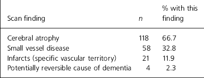
Table 2 summarises the results of 177 CT scans. The majority of scan reports made some reference to cerebral atrophy. There was a fairly high prevalence of both infarcts (11.9%) and small vessel disease (32.8%).
Table 2. Report from 177 consecutive cranial computed tomography scans

| Scan finding | n | % with this finding |
|---|---|---|
| Cerebral atrophy | 118 | 66.7 |
| Small vessel disease | 58 | 32.8 |
| Infarcts (specific vascular territory) | 21 | 11.9 |
| Potentially reversible cause of dementia | 4 | 2.3 |
Four scans showed potentially significant organic pathology (2.3%). The scan findings for these patients are summarised in Table 3. All four of these patients had features as set out by the Dietch criteria. Two of the four patients had indications for scanning as defined by the College criteria. Only in one case (patient 1) was treatment for organic pathology (steroids) instigated following the scan, and there were no cases of wholly reversible pathology.
Table 3. Potentially reversible causes of dementia

| Patient | Scan finding | Predicted by Dietch criteria? | Predicted by College criteria? | Outcome of case |
|---|---|---|---|---|
| 1 | Numberous ring enhancing masses most in keeping with multiple metastases | Yes: focal cerebral signs: hemianopia | Yes: atypical feature of hemianopia | Patient prescribed steroids but no other treatment |
| 2 | Enlarged ventricles out of proportion to sulcal size. Possible normal pressure hydrocephalus (NPH) | Yes: previous history of seizures and CVA | No: 2 year history, typical for vascular dementia | NPH not felt to be clinically likely. No further investigations |
| 3 | Single indeterminate white matter lesion | Yes: possible previous CVA | No | Neurological referral made, no further imaging. No specific treatment |
| 4 | Broad based extraaxial mass arising from right cerebello-pontine angle: probable meningioma | Yes: headaches, sudden onset of memory loss | Yes: sudden onset is atypical | Not felt to be clinically relevant. Patient under review for this lesion |
Discussion
This study demonstrates a relatively low prevalence of PRCs of dementia (2.3%). Most of the literature on the prevalence of PRCs comes from the 1980s. For example, Clarfield reported a 13.7% prevalence of PRCs in 1988. The only recent report from the UK literature was reviewing scans performed between 1994 and 1996 (Reference BrantonBranton, 1999), and this demonstrated a prevalence of 7.8%. The apparent decreasing prevalence of PRCs might reflect a lower threshold in requesting cranial CT as this investigation becomes more widely and routinely available.
In this cohort, no patient had any wholly reversible cause for cognitive impairment identified by CT. The study thus confirms that the prevalence of truly reversible causes of dementia identified by cranial CT is, in fact, extremely low (Reference Foster, Scott and PayneFoster et al, 1999).
All four patients with a PRC were correctly identified by the Dietch criteria. The Royal College of Psychiatrists guidelines correctly predicted two patients with the most significant intracranial pathology, namely patient 1 with multiple cerebral metastases and patient 4 with a probable meningioma. The College guidelines would not have predicted patients 2 and 3, however - these patients had fairly marginal imaging abnormalities, one with an indeterminate white matter lesion not thought to be clinically significant and another with enlarged ventricles, but in whom normal pressure hydrocephalus was felt clinically unlikely. In neither of these two cases did the scan findings result in a management change and in both cases, the scan result was thought to be of doubtful overall significance. Therefore, they should not be taken as evidence of a lack of sensitivity of the College criteria. This study, therefore, adds validity to both of the assessed clinical prediction rules.
Three of the scanned patients (2, 3 and 4) had abnormal CT reports raising the possibility of reversible organic pathology, but none of these patients had a treatment change resulting from the scan. It may be argued that the imaging findings in these cases are incidental and that the scans did not lead to any real benefit to the patients. In these cases, scanning adds cost to the management of the patient and generates more work, as well as provoking anxiety and uncertainty in the patient and their carers. Such problems are a well-recognised drawback to widely applying sensitive diagnostic tests to large populations of patients.
A notable finding of this study is the high prevalence of evidence of small (32.6%) and large (11.9%) vessel cerebrovascular disease in our study population (Table 2). A real benefit of scanning, therefore, is to raise the possibility of vascular dementia. In this respect, cranial CT may well add to the management of patients with cognitive impairment.


eLetters
No eLetters have been published for this article.