Hostname: page-component-745bb68f8f-mzp66 Total loading time: 0 Render date: 2025-01-15T14:31:45.009Z Has data issue: false hasContentIssue false

The Morphology, Histology, and Fine Structure of the Gut of the Green Peach Aphid, Myzus persicae (Sulzer) (Homoptera: Aphididae)

Published online by Cambridge University Press:  31 May 2012

A. R. Forbes*
Affiliation:
Research Station, Research Branch, Canada Department of Agriculture, 6660 N.W. Marine Drive, Vancouver8 B.C.
Get access

Abstract

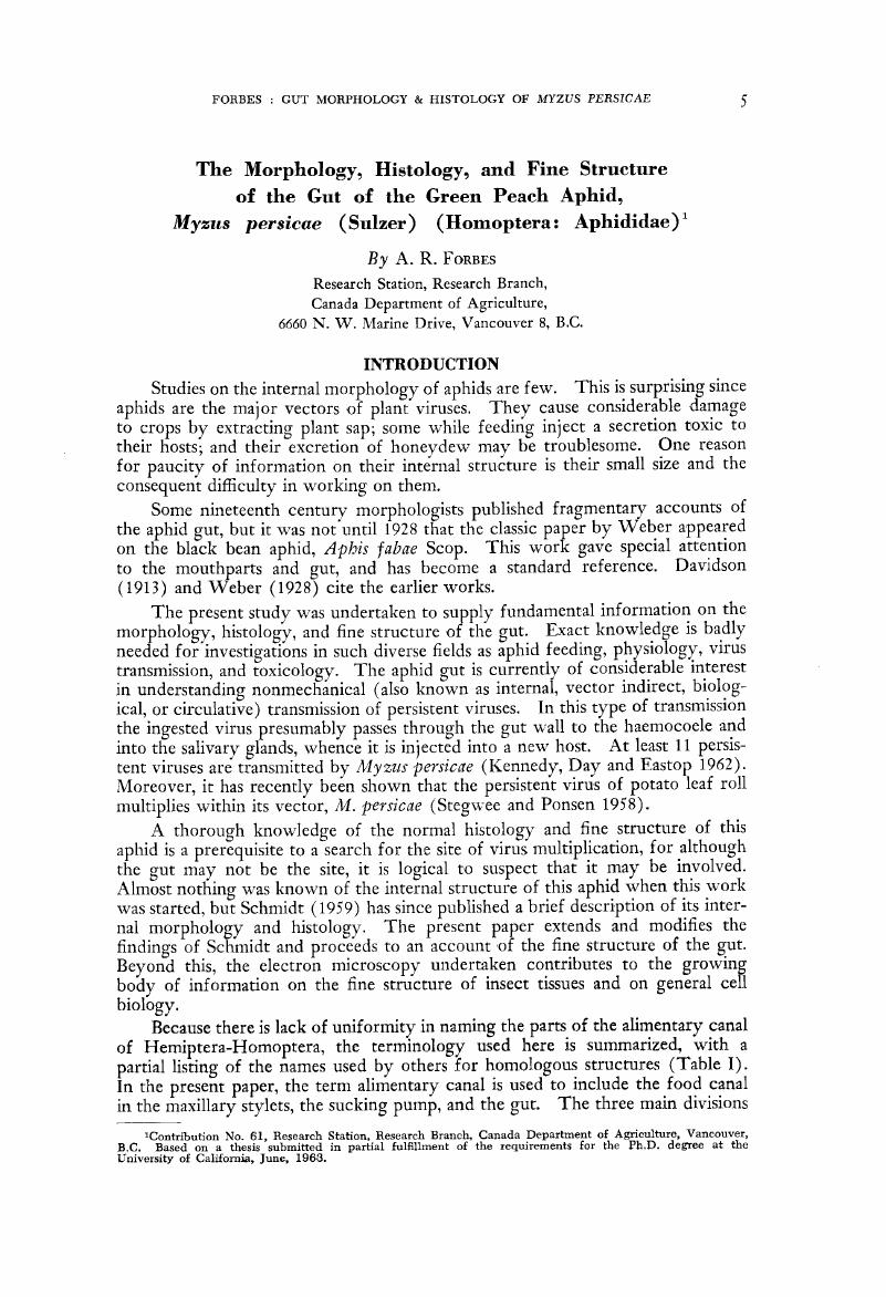
Image of the first page of this content. For PDF version, please use the ‘Save PDF’ preceeding this image.'
Type
Other
Copyright
Copyright © Entomological Society of Canada 1964

Access options

Get access to the full version of this content by using one of the access options below. (Log in options will check for institutional or personal access. Content may require purchase if you do not have access.)

Footnotes

1

Contribution No. 61, Research Branch, Canada Department of Agricluture, Vancouver, B.C, Based on a thesis submitted in partial fulfillment of the requirements for the Ph.D. degree at the University of California, June, 1963.

References

LITERATURE CITED

Anderson, E., and Beams, H. W.. 1956. Evidence from electron micrographs for the passage of material through pores of the nuclear membrane. J. biophys. biochem. Cytol. 2 Suppl.: 439444.Google Scholar
Auclair, J. L. 1963. Aphid feeding and nutrition. Annu. Rev. Ent. 8: 439490.Google Scholar
Baccetti, B. 1960. Ricerche sull'ultrastruttura dell'intestino degli insetti. I. L'ileo di un ortottero adulto. Redia 45: 263278.Google Scholar
Bahr, G. F., and Beerman, W.. 1954. The fine structure of the nuclear membrane in the larval salivary gland and midgut of Chironomus. Exp. Cell Res. 6: 519522.Google Scholar
Baker, A. C. 1915. The woolly apple aphis. U. S. Dep. Agric. Rep. 101, 56 pp.Google Scholar
Baker, J. R. 1942. The free border of the intestinal epithelial cell of vertebrates. Quart. J. micr. Sci. 84: 73104.Google Scholar
Baker, J. R. 1950. Cytological technique. Wiley, New York.3rd ed.Google Scholar
Barer, R. 1948. The structure of the striated muscle fibre. Biol. Rev. 23: 159200.Google Scholar
Bargmann, W., Knoop, A. and Schiebler, T. H.. 1955. Histologische, Cytochemische und elektronenmikroskopische Untersuchungen am Nephron (mit Berüchsichtigung der Mitochondrien). Z. Zellforsch. 42: 386422.Google Scholar
Beams, H. W., and Anderson, E.. 1957. Light and electron microscope studies on the striated border of the intestinal epithelial cells of insects, J. Morph. 100: 601619.Google Scholar
Beams, H. W., Tahmisian, T. N. and Devine, R. L.. 1955. Electron microscope studies on the cells of the Malpighian tubules of the grasshopper (Orthoptera, Acrididae). J. biophys. biochem. Cytol. 1: 197202.Google Scholar
Bennett, H. S. 1956. The concepts of membrane flow and membrane vesiculation as mechanisms for active transport and ion pumping, J. biophys. biochem. Cytol. 2 Suppl.: 99103.Google Scholar
Bennett, H. S. 1958. The cytology of striped muscle, pp. 343–380. In Palay, S. L. (ed.), Frontiers in cytology. Yale University Press, New Haven.Google Scholar
Bennett, H. S. 1960. The structure of striated muscle as seen by the electron microscope, pp. 137–181. In Bourne, G. H. (ed.), The structure and function of muscle, v.l. Academic Press, New York.Google Scholar
Berkaloff, A. 1958. Les grains de sécrétion des tubes de Malpighi de Gryllus domesticas (Orthoptère Gryllidae). C. R. Acad. Sci., Paris 246: 28072809.Google Scholar
Berkaloff, A. 1959. Variation de l'ultrastructure des tubes de Malpighi et leur fonctionnement chez Gryllus domesticus (Orthoptère Gryllidae). C. R. Acad. Sci., Paris 248: 466469.Google Scholar
Berkaloff, A. 1960. L'ultrastructure des tubes de Malpighi et le problème de leur fonctionnement chez les insects. Proc. 4th int. Conf. Electron Microscop. (1958) 2: 392396.Google Scholar
Bonhag, P. 1955. Histochemical studies of the ovarian nurse tissues and oöcytes of the milkweed bug, Oncopeltus fasciatus (Dallas). I. Cytology, nucleic acids, and carbohydrates. J. Morph. 96: 381441.Google Scholar
Bradfield, J. R. G. 1950. The localization of enzymes in cells. Biol. Rev. 25: 113157.Google Scholar
Bramstedt, F. 1948. Über die Verdauungsphysiologie der Aphiden. Z. Naturf. 3B: 1424.Google Scholar
Culling, C. F. A. 1957. Handbook of histopathological technique. Butterworth, London.Google Scholar
Dalton, A. J. 1951. Electron micrography of epithelial cells of the gastro-intestinal tract and pancreas. Amer. J. Anat. 89: 109133.Google Scholar
Dalton, A. J. 1961. Golgi apparatus and secretion granules, pp. 603619. In Brachet, J. and Mirsky, A. E. (ed.), The cell, v.2. Academic Press, New York.Google Scholar
Dalton, A. J., Kahler, H. and Lloyd, B.. 1951. The structure of the free surface of a series of epithelial cell types in the mouse as revealed by the electron microscope. Anat. Rec. 111: 6778.Google Scholar
Dalton, A. J. et al. , 1950. Finer structure of hepatic, intestinal and renal cells of the mouse as revealed by the electron microscope. J. nat. Cancer Inst. 11: 439461.Google Scholar
Davidson, J. 1913. The structure and biology of Schizoneura lanigera, Hausmann Part I. The apterous viviparous female. Quart. J. micr. Sci. 58: 653701.Google Scholar
Davidson, J. 1914. On the mouth-parts and mechanism of suction in Schizoneura lanigera, Hausmann, J. Linn. Soc. (Zool.) 32: 307330.Google Scholar
Day, M. F., and Waterhouse, D. F.. 1953a. Structure of the alimentary system, pp. 273298. In Roeder, K. D. (ed.), Insect physiology. Wiley, New York.Google Scholar
Day, M. F., and Waterhouse, D. F.. 1953b. The mechanism of digestion, pp. 311330. In Roeder, K. D. (ed.), Insect physiology. Wiley, New York.Google Scholar
Dempsey, E. W. 1958. Current concepts of cellular structure, pp. 9–18. In Palay, S. L. (ed.). Frontiers in cytology. Yale University Press, New Haven.Google Scholar
Dempsey, E. W., Wislocki, G. B. and Amoroso, E. C.. 1955. Electron microscopy of the pig's placenta with special reference to the cell membranes of the endometrium and chorion. Amer. J. Anat. 96: 65102.Google Scholar
Deuel, M. J. 1951. The lipids, their chemistry and biochemistry. Interscience Publishers, New York. 3 v.Google Scholar
Edwards, G. A. 1960. Insect micromorphology. Annu. Rev. Ent. 5: 1734.Google Scholar
Edwards, G. A., Ruska, H. and de Harven, E.. 1958. Neuromuscular junctions in flight and tymbal muscles of the cicada, J. biophys. biochem. Cytol. 4: 251256.Google Scholar
Edwards, G. A. et al. , 1956. Comparative cytophysiology of striated muscle with special reference to the role of the endoplasmic reticulum. J. biophys. biochem. Cytol. 2 Suppl.: 143156.Google Scholar
Fawcett, D. W. 1958. Structural specializations of the cell surface, pp. 1941. In Palay, S. L. (ed.), Frontiers in cytology. Yale University Press, New Haven.Google Scholar
Fawcett, D. W., and Porter, K. R.. 1954. A study of the fine structure of ciliated epithelia. J. Morph. 94: 221282.Google Scholar
Finck, H. 1958. An electron microscope study of basophile substances of frozen-dried rat liver. J. biophys. biochem. Cytol. 4: 291300.Google Scholar
Flax, M. H., and Himes, M. H.. 1952. Microspectrophotometric analysis of metachromatic staining of nucleic acids. Physiol. Zoöl. 25: 297311.Google Scholar
Forster, R. P. 1961. Kidney cells, pp. 89161. In Brachet, J. and Mirsky, A. E. (ed.), The cell, v. 5. Academic Press, New York.Google Scholar
Gabe, M., and Arvy, L.. 1961. Gland cells, pp. 188. In Brachet, J. and Mirsky, A. E. (ed.), The cell, v. 5. Academic Press, New York.Google Scholar
Gay, H. 1955. Nucleo-cytoplasmic relations in salivary-gland cells of Drosophila. Proc. nat. Acad. Sci., Wash. 41: 370375.Google Scholar
Gay, H. 1956. Nucleocytoplasmic relations in Drosophila. Cold Spr. Harb. Sym. quant. Biol. 21: 257269.Google Scholar
Gersch, M. 1942. Verteilung und Ausscheidung von Fluorescein bei Aphiden. II. Beitrag zur Exkretion bei Insekten. Z. vergl. Physiol. 29: 506531.Google Scholar
Granger, B., and Baker, R. F.. 1950. Electron microscope investigation of the striated border of intestinal epithelium. Anat. Rec. 107: 423441.Google Scholar
Groat, R. A. 1949. Initial and persisting power of solutions of iron-hematoxylin lake. Stain Tech. 24: 157163.Google Scholar
, H., A. J., 1962. Cytochemical progress in electron microscopy. Nature, Lond. 195: 11591160.Google Scholar
Haguenau, F. 1958. The ergastoplasm: its history, ultrastructure, and biochemistry. Int. Rev. Cytol. 7: 425483.Google Scholar
Hale, A. J. 1957. The histochemistry of polysaccharides. Int. Rev. Cytol. 6: 193263.Google Scholar
Hodge, A. J., Huxley, H. E. and Spiro, D.. 1954. Electron microscope studies on ultrathin sections of muscle. J. exp. Med. 99: 201206.Google Scholar
Hollande, M. A. 1927. La rénovation de l'épithélium de l'intestin moyen du Criquet égyptien Orthacanthaeris (Acridium) aegyptia L. C. R. Acad. Sci., Paris 184: 14761478.Google Scholar
Holmberg, A. 1955. Studies of the ultrastructure of the nonpigmented epithelium in the ciliary body. Acta ophthal., Kbh. 33: 377381.Google Scholar
Holter, H. 1959. Pinocytosis. Int. Rev. Cytol. 8: 481504.Google Scholar
Holtz, H. 1909. Von der Secretion und Absorption der Darmzellen bei Nematus. Anat. Hefte, Abt. I 39: 683694.Google Scholar
Hoof, H. A. van. 1957. On the mechanism of transmission of some plant viruses. Koninkl. Ned. Akad. Wetenschap. Proc. Ser. C 60: 314317.Google Scholar
Kennedy, J. S., Day, M. F. and Eastop, V. F.. 1962. A conspectus of aphids as vectors of plant viruses. Commonw. Inst. Ent. London.Google Scholar
Khan, M. R., and Ford, J. B.. 1962. Studies on digestive enzyme production and its relationship to the cytology of the midgut epithelium in Dysdercus fasciatus Sign. (Hemiptera, Pyrrhocoridae). J. Ins. Physiol. 8: 597608.Google Scholar
Knowlton, G. F. 1925. The digestive tract of Longistigma caryae (Harris). Ohio J. Sci. 25: 244252.Google Scholar
Leblond, C. P. 1950. Distribution of periodic acid-reactive carbohydrates in the adult rat. Amer. J. Anat. 86: 149.Google Scholar
Leonhardt, H. 1940. Beiträge zur Kenntnis der Lachniden, der wichtigsten Tannenhonigtauerzeuger. Z. angew. Ent. 27: 208272.Google Scholar
Lillie, R. D. 1954. Histopathologic technic and practical histochemistry. McGraw-Hill, New York.Google Scholar
Linder, H. J. 1956. Structure and histochemistry of the maxillary glands in the milkweed bug, Oncopeltus fasciatus (Hem.), J. Morph. 99: 575602.Google Scholar
Ludwig, E., and Richterich, R.. 1954. Morphologische und histochemische Untersuchungen am Stäbchensaum der Darmepithelzelle. Acta anat. 21: 168196.Google Scholar
Luft, J. H. 1961. Improvements in epoxy resin embedding methods. J. biophys. biochem. Cytol. 9: 409414.Google Scholar
Mercer, E. H., and Brunet, P. C. J.. 1959. The electron microscopy of the left colleterial gland of the cockroach. J. biophys. biochem. Cytol. 5: 257261.Google Scholar
Meyer, K. 1945. Mucoids and glycoproteins. Advanc. Protein Chem. 2: 249275.Google Scholar
Miller, F. W. 1932. The digestive epithelium of the aphid, Macrosiphum sanbornii. Proc. Penn. Acad. Sci. 6: 148151.Google Scholar
Mirsky, A. E., and Osawa, S.. 1961. The interphase nucleus, pp. 677770. In Brachet, J. and Mirsky, A. E. (ed.), The cell, v. 2 Academic Press, New York.Google Scholar
Mittler, T. E. 1957. Studies on the feeding and nutrition of Tuberolachnus salignus (Gmelin) (Homoptera, Aphididae) I. The uptake of phloem sap. J. exp. Biol. 34: 334341.Google Scholar
Mittler, T. E., and Dadd, R. H.. 1962. Artificial feeding and rearing of the aphid, Myzus persicae (Sulzer), on a completely defined synthetic diet. Nature, Lond. 195: 404.Google Scholar
Moog, F., and Wenger, E.. 1952. The occurrence of a neutral mucopolysaccharide at sites of high alkaline phosphatase activity. Amer. J. Anat. 90: 339377.Google Scholar
Newell, G. E., and Baxter, E. W.. 1936. On the nature of the free cell border of certain mid-gut epithelia. Quart. J. micr. Sci. 79: 123150.Google Scholar
Novikoff, A. B. 1961a. Mitochondria (Chondriosomes), pp. 299421. In Brachet, J. and Mirsky, A. E. (ed.), The cell, v. 2. Academic Press, New York.Google Scholar
Novikoff, A. B. 1961b. Lysosomes and related particles, pp. 423–488. In Brachet, J. and Mirsky, A. E. (ed.), The cell, v. 2. Academic Press, New York.Google Scholar
Ossiannilsson, F. 1961. Negative evidence against passage of whole plant virus particles through gut wall in aphid vectors. Nature, Lond. 190: 1222.Google Scholar
Painter, T. S. 1945. Nuclear phenomena associated with secretion in certain gland cells with especial reference to the origin of cytoplasmic nucleic acid. J. exp. Zool. 100: 523547.Google Scholar
Palade, G. E. 1952. A study of fixation for electron microscopy. J. exp. Med. 95: 285298.Google Scholar
Palade, G. E. 1955. A small particulate component of the cytoplasm, J. biophys. biochem. Cytol. 1: 5968.Google Scholar
Palade, G. E., and Siekevitz, P.. 1955. A correlated structural and chemical analysis of microsomes. (Abstract). Anat. Rec. 121: 347348.Google Scholar
Palay, S. L. 1958. The morphology of secretion, pp. 305342. In Palay, S. L. (ed.), Frontiers in cytology. Yale University Press, New Haven.Google Scholar
Pantin, C. F. A. 1948. Notes on microscopical technique for zoologists. Cambridge University Press, Cambridge.Google Scholar
Pearse, A. G. E. 1960. Histochemistry theoretical and applied. 2nd ed.J., and Churchill, A., London.Google Scholar
Pease, D. C. 1955a. The fine structure of the kidney seen by electron microscopy, J. Histochem. Cytochem. 3: 295309.Google Scholar
Pease, D. C. 1955b. Electron microscopy of the tubular cells of the kidney cortex. Anat. Rec. 121: 723743.Google Scholar
Pease, D. C. 1956. Infolded basal plasma membranes found in epithelia noted for their water transport. J. biophys. biochem. Cytol. 2 Suppl.: 203208.Google Scholar
Pease, D. C. 1960. Histological techniques for electron microscopy. Academic Press, New York.Google Scholar
Pelton, J. Z. 1938. The alimentary canal of the aphid Prociphilus tesselata Fitch. Ohio J. Sci. 38: 164169.Google Scholar
Pipa, R. L., and Cook, E. F.. 1958. The structure and histochemistry of the connective tissue of the sucking lice. J. Morph. 103: 353385.Google Scholar
Ponder, E. 1961. The cell membrane and its properties, pp. 184. In Brachet, J. and Mirsky, A. E. (ed.), The cell, v. 2. Academic Press, New York.Google Scholar
Porter, K. R. 1960. Problems in the study of nuclear fine structure. Proc. 4th int. Conf. Electron Microscop. (1958) 2: 186199.Google Scholar
Porter, K. R. 1961. The ground substance; observations from electron microscopy, pp. 621675. In Brachet, J. and Mirsky, A. E. (ed.), The cell, v. 2. Academic Press, New York.Google Scholar
Rhodin, J. 1954. Correlation of ultrastructural organization and function in normal and experimentally changed proximal convoluted cells of the mouse kidney. Thesis in Anatomy. Karolinska Inst. Stockh.Google Scholar
Rhodin, J. 1958. Anatomy of kidney tubules. Int. Rev. Cytol. 7: 485534.Google Scholar
Ruska, H. 1954. Histology of striated muscle. N. Y. St. Dep. Hlth. annu. Rep. Div. Lab. Res. 1954, 2425.Google Scholar
Schmidt, H. B. 1959. Beiträge zur Kenntnis der Ubertragung pflanzlicher Viren durch Aphiden. Biol. Zbl. 78: 889936.Google Scholar
Sjöstrand, F. S. 1956. Electron microscopy of cells and tissues, pp. 241298. In Oster, G. and Pollister, A. W. (ed.), Physical techniques in biological research, v. 3. Academic Press, New York.Google Scholar
Sjöstrand, F. S., and Rhodin, J.. 1953. The ultrastructure of the proximal convoluted tubules of the mouse kidney as revealed by high resolution microscopy. Exp. Cell Res. 4: 426456.Google Scholar
Smith, C. F. 1939. The digestive system of Macrosiphum solanifolii (Ash.) (Aphidae: Homoptera). Ohio J. Sci. 39: 5759.Google Scholar
Smith, D. S., and Littau, V. C.. 1960. Cellular specialization in the excretory epithelia of an insect, Macrosteles fascifrons Stål (Homoptera). J. biophys. biochem. Cytol. 8: 103133.Google Scholar
Snodgrass, R. E. 1935. Principles of insect morphology. McGraw-Hill, New York.Google Scholar
Sotelo, J. R., and Porter, K. R.. 1959. An electron microscope study of the rat ovum. J. biophys. biochem. Cytol. 5: 327342.Google Scholar
Stegwee, D., and Ponsen, M. B.. 1958. Multiplication of potato leafroll virus in the aphid Myzus persicae (Sulz.). Ent. exp. et appl. 1: 291300.Google Scholar
Steinbach, H. B. 1951. Permeability. Ann. Rev. Physiol. 13: 2140.Google Scholar
Sutton, M. F. 1951. On the food, feeding mechanism and alimentary canal of Corixidae (Hemiptera, Heteroptera). Proc. zool. Soc. Lond. 121: 465499.Google Scholar
Tsubo, I. 1961. Electron microscopy of the Malpighian tubule. J. Nara med. Ass. 12: 301317.Google Scholar
Tsubo, I., and Brandt, P. W.. 1962. An electron microscopic study of the Malipghian tubules of the grasshopper, Dissosteira carolina. J. Ultrastructure Res. 6: 2835.Google Scholar
Waterhouse, D. F. 1953. The occurrence and significance of the peritrophic membrane, with special reference to adult Lepidoptera and Diptera. Aust. J. Zool. 1: 299318.Google Scholar
Waterhouse, D. F., and Day, M. F.. 1953. Function of the gut in absorption, excretion, and intermediary metabolism, pp. 331349. In Roeder, K. D. (ed.), Insect physiology. Wiley, New York.Google Scholar
Waterhouse, D. F., and Wright, M.. 1960. The fine structure of the mosaic midgut epithelium of blowfly larvae. J. Ins. Physiol. 5: 230239.Google Scholar
Watson, M. L. 1955. The nuclear envelope — its structure and relation to cytoplasmic membranes. J. biophys. biochem. Cytol. 1: 257270.Google Scholar
Weber, H. 1928. Skelett, Muskulatur und Darm der schwarzen Blattlaus Aphis fabae Scop. mit besonderer Berücksichtigung der Funktion der Mundwerkzeuge und des Darms. Zoologica 28: 1120.Google Scholar
Weiss, J. M. 1953. The ergastoplasm — its fine structure and relation to protein synthesis as studied with the electron microscope in the pancreas of the Swiss albino mouse. J. exp. Med. 98: 607618.Google Scholar
Wigglesworth, V. B. 1953. The principles of insect physiology. 5th ed.Methuen, London.Google Scholar
Wigglesworth, V. B. 1956. The haemocytes and connective tissue formation in an insect, Rhodnius prolixus (Hemiptera). Quart. J. micr. Sci. 97: 8998.Google Scholar
Wigglesworth, V. B., and Salpeter, M. M.. 1962. Histology of the Malpighian tubules in Rhodnius prolixus Stål (Hemiptera). J. Ins. Physiol. 8: 299307.Google Scholar
Woodruff, B. H. 1933. Studies of the epithelium lining the caeca and mid-gut in the grasshopper. J. Morph. 55: 5379.Google Scholar
Zweigelt, F. 1914. Beiträge zur Kenntnis des Saugphänomens der Blattlause und der Reacktionen der Pflanzenzellen. Zbl. Bakt. (Abt. 2) 42: 265335.Google Scholar