Hostname: page-component-745bb68f8f-grxwn Total loading time: 0 Render date: 2025-01-10T21:15:43.117Z Has data issue: false hasContentIssue false

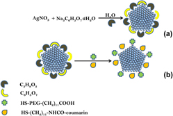
A size-controlled synthesis and characterization of mixed monolayer protected silver-S-(CH2)11-NHCO-coumarin nanoparticles and their Raman activities

Published online by Cambridge University Press:  08 June 2015

Mbuso Mlambo
Affiliation:
Molecular Sciences Institute, School of Chemistry, University of the Witwatersrand, Private Bag 3, Wits 2050, South Africa; and Advanced Materials Division, Nanotechnology Innovation Centre, Mintek, Private Bag X3015, Randburg 2125, South Africa
Phumlane S. Mdluli
Affiliation:
Durban University of Technology, Faculty of Applied Sciences, Department of Chemistry, Durban 4000, South Africa
Poslet Shumbula
Affiliation:
Advanced Materials Division, Nanotechnology Innovation Centre, Mintek, Private Bag X3015, Randburg 2125, South Africa
Amanda Skepu
Affiliation:
Advanced Materials Division, Nanotechnology Innovation Centre, Mintek, Private Bag X3015, Randburg 2125, South Africa
Robert Tshikhudo
Affiliation:
Advanced Materials Division, Nanotechnology Innovation Centre, Mintek, Private Bag X3015, Randburg 2125, South Africa
Nosipho Moloto*
Affiliation:
Molecular Sciences Institute, School of Chemistry, University of the Witwatersrand, Private Bag 3, Wits 2050, South Africa
*
a)Address all correspondence to this author. e-mail: [email protected]
Get access

Abstract

The study reports the functionalization of the size-controlled synthesized silver nanoparticles (AgNPs) with coumarin derivative. The size and the morphology of the as-synthesized AgNPs were obtained in the presence of glycerol and sodium citrate which acted as the reducing agent which led to nucleation of silver ions thus yielding different sizes of AgNPs. The obtained AgNPs were functionalized with different stoichiometric ratios of [HS-(CH2)11-NHCO-coumarin:HS-PEG-(CH2)11COOH] to form mixed monolayer protected silver clusters and their Raman activities were evaluated to determine the effect of particle size on the enhancement factor (EF). The functionalization and the stability of AgNPs were confirmed using a combination of techniques, namely UV–Visible spectroscopy, transmission electron microscopy, Zetasizer, and Raman spectroscopy. The obtained Raman spectra were used to calculate the EF of the HS-(CH2)11-NHCO-coumarin adsorbed on AgNPs, which was observed to increase with an increase in size of AgNPs from 16 to 30 nm. Increasing the particle size to 43 nm lowered the EF by 10-fold and hence an optimal size of ∼30 nm was achieved for the coumarin derivative adsorbate.

Type
Articles
Copyright
Copyright © Materials Research Society 2015 

Access options

Get access to the full version of this content by using one of the access options below. (Log in options will check for institutional or personal access. Content may require purchase if you do not have access.)

References

REFERENCES

Raman, C.V. and Krishnan, K.: A new type of secondary radiation. Nature 121, 501 (1928).CrossRefGoogle Scholar
Kneipp, K., Wang, Y., Kneipp, H., Perelman, L.T., Itzkan, I., Dasari, R.R., and Feld, M.S.: Single molecule detection using surface-enhanced Raman scattering (SERS). Phys. Rev. Lett. 78, 1667 (1997).Google Scholar
Fleischmann, M., Hendra, P.J., and Mcquillan, A.J.: Raman spectra of pyridine adsorbed at a silver electrode. Chem. Phys. Lett. 26, 163 (1974).Google Scholar
Jeanmaire, D.L. and Van Duyne, R.P.: Surface Raman spectroelectrochemistry: Part I. Heterocyclic, aromatic, and aliphatic amines adsorbed on the anodized silver electrode. J. Electroanal. Chem. Interfacial Electrochem. 84, 1 (1977).Google Scholar
Chang, K. and Furtak, T.E. eds.: Surface Enhanced Raman Scattering (Plenum Press, New York, 1982).Google Scholar
Moskovits, M.: Surface-enhanced spectroscopy. Rev. Mod. Phys. 57, 783 (1985).Google Scholar
Kneipp, K., Kneipp, H., Itzkan, I., and Dasari, R.R.: Surface-enhanced Raman scattering and biophysics. J. Phys. Condens. Matter 14, 597 (2002).CrossRefGoogle Scholar
Stiles, P.L., Dieringer, J.A., Shah, N.C., and Van Duyne, R.R.: Surface-enhanced Raman spectroscopy. Annu. Rev. Anal. Chem. 1, 601 (2008).Google Scholar
Joseph, V., Matschulat, A., Polte, J., Rolf, S., Emmerling, F., and Kneipp, J.: SERS enhancement of gold nanospheres of defined size. J. Raman Spectrosc. 42, 1742 (2011).Google Scholar
McFarland, A.D., Young, M.A., Dieringer, J.A., and Van Duyne, R.P.: Wavelength-scanned surface-enhanced Raman excitation spectroscopy. J. Phys. Chem. B 109, 11279 (2005).CrossRefGoogle ScholarPubMed
Emory, S.R., Haskins, W.E., and Nie, S.: Direct observation of size-dependent optical enhancement in single metal nanoparticles. J. Am. Chem. Soc. 120, 8009 (1998).Google Scholar
Kelly, K.L., Coronado, E., Zhao, L.L., and Schatz, G.C.: The optical properties of metal nanoparticles: The influence of size, shape, and dielectric environment. J. Phys. Chem. B 107, 668 (2003).Google Scholar
Le Ru, E.C. and Etchegoin, P.G.: Principles of Surface-Enhanced Raman Spectroscopy (Elsevier Science, Amsterdam, Netherlands, 2008).Google Scholar
Zhang, Q., Xie, J., Yu, Y., and Lee, J.Y.: Monodispersity control in the synthesis of monometallic and bimetallic quasi-spherical gold and silver nanoparticles. Nanoscale 2, 1962 (2010).Google Scholar
Lee, P.C. and Meisel, D.: Adsorption and surface-enhanced Raman of dyes on silver and gold sols. J. Phys. Chem. 86, 3391 (1982).CrossRefGoogle Scholar
Chreigton, J.A., Blatchford, C.G., and Albrecht, M.G.: Plasma resonance enhancement of Raman-scattering by pyridine adsorbed on silver of gold sol particles of size comparable to the excitation wavelength. J. Chem. Soc., Faraday Trans. 75, 790 (1979).CrossRefGoogle Scholar
Leopold, N. and Lendl, B.: A new method for fast preparation of highly surface-enhanced Raman scattering (SERS) active silver colloids at room temperature by reduction of silver nitrate with hydroxylamine hydrochloride. J. Phys. Chem. B 107, 5723 (2003).Google Scholar
Silvert, P-Y., Herrera-Urbina, R., Duvauchelle, N., Vijayakrishnan, V., and Elhsissen, K.T.: Preparation of colloidal silver dispersions by the polyol process. J. Mater. Chem. 6, 573 (1996).Google Scholar
Schutz, M., Steinigeweg, D., Salehi, M., Kompe, K., and Schlucker, S.: Hydrophilically stabilized gold nanostars as SERS labels for tissue imaging of the tumor suppressor p63 by immuno-SERS microscopy. Chem. Commun. 47, 4216 (2011).Google Scholar
Zhang, Q., Li, N., Goebl, J., Lu, Z., and Yin, Y.: A systematic study of the synthesis of silver nanoplates: Is citrate a “Magic” reagent? J. Am. Chem. Soc. 133, 18931 (2011).Google Scholar
Steinigeweg, D. and Schlücker, S.: Monodispersity and size control in the synthesis of 20-100 nm quasi-spherical silver nanoparticles by citrate and ascorbic acid reduction in glycerol-water mixtures. Chem. Commun. 48, 8682 (2012).Google Scholar
Templeton, A.C., Wuelfing, M.P., and Murray, R.W.: Monolayer protected cluster molecules. Acc. Chem. Res. 33, 27 (2000).Google Scholar
Steigerwald, M.L., Alivisatos, A.P., Gibson, J.M., Harris, T.D., Kortan, R., Muller, A.J., Thayer, A.M., Duncan, T.M., Douglass, D.C., and Brus, L.E.: Surface derivatization and isolation of semiconductor cluster molecules. J. Am. Chem. Soc. 110, 3046 (1998).Google Scholar
Mlambo, M., Mdluli, P.S., Shumbula, P., Mpelane, S., Moloto, N., Skepu, A., and Tshikhudo, R.: Synthesis and characterization of mixed monolayer protected gold nanorods and their Raman activities. Mater. Res. Bull. 48, 4181 (2013).Google Scholar
Pillay, J., Ozoemena, K.I., Tshikhudo, R.T., and Moutloali, R.M.: Monolayer-protected clusters of gold nanoparticles: Impacts of stabilizing ligands on the heterogeneous electron transfer dynamics and voltammetric detection. Langmuir 26, 9061 (2010).Google Scholar
Doty, R.C., Sigman, M.B. Jr., Stowell, C.A., Shah, P.S., Saunders, A.E., and Korgel, B.A.: Synthesis and Fabrication of Metal Nanocrystal Superlattices. In Semiconductor and Metal Nanocrystals: Synthesis and Electronic and Optical Properties, Vol. 87, Klimov, V.I. ed.; Marcel Dekker: New York, 2003; p. 387.Google Scholar
Gu, X., Nie, C., Lui, Y., and Lin, C.: Synthesis of silver nanorods and nanowires by tartrate-reduced route in aqueous solutions. Mater. Chem. Phys. 96, 217 (2006).Google Scholar
Joo, T.H., Kim, M.S., and Kim, K.: Surface-enhanced Raman scattering of benzenethiol in silver sol. J. Raman Spectrosc. 18, 57 (1987).Google Scholar
Stamplecoskie, K.G. and Scaiano, J.C.: Optimal size of silver nanoparticles for surface-enhanced Raman spectroscopy. J. Phys. Chem. C115, 1403 (2011).Google Scholar
Lin, W-C., Liao, L-S., Chen, Y-H., Chang, H-C., Tsai, D.P., and Chiang, H-P.: Size dependence of nanoparticle-SERS enhancement from silver film over nanosphere (AgFON) substrate. Plasmonics 6, 201 (2011).Google Scholar
Wang, H.H., Liu, C.Y., Wu, S.B., Liu, N.W., Peng, C.Y., Chan, T.H., Hsu, C.F., Wang, J.K., and Wang, Y.L.: Highly Raman-enhancing substrates based on silver nanoparticle arrays with tunable sub-10 nm gaps. Adv. Mater. 18, 491 (2006).Google Scholar
Lin, W.C., Huang, S.H., Chen, C.L., Chen, C.C., Tsai, D.P., and Chiang, H.P.: Controlling SERS intensity by tuning the size and height of a silver nanoparticle array. Appl. Phys. A 101, 185 (2010).Google Scholar
Fang, P., Li, J., Yang, Z., Li, L., Ren, B., and Tian, Z.: Optimization of SERS activities of gold nanoparticles and gold-core–palladium-shell nanoparticles by controlling size and shell thickness. J. Raman Spectrosc. 39, 1679 (2008).Google Scholar
Meier, M. and Wokaun, A.: Enhanced fields on large metal particles: Dynamic depolarization. Opt. Lett. 8, 581 (1983).Google Scholar
Supplementary material: File

Mlambo et al. supplementary material

Supplementary figures

Download Mlambo et al. supplementary material(File)
File 825 KB