Hostname: page-component-745bb68f8f-cphqk Total loading time: 0 Render date: 2025-01-26T23:22:53.412Z Has data issue: false hasContentIssue false

Silicon nanowire/polycaprolactone composites and their impact on stromal cell function

Published online by Cambridge University Press:  09 August 2012

Ke Jiang
Affiliation:
Department of Chemistry, Texas Christian University, Fort Worth, Texas 76129
Jeffery L. Coffer*
Affiliation:
Department of Chemistry, Texas Christian University, Fort Worth, Texas 76129
Giridhar R. Akkaraju
Affiliation:
Department of Biology, Texas Christian University, Fort Worth, Texas 76129
*
a)Address all correspondence to this author. e-mail: [email protected]
Get access

Abstract

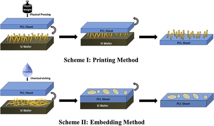
In this study, silicon nanowire (SiNW)/polycaprolactone composites with different surface topographies were fabricated by straightforward embedding or printing methods and their cytocompatibility was evaluated with a bone-relevant cell line derived from mouse stroma. The incorporation of biocompatible polymers with semiconducting SiNWs can ideally provide an enhanced environment to support proliferation and differentiation functions of bone cells. Cell/composite interactions were assessed with suitable assays including viability and alkaline phosphatase activity, while scanning electron microscopy characterization was used to study the morphology of cells grown on composites. Such results suggest that for nanowires in a vertical array, the presence of the polymer improves cellular attachment and overall viability relative to the nanowire-only system.

Type
Articles
Copyright
Copyright © Materials Research Society 2012

Access options

Get access to the full version of this content by using one of the access options below. (Log in options will check for institutional or personal access. Content may require purchase if you do not have access.)

References

REFERENCES

Choi, C.H., Hagvall, S.H., Wu, B.M., Dunn, J.C.Y., Beygui, R.E., and Kim, C.J.: Cell interaction with three-dimensional sharp-tip nanotopography. Biomaterials 28, 16721679 (2007).CrossRefGoogle ScholarPubMed
Garibaldi, S., Brunelli, C., Bavastrello, V., Ghigliotti, G., and Nicolini, C.: Carbon nanotube biocompatibility with cardiac muscle cells. Nanotechnology 17, 391397 (2006).CrossRefGoogle Scholar
Dably, M.J., Riehle, M.O., Johnstone, H., Affrossman, S., and Curtis, A.S.G.: In vitro reaction of endothelial cells to polymer demixed nanotopography. Biomaterials 23, 29452954 (2002).Google Scholar
Ishaug, S.L., Crane, G.M., Miller, M.J., Yasko, A.W., Yaszemski, M.J., and Mikos, A.G.: Bone formation by three-dimensional stromal osteoblast culture in biodegradable polymer scaffolds. J. Biomed. Mater. Res. 36, 1728 (1997).3.0.CO;2-O>CrossRefGoogle ScholarPubMed
Shwartz, Z. and Boyan, B.D.: Underlying mechanisms at the bone–biomaterial interface. J. Cell. Biochem. 56, 340347 (1994).CrossRefGoogle Scholar
Lee, J., Kang, B.S., Hicks, B., Chancellor, T.F. Jr., Chu, B.H., Wang, H.T., Keselowsky, B.G., Ren, F., and Lele, T.P.: The control of cell adhesion and viability by zinc oxide nanorods. Biomaterials 29, 37433749 (2008).CrossRefGoogle ScholarPubMed
Cui, Y., Wei, Q., Park, H., and Lieber, C.M.: Nanowire nanosensors for highly sensitive and selective detection of biological and chemical species. Science 293, 12891292 (2001).CrossRefGoogle ScholarPubMed
Shao, M.W., Yao, H., Zhang, M.L., Wong, N.B., Shan, Y.Y., and Lee, S.T.: Fabrication and application of long strands of silicon nanowires as sensors for bovine serum albumin detection. Appl. Phys. Lett. 87, 183106 (2005).CrossRefGoogle Scholar
Gudiksen, M.S., Lauhon, L., Wang, L., Smith, D., and Lieber, C.M.: Growth of nanowire superlattice structures for nanoscale photonics and electronics. Nature 415, 617620 (2002).CrossRefGoogle ScholarPubMed
Cui, Y. and Lieber, C.M.: Functional nanoscale electronic devices assembled using silicon nanowire building blocks. Science 291, 851853 (2001).CrossRefGoogle ScholarPubMed
Popat, K.C., Daniels, R.H., Dubrow, R.S., Hardev, V., and Desai, T.A.: Nanostructured surfaces for bone biotemplating applications. J. Orthop. Res. 24, 619627 (2006).CrossRefGoogle ScholarPubMed
Kwon, H., Beaux, M.F. II, Ebert, C., Wang, L., Lassiter, B.E., Park, Y.H., Mcllroy, D.N., Hovde, C.J., and Bohach, G.A.: Nanowire-based delivery Escherichia coli O157 Shiga toxin 1 A subunit into human bovine cells. Nano Lett. 7, 27182723 (2007).CrossRefGoogle ScholarPubMed
Brammer, K.S., Choi, C., Oh, S., Cobb, C.J., Connelly, L.S., Loya, M.S., Kong, D., and Jin, S.: Antibiofouling, sustained antibiotic release by Si nanowire templates. Nano Lett. 9, 35703574 (2009).CrossRefGoogle ScholarPubMed
Qi, S., Yi, C., Chen, W., Fong, C.C., Lee, S.T., and Yang, M.: Effects of silicon nanowires on HepG2 cell adhesion and spreading. ChemBioChem 8, 11151118 (2007).CrossRefGoogle ScholarPubMed
Qi, S., Yi, C., Ji, S., Fong, C.C., and Yang, M.: Cell adhesion and spreading behavior on vertically aligned silicon nanowire arrays. ACS Appl. Mater. Interfaces 1, 3034 (2009).CrossRefGoogle ScholarPubMed
Piret, G., Galopin, E., Coffinier, Y., Boukherroub, R., Legrand, D., and Slomianny, C.: Culture of Mammalian Cells on Patterned Superhydrophilic/Superhydrophobic Silicon Nanowire Arrays. Soft Matter 7, 86428649 (2011).CrossRefGoogle Scholar
Nagesha, D.K., Whitehead, M.A., and Coffer, J.L.: Biorelevant calcification and non-cytotoxic behavior in silicon nanowires. Adv. Mater. 17, 921924 (2005).CrossRefGoogle Scholar
Jiang, K., Fan, D., Belabassi, Y., Akkaraju, G.R., Montchamp, J-L., and Coffer, J.L.: Medicinal surface modification of silicon nanowires: Impact on calcification and stromal cell proliferation. ACS Appl. Mater. Interfaces 1, 266269 (2009).CrossRefGoogle ScholarPubMed
Carlisle, E.M.: In vivo requirement for silicon in articular cartilage and connective tissue formation in the chick. J. Nutr. 106, 478484 (1976).CrossRefGoogle ScholarPubMed
Carlisle, E.M.: Biochemical and morphological changes associated with long bone abnormalities in silicon deficiency. J. Nutr. 110, 10461056 (1980).CrossRefGoogle ScholarPubMed
Burg, K.J.L., Porter, S., and Kellam, J.F.: Biomaterial developments for bone tissue engineering. Biomaterials 21, 23472359 (2000).CrossRefGoogle ScholarPubMed
Snyder, E.J., Chideme, J., and Craig, G.S.W.: Fluidic self-assembly of semiconductor devices: A promising new method of mass-producing flexible circuitry. Jpn. J. Appl. Phys. 41, 43664369 (2002).CrossRefGoogle Scholar
Duan, X., Niu, C., Sahi, V., Chen, J., Parce, J.W., Empedocles, S., and Goldman, J.L.: High-performance thin-film transistors using semiconductor nanowires and nanoribons. Nature 425, 274278 (2003).CrossRefGoogle Scholar
Ahn, J.H., Kim, H.S., Lee, K.J., Jeon, S., Kang, S.J., Sun, Y., Nuzzo, R.G., and Rogers, J.A.: Heterogeneous three-dimensional electronics by use of printed semiconductor nanomaterials. Science 314, 17541757 (2006).CrossRefGoogle ScholarPubMed
Sun, Y., Kim, S., Adesida, I., and Rogers, J.A.: Bendable GaAs metal-semiconductor field-effect transistors formed with printed GaAs wire arrays on plastic substrates. Appl. Phys. Lett. 87, 083501 (2005).CrossRefGoogle Scholar
Lee, K.J., Lee, J., Hwang, H., Reitmeier, Z.J., Davis, R.F., Rogers, J.A., and Nuzzo, R.G.: A printable form of single-crystalline gallium nitride for flexible optoelectronic systems. Small 1, 11641168 (2005).CrossRefGoogle ScholarPubMed
Lam, C.X.F., Hutmacher, D.W., Schantz, J.T., Woodruff, M.A., and Teoh, S.H.: Evaluation of polycaprolactone scaffold degradation for 6 months in vitro and in vivo. J. Biomed. Mater. Res. 90A, 906919 (2009).CrossRefGoogle Scholar
Tuan, R.S., Boland, G., and Tuli, R.: Adult mesenchymal stem cells and cell-based tissue engineering. Arthritis Res. Ther. 5, 3245 (2003).CrossRefGoogle ScholarPubMed
Kolb, F.M., Hofmeister, H., Scholz, R., Zacharias, M., Gosele, U., Ma, D.D., and Lee, S.T.: Analysis of silicon nanowires grown by combining SiO evaporation with the VLS mechanism. J. Electrochem. Soc. 151, G472G475 (2004).CrossRefGoogle Scholar
Bershadsky, A., Kozlov, M., and Geiger, B.: Adhesion-mediated mechanosensitivity: A time to experiment, and a time to theorize. Curr. Opin. Cell Biol. 8, 472481 (2006).CrossRefGoogle Scholar
Spatz, J.P. and Geiger, B.: Molecular engineering of cellular environments: Cell adhesion to nano-digital surfaces. Methods Cell Biol. 83, 89111 (2007).CrossRefGoogle ScholarPubMed
Arnold, M., Cavalcanti-Adam, E.A., Glass, R., Blummel, J., Eck, W., Kantlehner, M., Kessler, H.J., and Spatz, P.: Activation of integrin function by nanopatterned adhesive interfaces. ChemPhysChem 5, 383388 (2004).CrossRefGoogle ScholarPubMed
Cavalcanti-Adam, E.A., Volberg, T., Micoulet, A., Kessler, H., Geiger, B., and Spatz, J.P.: Cell spreading and focal adhesion dynamics are regulated by spacing of integrin ligands. Biophys. J. 92, 29642974 (2007).CrossRefGoogle ScholarPubMed
Woong, K., Ng, J.K., Kunitake, M.E., Conklin, B.R., and Yang, P.D.: Interfacing silicon nanowires with mammalian cells. J. Am. Chem. Soc. 129, 72287229 (2007).Google Scholar
Shalek, A.K., Robinson, J.T., Karp, E.S., Lee, J.S., Ahn, D.R., Yoon, M.H., Sutton, A., Jorgolli, M., Gertner, R.S., Gujral, T.S., MacBeath, G., Yang, E.G., and Park, H.: Vertical silicon nanowires as a universal platform for delivering biomolecules into living cells. Proc. Natl. Acad. Sci. U.S.A. 107, 18701875 (2010).CrossRefGoogle ScholarPubMed
Mosmann, T.: Rapid colorimetric assay for cellular growth and survival: Application to proliferation and cytotoxicity assays. J. Immunol. Meth. 65, 5563 (1983).CrossRefGoogle ScholarPubMed
Bessey, O.A., Lowry, O.H., and Brock, M.J.: A method for the rapid determination of alkaline phosphatase with five cubic millimeters of serum. J. Biol. Chem. 164, 321329 (1946).CrossRefGoogle ScholarPubMed
Egusa, H., Kaneda, Y., Akashi, Y., Hamada, Y., Matsumoto, T., Saeki, M., Thakor, D.K., Tabata, Y., Matsuura, N., and Yatani, H.: Enhanced bone regeneration via multimodal actions of synthetic peptide SVVYGLR on osteoprogenitors and osteoclasts. Biomaterials 30, 46764686 (2009).CrossRefGoogle ScholarPubMed
Ripamonti, U. and Duneas, N.: Tissue morphogenesis and regeneration by bone morphogenetic proteins. Plast. Reconstr. Surg. 101, 227239 (1998).CrossRefGoogle ScholarPubMed
Bostrom, M.P. and Camacho, N.P.: Potential role of bone morphogenetic proteins in fracture healing. Clin. Orthop. Relat. Res. 355, S274S282 (1998).CrossRefGoogle Scholar
Sampath, T.K., Maliakal, J.C., Hauschka, P.V., Jones, W.K., Sasak, H., Tucker, R.F., White, K.H., Coughlin, J.E., Tucker, M.M., Pang, R.H.L., Corbett, C., Ozkaynak, E., Oppermann, H., and Rueger, D.C.: Recombinant human osteogenic protein-1 (hOP-1) induces new bone formation in vivo with a specific activity comparable with natural bovine osteogenic protein and stimulates osteoblast proliferation and differentiation in vitro. J. Biol. Chem. 267, 2035220362 (1992).CrossRefGoogle ScholarPubMed
Supplementary material: File

Jiang Supplementary Material

Figures

Download Jiang Supplementary Material(File)
File 3 MB