Hostname: page-component-cd9895bd7-lnqnp Total loading time: 0 Render date: 2024-12-23T11:28:27.589Z Has data issue: false hasContentIssue false

Relationship between the glutathione-responsive degradability of thiol-organosilica nanoparticles and the chemical structures

Published online by Cambridge University Press:  06 February 2019

Tomohiro Doura
Affiliation:
Department of Organ Anatomy and Nanomedicine, Yamaguchi University Graduate School of Medicine, Ube, Yamaguchi 755-8505, Japan
Tadashi Nishio
Affiliation:
Department of Organ Anatomy and Nanomedicine, Yamaguchi University Graduate School of Medicine, Ube, Yamaguchi 755-8505, Japan
Fuyuhiko Tamanoi
Affiliation:
Department of Microbiology, Immunology, and Molecular Genetics, University of California, Los Angeles, California 90095, USA
Michihiro Nakamura*
Affiliation:
Department of Organ Anatomy and Nanomedicine, Yamaguchi University Graduate School of Medicine, Ube, Yamaguchi 755-8505, Japan
*
a)Address all correspondence to this author. e-mail: [email protected]
Get access

Abstract

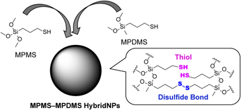
Stimuli-responsive degradable silica nanoparticles (NPs) are active topics of nanomaterial research, because they are expected to be low health-risk nanocarriers capable of controlled release of drugs. Among various stimuli-responsive silica NPs, disulfide bond-containing NPs show degradability by glutathione reduced form (GSH). Here, we synthesized and characterized three kinds of thiol-organosilica NPs made from 3-mercaptopropyltrimethoxysilane (MPMS) and 3-mercaptopropyl(dimethoxy)methylsilane (MPDMS). MPMS NPs, MPDMS NPs, and MPMS–MPDMS hybrid NPs revealed that the abundance ratio of disulfide bonds to thiols increased with the increase in content rate of MPDMS in thiol-organosilica NPs. We also revealed that thiol-organosilica NPs, which have disulfide bonds, are GSH-responsive degradable silica NPs using an electron microscopy and Ellman’s tests. Furthermore, we synthesized fluorescent MPMS–MPDMS NPs, including rhodamine B, and demonstrated the GSH-responsive release of dye from the NPs. These experiments indicate the potential of thiol-organosilica NPs, which have disulfide bonds as a GSH-responsive drug carrier.

Type
Article
Copyright
Copyright © Materials Research Society 2019 

Access options

Get access to the full version of this content by using one of the access options below. (Log in options will check for institutional or personal access. Content may require purchase if you do not have access.)

References

Gagner, J.E., Shrivastava, S., Qian, X., Dordick, J.S., and Siegel, R.W.: Engineering nanomaterials for biomedical applications requires understanding the nano-bio interface: A perspective. J. Phys. Chem. Lett. 3, 3149 (2012).CrossRefGoogle ScholarPubMed
Cheng, L-C., Jiang, X., Wang, J., Chen, C., and Liu, R-S.: Nano-bio effects: Interaction of nanomaterials with cells. Nanoscale 5, 3547 (2013).CrossRefGoogle ScholarPubMed
Bitar, A., Ahmad, N.M., Fessi, H., and Elaissari, A.: Silica-based nanoparticles for biomedical applications. Drug Discov. Today 17, 1147 (2012).CrossRefGoogle ScholarPubMed
Tang, L. and Cheng, J.: Nonporous silica nanoparticles for nanomedicine application. Nano Today 8, 290 (2013).CrossRefGoogle ScholarPubMed
Wang, Y., Zhao, Q., Han, N., Bai, L., Li, J., Liu, J., Che, E., Hu, L., Zhang, Q., Jiang, T., and Wang, S.: Mesoporous silica nanoparticles in drug delivery and biomedical applications. Nanomedicine 11, 313 (2015).CrossRefGoogle ScholarPubMed
Tiwari, P.M., Vig, K., Dennis, V.A., and Singh, S.R.: Functionalized gold nanoparticles and their biomedical applications. Nanomaterials 1, 31 (2011).CrossRefGoogle ScholarPubMed
Ali, A., Zafar, H., Zia, M., Haq, I.U., Phull, A.R., Ali, J.S., and Hussain, A.: Synthesis, characterization, applications, and challenges of iron oxide nanoparticles. Nanotechnol. Sci. Appl. 9, 49 (2016).CrossRefGoogle ScholarPubMed
Zhang, Y., Nayak, T.R., Hong, H., and Cai, W.: Biomedical applications of zinc oxide nanomaterials. Curr. Mol. Med. 13, 1633 (2013).CrossRefGoogle ScholarPubMed
Chimene, D., Alge, D.L., and Gaharwar, A.K.: Two-dimensional nanomaterials for biomedical applications: Emerging trends and future prospects. Adv. Mater. 27, 7261 (2015).CrossRefGoogle ScholarPubMed
Halas, N.J.: Nanoscience under glass: The versatile chemistry of silica nanostructures. ACS Nano 2, 179 (2008).CrossRefGoogle ScholarPubMed
Nishimori, H., Kondoh, M., Isoda, K., Tsunoda, S., and Tsutsumi, Y.: Histological analysis of 70-nm silica particles-induced chronic toxicity in mice. Eur. J. Pharm. Biopharm. 72, 626 (2009).CrossRefGoogle ScholarPubMed
Xie, G., Sun, J., Zhong, G., Shi, L., and Zhang, D.: Biodistribution and toxicity of intravenously administered silica nanoparticles in mice. Arch. Toxicol. 84, 183 (2010).CrossRefGoogle ScholarPubMed
Yu, Y., Duan, J., Li, Y., Li, Y., Jing, L., Yang, M., Wang, J., and Sun, Z.: Silica nanoparticles induce liver fibrosis via TGF-β1/Smad3 pathway in ICR mice. Int. J. Nanomed. 12, 6045 (2017).CrossRefGoogle ScholarPubMed
Liu, T., Li, L., Teng, X., Huang, X., Liu, H., Chen, D., Ren, J., He, J., and Tang, F.: Single and repeated dose toxicity of mesoporous hollow silica nanoparticles in intravenously exposed mice. Biomaterials 32, 1657 (2011).CrossRefGoogle ScholarPubMed
Lin, T., Li, L., Fu, C., Liu, H., Chen, D., and Tang, F.: Pathological mechanisms of liver injury caused by continuous intraperitoneal injection of silica nanoparticles. Biomaterials 33, 2399 (2012).Google Scholar
Mekaru, H., Lu, J., and Tamanoi, F.: Development of mesoporous silica-based nanoparticles with controlled release capability for cancer therapy. Adv. Drug Deliv. Rev. 95, 40 (2015).CrossRefGoogle ScholarPubMed
Moreira, A.F., Dias, D.R., and Correia, I.J.: Stimuli-responsive mesoporous silica nanoparticles for cancer therapy. Microporous Mesoporous Mater. 236, 141 (2016).CrossRefGoogle Scholar
Karimi, M., Zangabad, P.S., Baghaee-Ravari, S., Ghazadeh, M., Mirshekari, H., and Hamblin, M.R.: Smart nanostructures for cargo delivery: Uncaging and activating by light. J. Am. Chem. Soc. 139, 4584 (2017).CrossRefGoogle Scholar
Baeza, A., Guisasola, E., Ruiz-Hernández, E., and Vallet-Regí, M.: Magnetically triggered multidrug release by hybrid mesoporous silica nanoparticles. Chem. Mater. 24, 517 (2012).CrossRefGoogle Scholar
Saint-Cricq, P., Deshayes, S., Zink, J.I., and Kasko, A.M.: Magnetic field activated drug delivery using thermodegradable azo-functionalised PEG-coated core–shell mesoporous silica nanoparticles. Nanoscale 7, 13168 (2015).CrossRefGoogle ScholarPubMed
Lee, C-H., Cheng, S-H., Huang, I-P., Souris, J.S., Yang, C-S., Mou, C-Y., and Lo, L-W.: Intracellular pH-responsive mesoporous silica nanoparticles for the controlled release of anticancer chemotherapeutics. Angew. Chem., Int. Ed. 49, 8214 (2010).CrossRefGoogle ScholarPubMed
Meng, H., Xue, M., Xia, T., Zhao, Y-L., Tamanoi, F., Stoddart, J.F., Zink, J.I., and Nel, A.E.: Autonomous in vitro anticancer drug release from mesoporous silica nanoparticles by pH-sensitive nanovalves. J. Am. Chem. Soc. 132, 12690 (2010).CrossRefGoogle ScholarPubMed
Zhao, Y-L., Li, Z., Kabehie, S., Botros, Y.Y., Stoddart, J.F., and Zink, J.I.: pH-operated nanopistons on the surfaces of mesoporous silica nanoparticle. J. Am. Chem. Soc. 132, 13016 (2010).CrossRefGoogle Scholar
Zhou, S., Wu, D., Yin, X., Jin, X., Zhang, X., Zheng, S., Wang, C., and Liu, Y.: Intracellular pH-responsive and rituximab-conjugated mesoporous silica nanoparticles for targeted drug delivery to lymphoma B cells. J. Exp. Clin. Canc. Res. 36, 24 (2017).CrossRefGoogle ScholarPubMed
Giri, S., Trewyn, B.G., Stellmaker, M.P., and Li, V.S-Y.: Stimuli-responsive controlled-release delivery system based on mesoporous silica nanorods capped with magnetic nanoparticles. Angew. Chem., Int. Ed. 44, 5038 (2005).CrossRefGoogle ScholarPubMed
Cui, Y., Dong, H., Cai, X., Wang, D., and Li, Y.: Mesoporous silica nanoparticles capped with disulfide-linked PEG gatekeepers for glutathione-mediated controlled release. ACS Appl. Mater. Interfaces 4, 3177 (2012).CrossRefGoogle ScholarPubMed
Zhang, Q., Liu, F., Nguyen, K.T., Ma, X., Wang, X., Xing, B., and Zhao, Y.: Multifunctional mesoporous silica nanoparticles for cancer-targeted and controlled drug delivery. Adv. Funct. Mater. 22, 5144 (2012).CrossRefGoogle Scholar
Yang, Y., Wan, J., Niu, Y., Gu, Z., Zhang, J., Yu, M., and Yu, C.: Structure-dependent and glutathione-responsive biodegradable dendritic mesoporous organosilica nanoparticles for safe protein delivery. Chem. Mater. 28, 9008 (2016).CrossRefGoogle Scholar
Prasetyanto, E.A., Bertucci, A., Septiadi, D., Corradini, R., Castro-Hartmann, P., and De Cola, L.: Breakable hybrid organosilica nanoparticles for protein delivery. Angew. Chem., Int. Ed. 55, 3323 (2016).CrossRefGoogle ScholarPubMed
Hayashi, K., Maruhashi, T., Nakamura, M., Sakamoto, W., and Yogo, T.: One-pot synthesis of dual stimuli-responsive degradable hollow hybrid nanoparticles for image-guided trimodal therapy. Adv. Funct. Mater. 26, 8613 (2016).CrossRefGoogle Scholar
Zhou, M., Du, X., Li, W., Li, X., Huang, H., Liao, Q., Shi, B., Zhang, X., and Zhang, M.: One-pot synthesis of redox-triggered biodegradable hybrid nanocapsules with a disulfide-bridged silsesquioxane framework for promising drug delivery. J. Mater. Chem. B 5, 4455 (2017).CrossRefGoogle Scholar
Mondragón, L., Mas, N., Ferragud, V., de la Torre, C., Agostini, A., Martínez-Máñez, R., Sancenón, F., Amorós, P., Pérez-Payá, E., and Orzáez, M.: Enzyme-responsive intracellular-controlled release using silica mesoporous nanoparticles capped with ε-poly-L-lysine. Chem. – Eur. J. 20, 5271 (2014).CrossRefGoogle ScholarPubMed
Liu, J., Zhang, B., Luo, Z., Ding, X., Li, J., Dai, L., Zhou, J., Zhao, X., Ye, J., and Cai, K.: Enzyme responsive mesoporous silica nanoparticles for targeted tumor therapy in vitro and in vivo. Nanoscale 7, 3614 (2015).CrossRefGoogle ScholarPubMed
van Rijt, S.H., Bölükbas, D.A., Argyo, C., Datz, S., Lindner, M., Eickelberg, O., Königshoff, M., Bein, T., and Meiners, S.: Protease-mediated release of chemotherapeutics from mesoporous silica nanoparticles to ex vivo human and mouse lung tumors. ACS Nano 9, 2377 (2015).CrossRefGoogle ScholarPubMed
Circu, M.L. and Aw, T.Y.: Glutathione and apoptosis. Free Radic. Res. 42, 689 (2008).CrossRefGoogle ScholarPubMed
Forman, H.J., Zhang, H., and Rinna, A.: Glutathione: Overview of its protective roles, measurement, and biosynthesis. Mol. Aspect. Med. 30, 1 (2009).CrossRefGoogle ScholarPubMed
Nakamura, M. and Ishimura, K.: One-pot synthesis and characterization of three kinds of thiol-organosilica nanoparticles. Langmuir 24, 5099 (2008).CrossRefGoogle ScholarPubMed
Nakamura, M., Ozaki, S., Abe, M., Doi, H., Matsumoto, T., and Ishimura, K.: Size-controlled synthesis, surface functionalization, and biological applications of thiol-organosilica particles. Colloids Surf., B 79, 19 (2010).CrossRefGoogle ScholarPubMed
Doura, T., Tamanoi, F., and Nakamura, M.: Miniaturization of thiol-organosilica nanoparticles induced by an anionic surfactant. J. Colloid Interface Sci. 526, 51 (2018).CrossRefGoogle ScholarPubMed
Nakamura, M., Awaad, A., Hayashi, K., Ochiai, K., and Ishimura, K.: Thiol-organosilica particles internally functionalized with propidium iodide as a multicolor fluorescence and X-ray computed tomography probe and application for non-invasive functional gastrointestinal tract imaging. Chem. Mater. 24, 3772 (2012).CrossRefGoogle Scholar
Nakamura, M., Hayashi, K., Nakano, M., Kanadani, T., Miyamoto, K., Kori, T., and Horikawa, K.: Identification of polyethylene glycol-resistant macrophages on stealth imaging in vitro using fluorescent organosilica nanoparticles. ACS Nano 9, 1058 (2015).CrossRefGoogle ScholarPubMed
Nakamura, M., Hayashi, K., Kubo, H., Kanadani, T., Harada, M., and Yogo, T.: Relaxometric property of organosilica nanoparticles internally functionalized with iron oxide and fluorescent dye for multimodal imaging. J. Colloid Interface Sci. 492, 127 (2017).CrossRefGoogle ScholarPubMed
Nakamura, M., Hayashi, K., Kubo, H., Harada, M., Izumi, K., Tsuruo, Y., and Yogo, T.: Mesoscopic multimodal imaging provides new insight to tumor tissue evaluation: An example of macrophage imaging of hepatic tumor using organosilica nanoparticles. Sci. Rep. 7, 3953 (2017).CrossRefGoogle ScholarPubMed
Blanco, E., Shen, H., and Ferrari, M.: Principles of nanoparticle design for overcoming biological barriers to drug delivery. Nat. Biotechnol. 33, 941 (2015).CrossRefGoogle ScholarPubMed
Svenson, S.: Theranostics: Are we there yet? Mol. Pharm. 10, 848 (2013).CrossRefGoogle ScholarPubMed
Irmukhametova, G.S., Mun, G.A., and Khutoryanskiy, V.V.: Thiolated mucoadhesive and PEGylated nonmucoadhesive organosilica nanoparticles from 3-mercaptopropyltrimethoxysilane. Langmuir 27, 9551 (2011).CrossRefGoogle ScholarPubMed
Irmukhametova, G.S., Fraser, B.J., Keddie, J.L., Mun, G.A., and Khutoryanskiy, V.V.: Hydrogen-bonding-driven self-assembly of PEGylated organosilica nanoparticles with poly(acrylic acid) in aqueous solutions and in layer-by-layer deposition at solid surface. Langmuir 28, 299 (2012).CrossRefGoogle Scholar
Mahrooqi, J.H.A., Mun, E.A., Williams, A.C., and Khutoryanskiy, V.V.: Controlling the size of thiolated organosilica nanoparticles. Langmuir 34, 8347 (2018).CrossRefGoogle ScholarPubMed
Mansfield, E.D.H., Sillence, K., Hole, P., Williams, A.C., and Khutoryanskiy, V.V.: POZylation: A new approach to enhance nanoparticle diffusion through mucosal barriers. Nanoscale 7, 13671 (2015).CrossRefGoogle ScholarPubMed
Mansfield, E.D.H., de la Rosa, V.R., Kowalczyk, R.M., Grillo, I., Hoogenboom, R., Sillence, K., Hole, P., Williams, A.C., and Khutoryanskiy, V.V.: Side chain variations radically alter the diffusion of poly(2-alkyl-2-oxazoline) functionalized nanoparticles through a mucosal barrier. Biomater. Sci. 4, 1318 (2016).CrossRefGoogle Scholar
Quignard, S., Masse, S., Laurent, G., and Coradin, T.: Introduction of disulfide bridges within silica nanoparticles to control their intra-cellular degradation. Chem. Commun. 49, 3410 (2013).CrossRefGoogle ScholarPubMed
Bazylewski, P., Divigalpitiya, R., and Fanchini, G.: In situ Raman spectroscopy distinguishes between reversible and irreversible thiol modifications in L-cysteine. RSC Adv. 7, 2964 (2017).CrossRefGoogle Scholar
Van Wart, H.E. and Scherag, H.A.: Agreement with the disulfide stretching frequency-conformation correlation of Sugeta, Go, and Miyazawa. Proc. Natl. Acad. Sci. U. S. A 83, 3064 (1986).CrossRefGoogle ScholarPubMed
Pawlukojć, A., Leciejewicz, J., Ramirez-Cuesta, A.J., and Nowicka-Scheibe, J.: L-Cysteine: Neutron spectroscopy, Raman, IR and ab initio study. Spectrochim. Acta, Part A 61, 2474 (2005).CrossRefGoogle ScholarPubMed
Supplementary material: File

Doura et al. supplementary material

Doura et al. supplementary material 1

Download Doura et al. supplementary material(File)
File 820.2 KB
Supplementary material: Image

Doura et al. supplementary material

Doura et al. supplementary material 2

Download Doura et al. supplementary material(Image)
Image 7.9 MB
Supplementary material: Image

Doura et al. supplementary material

Doura et al. supplementary material 3

Download Doura et al. supplementary material(Image)
Image 5 MB