Hostname: page-component-586b7cd67f-r5fsc Total loading time: 0 Render date: 2024-11-26T00:15:54.006Z Has data issue: false hasContentIssue false

In vivo performance analysis of silanized and coated nitinol wires in biological environment

Published online by Cambridge University Press:  19 May 2020
Sarmita Sinha
Affiliation:
Department of Mechanical Engineering, Jadavpur University, Kolkata 700032, India
Jyotsana Priyadarshani
Affiliation:
School of Medical Science and Technology, Indian Institute of Technology-Kharagpur, Kharagpur 721302, India
Karuppasamy Bavya Devi
Affiliation:
Department of Metallurgical and Materials Engineering, Indian Institute of Technology-Kharagpur, Kharagpur 721302, India
Anyam Vijay Kishore
Affiliation:
Department of Veterinary Surgery and Radiology, West Bengal University of Animal and Fishery Sciences, Kolkata 700037, India
Piyali Das
Affiliation:
Department of Veterinary Surgery and Radiology, West Bengal University of Animal and Fishery Sciences, Kolkata 700037, India
Abhijit Chanda*
Affiliation:
Department of Mechanical Engineering, Jadavpur University, Kolkata 700032, India
Soumen Das
Affiliation:
School of Medical Science and Technology, Indian Institute of Technology-Kharagpur, Kharagpur 721302, India
Mangal Roy
Affiliation:
Department of Metallurgical and Materials Engineering, Indian Institute of Technology-Kharagpur, Kharagpur 721302, India
Samit Kumar Nandi*
Affiliation:
Department of Veterinary Surgery and Radiology, West Bengal University of Animal and Fishery Sciences, Kolkata 700037, India
*
a)Address all correspondence to these authors. e-mail: [email protected]
Get access

Abstract

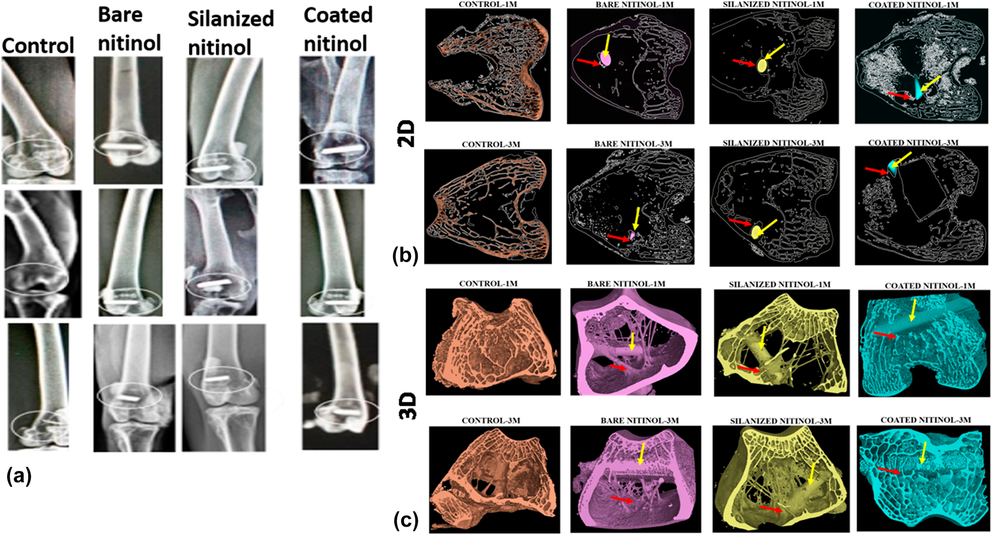
Some interesting properties such as superelasticity, shape memory effect, kink resistance, good biocompatibility, biomechanical properties, and corrosion resistance made nitinol a popular biomaterial as stent and orthopedic implants. But surface modification is needed to control nickel leaching from its surface, making safe for human body. The aim of this study was to modify the nitinol surface by the silanization technique and electrophoretically deposited hydroxyapatite coating, and to conduct a detailed in vitro and in vivo investigation. Detailed in vitro investigation involved MTT assay with the human osteoblastic cells (MG63 cell) over a period of 5 days and confocal image study. In case of in vivo study, histological study, fluorochrome labeling study, and Micro-Ct study were conducted. The overall in vitro and in vivo results indicate that silanized nitinol samples are showing slightly better level of performance, but both the surface-modified samples are suitable as the potential bio-implant for orthopedic purpose.

Type
Article
Copyright
Copyright © Materials Research Society 2020

Access options

Get access to the full version of this content by using one of the access options below. (Log in options will check for institutional or personal access. Content may require purchase if you do not have access.)

Footnotes

c)

Present address: Department of Chemistry, Nims Institute of Engineering & Technology, Nims University Rajasthan, Jaipur-Delhi Highway (NH-11C), Nims Institute of Engineering & Technology, Jaipur-303121, Rajasthan, India.

References

Fu, C.H., Sealy, P.M., Guo, Y.B., and Wei, X.T.: Austenite–martensite phase transformation of biomedical nitinol by ball burnishing. J. Mater. Process. Technol. 214, 3122 (2014).10.1016/j.jmatprotec.2014.07.019CrossRefGoogle Scholar
Nie, F.L., Zheng, Y.F., Cheng, Y., Wei, S.C., and Valiev, R.Z.: In vitro corrosion and cytotoxicity on microcrystalline, nanocrystalline, and amorphous NiTi alloy fabricated by high pressure torsion. Mater. Lett. 64, 983 (2010).10.1016/j.matlet.2010.01.081CrossRefGoogle Scholar
Gotman, I., Ben-David, D., Unger, R.E., Böse, T., Gutmanas, E.Y., and Kirkpatrick, C.J.: Mesenchymal stem cell proliferation and differentiation on load-bearing trabecular nitinol scaffolds. Acta Biomater. 9, 8440 (2013).10.1016/j.actbio.2013.05.030CrossRefGoogle ScholarPubMed
Akmal, M., Raza, A., Khan, M.M., Khan, M.I., and Hussain, M.A.: Effect of nano-hydroxyapatite reinforcement in mechanically alloyed NiTi composites for biomedical implant. Mater. Sci. Eng., C 68, 30 (2016).10.1016/j.msec.2016.05.092CrossRefGoogle ScholarPubMed
Kamali, S., Shemshad, S., Khavandi, A., and Azari, S.: Construction novel hydroxyapatite-nitinol nanocomposite for hard tissue applications. Mater. Chem. Phys. 220, 331 (2018).10.1016/j.matchemphys.2018.08.077CrossRefGoogle Scholar
Khalil-Allafi, J., Amin-Ahmadi, B., and Zare, M.: Biocompatibility and corrosion behavior of the shape memory NiTi alloy in the physiological environments simulated with body fluids for medical applications. Mater. Sci. Eng., C 30, 1112 (2010).10.1016/j.msec.2010.06.007CrossRefGoogle Scholar
Shabalovskaya, S.A., Tian, H., Anderegg, J.W., Schryvers, D.U., Carroll, W.U., and Humbeeck, J.V.: The influence of surface oxides on the distribution and release of nickel from nitinol wires. Biomaterials 30, 468 (2009).10.1016/j.biomaterials.2008.10.014CrossRefGoogle ScholarPubMed
Shabalovskaya, S.A., Anderegg, J., Laab, F., Thiel, P.A., and Rondelli, G.: Surface conditions of nitinol wires, tubing, and as-cast alloys. The effect of chemical etching, aging in boiling water, and heat treatment. J. Biomed. Mater. Res., Part B 65, 193 (2003).10.1002/jbm.b.10001CrossRefGoogle ScholarPubMed
Singh, R. and Dahotre, N.B.: Corrosion degradation and prevention by surface modification of biometallic materials. J. Mater. Sci.: Mater. Med. 18, 725 (2007).Google ScholarPubMed
Sun, F., Pang, X., and Zhitomirsky, I.: Electrophoretic deposition of composite hydroxyapatite–chitosan–heparin coatings. J. Mater. Process. Technol. 209, 1597 (2009).10.1016/j.jmatprotec.2008.04.007CrossRefGoogle Scholar
Dudek, K., Plawecki, M., Dulski, M., and Kubacki, J.: Multifunctional layers formation on the surface of NiTi SMA during β-tricalcium phosphate deposition. Mater. Lett. 157, 295 (2015).10.1016/j.matlet.2015.05.079CrossRefGoogle Scholar
Tsui, Y.C., Doyle, C., and Clyne, T.W.: Plasma sprayed hydroxyapatite coatings on titanium substrates. Part 2: Optimisation of coating properties. Biomaterials 19, 2031 (1998).10.1016/S0142-9612(98)00104-5CrossRefGoogle ScholarPubMed
Hsieh, M.F., Perng, L.H., Chin, T.S., and Perng, H.G.: Phase purity of sol–gel-derived hydroxyapatite ceramic. Biomaterials 22, 2601 (2001).10.1016/S0142-9612(00)00448-8CrossRefGoogle ScholarPubMed
Venugopal, J., Prabhakaran, M.P., Zhang, Y., Low, S., Choon, A.T., and Ramakrishna, S.: Biomimetic hydroxyapatite-containing composite nanofibrous substrates for bone tissue engineering. Philos. Trans. R. Soc., A 368, 2065 (2010).10.1098/rsta.2010.0012CrossRefGoogle ScholarPubMed
Cui, F.Z., Luo, Z.S., and Feng, Q.L.: Highly adhesive hydroxyapatite coatings on titanium alloy formed by ion beam assisted deposition. J. Mater. Sci.: Mater. Med. 8, 403 (1997).Google ScholarPubMed
Etminanfar, M.R. and Khalil-Allafi, J.: On the electrodeposition of Ca–P coatings on nitinol alloy: A comparison between different surface modification methods. J. Mater. Eng. Perform. 25, 466 (2016).10.1007/s11665-015-1876-4CrossRefGoogle Scholar
Es-Souni, M., Es-Souni, M., and Fischer-Brandies, H.: On the properties of two binary NiTi shape memory alloys. Effects of surface finish on the corrosion behaviour and in vitro biocompatibility. Biomaterials 23, 2887 (2002).10.1016/S0142-9612(01)00416-1CrossRefGoogle ScholarPubMed
Li, Y., Zhao, T., Wei, S., Xiang, Y., and Chen, H.: Effect of Ta2O5/TiO2 thin film on mechanical properties, corrosion, and cell behavior of the NiTi alloy implanted with tantalum. Mater. Sci. Eng., C 30, 1227 (2010).10.1016/j.msec.2010.07.001CrossRefGoogle Scholar
Bernard, S.A., Balla, V.K., Davies, N.M., Bose, S., and Bandyopadhyay, A.: Bone cell–materials interactions and Ni ion release of anodized equiatomic NiTi alloy. Acta Biomater. 7, 1902 (2011).10.1016/j.actbio.2011.01.004CrossRefGoogle ScholarPubMed
Yeh, H.Y. and Lin, J.C.: Bioactivity and platelet adhesion study of a human thrombomodulin-immobilized nitinol surface. J. Biomater. Sci. Polym. Ed. 20, 807 (2009).10.1163/156856209X426952CrossRefGoogle ScholarPubMed
Yu, H., Yan, J., Ma, H., Zeng, X., Liu, Y., and Zhao, X.: Creating poly(ethylene glycol) film on the surface of NiTi alloy by gamma irradiation. Radiat. Phys. Chem. 112, 199 (2015).CrossRefGoogle Scholar
Zhao, C., Pandit, S., Fu, Y., Mijakovic, I., Jesorka, A., and Liu, J.: Graphene oxide based coatings on nitinol for biomedical implant applications: Effectively promote mammalian cell growth but kill bacteria. RSC Adv. 6, 38124 (2016).CrossRefGoogle Scholar
Simsekyilmaz, S., Liehn, E.A., Weinandy, S., Schreiber, F., Megens, R.T.A., Theelen, W., Smeets, R., Jockenhövel, S., Gries, T., Möller, M., Klee, D., Weber, C., and Zenecke, A.: Targeting in-stent-stenosis with RGD- and CXCL1-coated mini-stents in mice. PLoS One 11, 155829 (2016).CrossRefGoogle ScholarPubMed
Gorgin Karaji, Z., Speir, M., Dadbakhsh, S., Kruth, J.P., Weinans, H., Zadpoor, A.A., and Yavari, S.A.: Additively manufactured and surface biofunctionalized porous nitinol. ACS Appl. Mater. Interfaces 9, 1293 (2017).CrossRefGoogle ScholarPubMed
Sinha, S., Begam, H., Kumar, V., Nandi, S.K., Kubacki, J., and Chanda, A.: Improved performance of the functionalized nitinol as a prospective bone implant material. J. Mater. Res. 33, 2554 (2018).CrossRefGoogle Scholar
Rodella, L.F., Bonazza, V., Labanca, M., Lonati, C., and Rezzani, R.A.: Review of the effects of dietary silicon intake on bone homeostasis and regeneration. J. Nutr. Health Aging 18, 820 (2014).10.1007/s12603-014-0555-8CrossRefGoogle Scholar
Devi, B.K., Tripathy, B., Roy, A., Lee, B., Kumta, P.N., Nandi, S.K., and Roy, M.: In vitro biodegradation and in vivo biocompatibility of forsterite bio-ceramics: Effects of strontium substitution. ACS Biomater. Sci. Eng. 5, 530 (2019).10.1021/acsbiomaterials.8b00788CrossRefGoogle Scholar
Hing, K.A., Revell, P.A., Smith, N., and Buckland, T.: Effect of silicon level on rate, quality, and progression of bone healing within silicate-substituted porous hydroxyapatite scaffolds. Biomaterials 27, 5014 (2006).CrossRefGoogle ScholarPubMed
Shi, M., Zhou, Y., Shao, J., Chen, Z., Song, B., Chang, J., Wu, C., and Xiao, Y.: Stimulation of osteogenesis and angiogenesis of HBMSCs by delivering Si ions and functional drug from mesoporous silica nanospheres. Acta Biomater. 21, 178 (2015).10.1016/j.actbio.2015.04.019CrossRefGoogle ScholarPubMed
Kim, B.S., Yang, S.S., Yoon, J.H., and Lee, J.: Enhanced bone regeneration by silicon-substituted hydroxyapatite derived from cuttlefish bone. Clin. Oral Implants Res. 28, 49 (2017).CrossRefGoogle ScholarPubMed
Price, C.T., Koval, K.J., and Langford, J.R.: Silicon: A review of its potential role in the prevention and treatment of postmenopausal osteoporosis. Int. J. Endocrinol. 2013, 316783 (2013).CrossRefGoogle ScholarPubMed