Hostname: page-component-745bb68f8f-l4dxg Total loading time: 0 Render date: 2025-01-10T20:49:26.844Z Has data issue: false hasContentIssue false

Graphene oxide coated popcorn-like Ag nanoparticles for reliable sensitive surface-enhanced Raman scattering detection of drug residues

Published online by Cambridge University Press:  12 March 2019

Maofeng Zhang*
Affiliation:
School of Materials Science and Engineering, Hefei University of Technology, Hefei 230009, People’s Republic of China
Zhexue Chen
Affiliation:
School of Materials Science and Engineering, Hefei University of Technology, Hefei 230009, People’s Republic of China
Zhuoer Wang*
Affiliation:
School of Materials Science and Engineering, Hefei University of Technology, Hefei 230009, People’s Republic of China
Zhiyuan Zheng
Affiliation:
School of Materials Science and Engineering, Hefei University of Technology, Hefei 230009, People’s Republic of China
Dapeng Wang*
Affiliation:
Institute of Intelligent Machines, Chinese Academy of Sciences, Hefei 230031, People’s Republic of China
*
a)Address all correspondence to these authors. e-mail: [email protected]
Get access

Abstract

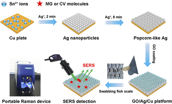
Conventional methods for determination of trace drug residues are either time consuming or labor intensive or require large specialized instruments, which hamper their practical applications in field analysis. Here, we present a rapid and quantitative surface-enhanced Raman scattering (SERS) detection method coupled with a portable Raman spectrometer for determination of trace drug residues on fish surface. Graphene oxide (GO) decorated popcorn-like Ag nanoparticles (NPs) on Cu plate (GO/AgNPs/Cu) were fabricated by a facile approach and directly employed as a robust SERS detection substrate. For practical SERS detections, trace-level residues of crystal violet (10−8 M, 4.1 ng/g) and malachite green (10−8 M, 3.6 ng/g) could be readily detected by simply swabbing the contaminated fish scale surface with the SERS substrate. Importantly, SERS detection was quantitatively realized in the broad linear concentrations. Compared with lab-based Raman spectrometer with large footprints, our method has potential applications in practical rapid, accurate, and on-site SERS determination.

Type
Article
Copyright
Copyright © Materials Research Society 2019 

Access options

Get access to the full version of this content by using one of the access options below. (Log in options will check for institutional or personal access. Content may require purchase if you do not have access.)

References

Jia, J., Yan, S., Lai, X.X., Xu, Y.Z., Liu, T., and Xiang, Y.H.: Colorimetric aptasensor for detection of malachite green in fish sample based on RNA and gold nanoparticles. Food Anal. Methods 11, 16681676 (2018).CrossRefGoogle Scholar
Bergwerff, A.A. and Scherpenisse, P.: Determination of residues of malachite green in aquatic animals. J. Chromatogr. B: Anal. Technol. Biomed. Life Sci. 788, 351359 (2003).CrossRefGoogle ScholarPubMed
Arroyo, D., Ortiz, M.C., Sarabia, L.A., and Palacios, F.: Advantages of PARAFAC calibration in the determination of malachite green and its metabolite in fish by liquid chromatography–tandem mass spectrometry. J. Chromatogr. A 1187, 110 (2008).CrossRefGoogle ScholarPubMed
Wang, Y.L., Liao, K.R., Huang, X.J., and Yuan, D.X.: Simultaneous determination of malachite green, crystal violet and their leuco-metabolites in aquaculture water samples using monolithic fiber-based solid-phase microextraction coupled with high performance liquid chromatography. Anal. Methods 7, 81388145 (2015).CrossRefGoogle Scholar
Shalaby, A.R., Emam, W.H., and Anwar, M.M.: Mini-column assay for rapid detection of malachite green in fish. Food Chem. 226, 813 (2017).CrossRefGoogle Scholar
Stolker, A.A.M., Zuidema, T., and Nielen, M.W.F.: Residue analysis of veterinary drugs and growth promoting agents. Trends Anal. Chem. 26, 967979 (2007).CrossRefGoogle Scholar
Zhang, Y.Y., Yu, W.S., Pei, L., Lai, K.Q., Rasco, B.A., and Huang, Y.Q.: Rapid analysis of malachite green and leucomalachite green in fish muscles with surface-enhanced resonance Raman scattering. Food Chem. 169, 8084 (2015).CrossRefGoogle ScholarPubMed
Dowling, G., Mulder, P.P.J., Duffy, C., Regan, L., and Smyth, M.R.: Confirmatory analysis of malachite green, leucomalachite green, crystal violet and leucocrystal violet in salmon by liquid chromatography–tandem mass spectrometry. Anal. Chim. Acta 586, 411419 (2007).CrossRefGoogle ScholarPubMed
Singh, G., Koerner, T., Gelinas, J.M., Abbott, M., Brady, B., Huet, A.C., Charlier, C., Delahaut, P., and Godefroy, S.B.: Design and characterization of a direct ELISA for the detection and quantification of leucomalachite green. Food Addit. Contam., Part A 28, 731739 (2011).CrossRefGoogle ScholarPubMed
Guo, Z., Gai, P., Hao, T., Duan, J., and Wang, S.: Determination of malachite green residues in fish using a highly sensitive electrochemiluminescence method combined with molecularly imprinted solid phase extraction. J. Agric. Food Chem. 59, 52575262 (2011).CrossRefGoogle ScholarPubMed
Mitroska, K., Posyniak, A., Zmudzki, J., and Chromatogr, J.: Determination of malachite green and leucomalachite green in carp muscle by liquid chromatography with visible and fluorescence detection. J. Chromatogr. A 1089, 187192 (2005).CrossRefGoogle Scholar
Xia, X., Zeng, J., McDearmon, B., Zheng, Y., Li, Q., and Xia, Y.: Silver nanocrystals with concave surfaces and their optical and surface-enhanced Raman scattering properties. Angew. Chem. 50, 1254212546 (2011).CrossRefGoogle ScholarPubMed
Liu, K.K., Tadepalli, S., Tian, L.M., and Singamaneni, S.: Size-dependent surface enhanced Raman scattering activity of plasmonic nanorattles. Chem. Mater. 27, 56785684 (2015).CrossRefGoogle Scholar
Chen, J.M., Huang, Y.J., Kannan, P., Zhang, L., Lin, Z.Y., Zhang, J.W., Chen, T., and Guo, L.H.: Flexible and adhesive surface enhance Raman scattering active tape for rapid detection of pesticide residues in fruits and vegetables. Anal. Chem. 88, 21492155 (2016).CrossRefGoogle ScholarPubMed
Sharma, B., Frontiera, R.R., Henry, A., Ringe, E., and Van Duyne, R.P.: SERS: Materials, applications, and the future. Mater. Today 15, 1625 (2012).CrossRefGoogle Scholar
Zhang, X.Y., Zheng, Y.H., Liu, X., Lu, W., Dai, J.Y., Lei, D.Y., and MacFarlane, D.R.: Hierarchical porous plasmonic metamaterials for reproducible ultrasensitive surface‐enhanced Raman spectroscopy. Adv. Mater. 27, 10901096 (2015).CrossRefGoogle ScholarPubMed
Li, C.Y., Huang, Y.Q., Lai, K.Q., Rasco, B.A., and Fan, Y.X.: Analysis of trace methylene blue in fish muscles using ultra-sensitive surface-enhanced Raman spectroscopy. Food Control 65, 99105 (2016).CrossRefGoogle Scholar
Chen, X.W., Nguyen, T.H.D., Gu, L.Q., and Lin, M.S.: Use of standing gold nanorods for detection of malachite green and crystal violet in fish by SERS. J. Food Sci. 82, 16401646 (2017).CrossRefGoogle ScholarPubMed
Müller, C., David, L., Chis, V., and Pînzaru, S.C.: Detection of thiabendazole applied on citrus fruits and bananas using surface enhanced Raman scattering. Food Chem. 145, 814820 (2014).CrossRefGoogle ScholarPubMed
Zhou, N.N., Meng, G.W., Zhu, C.H., Chen, B., Zhou, Q.T., Ke, Y., and Huo, D.X.: A silver-grafted sponge as an effective surface-enhanced Raman scattering substrate. Sens. Actuators, B 258, 5663 (2018).CrossRefGoogle Scholar
Yu, X., Cai, H., Zhang, W., Li, X., Pan, N., Luo, Y., Wang, X., and Hou, J.: Tuning chemical enhancement of SERS by controlling the chemical reduction of graphene oxide nanosheets. ACS Nano 5, 952958 (2011).CrossRefGoogle ScholarPubMed
Chi, H., Liu, Y.J., Wang, F., and He, C.: Highly sensitive and fast response colorimetric humidity sensors based on graphene oxide film. ACS Appl. Mater. Interfaces 7, 1988219886 (2015).CrossRefGoogle Scholar
Jiang, T., Wang, X., Tang, S., Zhou, J., Gu, C., and Tang, J.: Seed-mediated synthesis and SERS performance of graphene oxide-wrapped Ag nanomushroom. Sci. Rep. 7, 97959804 (2017).CrossRefGoogle ScholarPubMed
Lu, Z.Y., Liu, Y.J., Wang, M.H., Zhang, C., Li, Z., Huo, Y.Y., Li, Z., Xu, S.C., Man, B.Y., and Jiang, S.Z.: A novel natural surface-enhanced Raman spectroscopy (SERS) substrate based on graphene oxide-Ag nanoparticles-Mytilus coruscus hybrid system. Sens. Actuators, B 261, 110 (2018).CrossRefGoogle Scholar
Zhao, X.F., Yu, J., Zhang, C., Chen, C.S., Xu, S.C., Li, C.H., Li, Z., Zhang, S.Z., Liu, A.H., and Man, B.Y.: Flexible and stretchable SERS substrate based on a pyramidal PMMA structure hybridized with graphene oxide assivated AgNPs. Appl. Surf. Sci. 455, 11711178 (2018).CrossRefGoogle Scholar
Hutter, E. and Fendler, J.H.: Exploitation of localized surface plasmon resonance. Adv. Mater. 16, 16851706 (2004).CrossRefGoogle Scholar
Shuford, K.L., Lee, J., Odom, T.W., and Schatz, G.C.: Optical properties of gold pyramidal shells. J. Phys. Chem. C 112, 66626666 (2008).CrossRefGoogle Scholar
Zhang, M.F., Meng, J.T., Wang, D.P., Tang, Q., Chen, T., Rong, S.Z., Liu, J.Q., and Wu, Y.C.: Biomimetic synthesis of hierarchical 3D Ag butterfly wing scales arrays/graphene composites as ultrasensitive SERS substrates for efficient trace chemical detection. J. Mater. Chem. C 6, 19331943 (2018).CrossRefGoogle Scholar
Zrimsek, A.B., Wong, N.L., and Van Duyne, R.P.: Single molecule surface-enhanced Raman spectroscopy: A critical analysis of the bianalyte versus isotopologue proof. J. Phys. Chem. C 120, 51335142 (2016).CrossRefGoogle Scholar
Bibikova, O., Haas, J., López-Lorente, A.I., Popov, A., Kinnunen, M., Meglinski, I., and Mizaikoff, B.: Towards enhanced optical sensor performance: SEIRA and SERS with plasmonic nanostars. Analyst 142, 951958 (2017).CrossRefGoogle ScholarPubMed
Zhang, D.H., Liu, X.H., and Wang, X.: Synthesis of single-crystal silver slices with predominant (111) facet and their SERS effect. J. Mol. Struct. 985, 8285 (2011).CrossRefGoogle Scholar
Sanci, R. and Volkan, M.: Surface-enhanced Raman scattering (SERS) studies on silver nanorod substrates. Sens. Actuators, B 139, 150155 (2009).CrossRefGoogle Scholar
Yang, Y., Matsubara, S., Xiong, L.M., Hayakawa, T., and Nogami, M.: Solvothermal synthesis of multiple shapes of silver nanoparticles and their SERS properties. J. Phys. Chem. C 111, 90959104 (2007).CrossRefGoogle Scholar
Jung, N., Crowther, A.C., Kim, N., Kim, P., and Brus, L.: Raman enhancement on graphene: Adsorbed and intercalated molecular species. ACS Nano 4, 70057013 (2010).CrossRefGoogle ScholarPubMed
Qu, L.L., Liu, Y.Y., Liu, M.K., Yang, G.H., Li, D.W., and Li, H.T.: Highly reproducible Ag NPs/CNT-intercalated GO membranes for enrichment and SERS detection of antibiotics. ACS Appl. Mater. Interfaces 8, 2818028186 (2016).CrossRefGoogle Scholar
Cai, W.B., Ren, B., Li, X.Q., She, C.X., Liu, F.M., Cai, X.W., and Tian, Z.Q.: Investigation of surface-enhanced Raman scattering from platinum electrodes using a confocal Raman microscope: Dependence of surface roughening pretreatment. Surf. Sci. 406, 922 (1998).CrossRefGoogle Scholar
Kang, L., Chu, J., Zhao, H., Xu, P., and Sun, M.: Recent progress in the applications of graphene in surface-enhanced Raman scattering and plasmon-induced catalytic reactions. J. Mater. Chem. C 3, 90249037 (2015).CrossRefGoogle Scholar
Zhou, Q., Li, X., Fan, Q., Zhang, X., and Zheng, J.: Charge transfer between metal nanoparticles interconnected with a functionalized molecule probed by surface-enhanced Raman spectroscopy. Angew. Chem., Int. Ed. 45, 39703973 (2006).CrossRefGoogle ScholarPubMed
Xie, L., Ling, X., Fang, Y., Zhang, J., and Liu, Z.: Graphene as a substrate to suppress fluorescence in resonance Raman spectroscopy. J. Am. Chem. Soc. 131, 98909891 (2009).CrossRefGoogle ScholarPubMed
Hu, L.T., Liu, Y.J., Han, Y.S., Chen, P.X., Zhang, C., Li, C.H., Lu, Z.Y., Luo, D., and Jiang, S.Z.: Graphene oxide-decorated silver dendrites for high-performance surface-enhanced Raman scattering applications. J. Mater. Chem. C 5, 39083915 (2017).CrossRefGoogle Scholar
Kaplan, M., Olgun, E.O., and Karaoglu, O.: A rapid and simple method for simultaneous determination of triphenyl-methane dye residues in rainbow trouts by liquid chromatography–tandem mass spectrometry. J. Chromatogr. A 1349, 3743 (2014).CrossRefGoogle Scholar
Supplementary material: File

Zhang et al. supplementary material

Zhang et al. supplementary material 1

Download Zhang et al. supplementary material(File)
File 348.4 KB