Hostname: page-component-586b7cd67f-vdxz6 Total loading time: 0 Render date: 2024-11-25T23:51:18.875Z Has data issue: false hasContentIssue false

Fabrication and characterization of CdSe/ZnS quantum dots-doped polystyrene microspheres prepared by self-assembly

Published online by Cambridge University Press:  17 October 2012
Yu-Hsiang Lee*
Affiliation:
Graduate Institute of Biomedical Engineering, National Central University, Taoyuan 32001, Taiwan, Republic of China
Ching-Shiow Tseng
Affiliation:
Graduate Institute of Biomedical Engineering, National Central University, Taoyuan 32001, Taiwan, Republic of China; and Department of Mechanical Engineering, National Central University, Taoyuan 32001, Taiwan, Republic of China
Yen-Lin Wei
Affiliation:
Graduate Institute of Biomedical Engineering, National Central University, Taoyuan 32001, Taiwan, Republic of China
*
a)Address all correspondence to this author. e-mail: [email protected]
Get access

Abstract

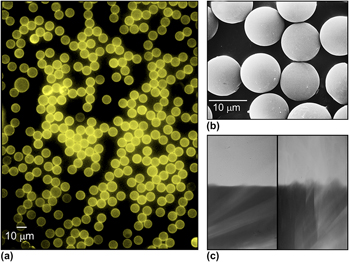
Semiconductor quantum dots (QDs)-doped polystyrene (PS) microspheres with high luminescence were prepared using a self-assembly approach. Hydrophobic CdSe/ZnS QDs were first carboxylized by ligand exchange using mercaptocarboxylic acid. PS microspheres were separately encapsulated with polyethyleneimine via electrostatic interactions and then adsorbed with the carboxyl QDs to form QDs-doped microspheres. We then characterized the combinations using optical, electrical, and mechanical approaches and obtained the following findings: (i) microspheres can be fully coated by QD nanoparticles with a coverage rate of 1.0 pmole/cm2, in which QDs were evenly distributed on the surfaces; (ii) the anchored QDs exhibited similar optical property as they performed in isolated suspension; and (iii) the fluorescence of QDs-doped microspheres remained intact after stressed by ultrasound-induced cavitation, demonstrating the robustness of interactions between QDs and microspheres. The self-assembly approach developed in this study offered a facile and controllable strategy for preparation of QDs-encoded microparticles with high luminescence and stability.

Type
Articles
Copyright
Copyright © Materials Research Society 2012

Access options

Get access to the full version of this content by using one of the access options below. (Log in options will check for institutional or personal access. Content may require purchase if you do not have access.)

References

REFERENCES

Yang, J., Sena, M.P., and Gao, X.: Quantum dot-encoded fluorescent beads for biodetection and imaging. Rev. Fluoresc. 4, 139 (2009).CrossRefGoogle Scholar
Rogach, A., Susha, A., Caruso, F., Sukhorukov, G., Kornowski, A., Kershaw, S., Möhwald, H., Eychmüller, A., and Weller, H.: Nano- and microengineering: Three-dimensional colloidal photonic crystals prepared from submicrometer-sized polystyrene latex spheres pre-coated with luminescent polyelectrolyte/nanocrystal shells. Adv. Mater. 12, 333 (2000).3.0.CO;2-X>CrossRefGoogle Scholar
Rogach, A.L., Nagesha, D., Ostrander, J.W., Giersig, M., and Kotov, N.A.: “Raisin bun”-type composite spheres of silica and semiconductor nanocrystals. Chem. Mater. 12, 2676 (2000).CrossRefGoogle Scholar
Gao, X. and Nie, S.: Doping mesoporous materials with multicolor quantum dots. J. Phys. Chem. B 107, 11575 (2003).CrossRefGoogle Scholar
Gong, Y., Gao, M., Wang, D., and Mohwald, H.: Incorporating fluorescent CdTe nanocrystals into a hydrogel via hydrogen bonding: Toward fluorescent microspheres with temperature-responsive properties. Chem. Mater. 17, 2648 (2005).CrossRefGoogle Scholar
Gaponik, N., Radtchenko, I.L., Sukhorukov, G.B., Weller, H., and Rogach, A.L.: Toward encoding combinatorial libraries: Charge-driven microencapsulation of semiconductor nanocrystals luminescing in the visible and near IR. Adv. Mater. 14, 879 (2002).3.0.CO;2-A>CrossRefGoogle Scholar
Joumaa, N., Lansalot, M., Théretz, A., Elaissari, A., Sukhanova, A., Artemyev, M., Nabiev, I., and Cohen, J.H.: Synthesis of quantum dot-tagged submicrometer polystyrene particles by miniemulsion polymerization. Langmuir 22, 1810 (2006).CrossRefGoogle ScholarPubMed
Li, Y., Liu, E.C.Y., Pickett, N., Skabara, P.J., Cummins, S.S., Ryley, S., Sutherland, A.J., and O’Brien, P.: Synthesis and characterization of CdS quantum dots in polystyrene microbeads. J. Mater. Chem. 15, 1238 (2005).Google Scholar
Allen, C.N., Lequeux, N., Chassenieux, C., Tessier, G., and Dubertret, B.: Optical analysis of beads encoded with quantum dots coated with a cationic polymer. Adv. Mater. 19, 4420 (2007).CrossRefGoogle Scholar
Pan, X., Lu, M., Wu, D., and Gai, L.: Synthesis and characterization of double-layer quantum-dots-tagged microspheres. IEEE Trans. Nanobiosci. 8, 13 (2009).Google ScholarPubMed
Han, M., Gao, X., Su, J., and Nie, S.: Quantum-dot-tagged microbeads for multiplexed optical coding of biomolecules. Nat. Biotech. 19, 631 (2001).CrossRefGoogle ScholarPubMed
Buranda, T., Wu, Y., and Sklar, L.A.: Quantum dots for quantitative flow cytometry. Methods Mol. Biol. 699, 67 (2011).CrossRefGoogle ScholarPubMed
Wu, Y., Campos, S.K., Lopez, G.P., Ozbun, M.A., Sklar, L.A., and Buranda, T.: The development of quantum dot calibration beads and quantitative multicolor bioassays in flow cytometry and microscopy. Anal. Biochem. 364, 180 (2007).CrossRefGoogle ScholarPubMed
Xiang, Y., Zhang, H., Jiang, B., Chai, Y., and Yuan, R.: Quantum dot layer-by-layer assemblies as signal amplification labels for ultrasensitive electronic detection of uropathogens. Anal. Chem. 83, 4302 (2011).CrossRefGoogle ScholarPubMed
Sun, L., Yu, X., Sun, M., Wang, H., Xu, S., Dixon, J.D., Wang, Y.A., Li, Y., Yang, Q., and Xu, X.: Preparation of quantum dots encoded microspheres by electrospray for the detection of biomolecules. J. Colloid Interface Sci. 358, 73 (2011).CrossRefGoogle ScholarPubMed
Sukhanova, A. and Nabiev, I.: Fluorescent nanocrystal-encoded microbeads for multiplexed cancer imaging and diagnosis. Crit. Rev. Oncol. Hematol. 68, 39 (2008).CrossRefGoogle ScholarPubMed
Stroh, M., Zimmer, J.P., Duda, D.G., Levchenko, T.S., Cohen, K.S., Brown, E.B., Scadden, D.T., Torchilin, V.P., Bawendi, M.G., Fukumura, D., and Jain, R.K.: Quantum dots spectrally distinguish multiple species within the tumor milieu in vivo. Nat. Med. 11, 678 (2005).CrossRefGoogle ScholarPubMed
Di Corato, R., Bigall, N.C., Ragusa, A., Dorfs, D., Genovese, A., Marotta, R., Manna, L., and Pellegrino, T.: Multifunctional nanobeads based on quantum dots and magnetic nanoparticles: Synthesis and cancer cell targeting and sorting. ACS Nano. 5, 1109 (2011).CrossRefGoogle ScholarPubMed
Chan, Y., Zimmer, J.P., Stroh, M., Steckel, J.S., Jain, R.K., and Bawendi, M.G.: Incorporation of luminescent nanocrystals into monodisperse core-shell silica microspheres. Adv. Mater. 16, 2092 (2004).CrossRefGoogle Scholar
Graf, C., Dembski, S., Hofmann, A., and Ruhl, E.: A general method for the controlled embedding of nanoparticles in silica colloids. Langmuir 22, 5604 (2006).CrossRefGoogle ScholarPubMed
Kuang, M., Wang, D., Bao, H., Gao, M.Y., Möhwald, H., and Jiang, M.: Fabrication of multicolor-encoded microspheres by tagging semiconductor nanocrystals to hydrogel spheres. Adv. Mater. 17, 267 (2005).CrossRefGoogle Scholar
Chan, W.C. and Nie, S.: Quantum dot bioconjugates for ultrasensitive nonisotopic detection. Science 281, 2016 (1998).CrossRefGoogle ScholarPubMed
Yu, W.W., Qu, L., Guo, W., and Peng, X.: Experimental determination of the extinction coefficient of CdTe, CdSe, and CdS nanocrystals. Chem. Mater. 15, 2854 (2003).CrossRefGoogle Scholar
Gómez, D.E., Pastoriza-Santos, I., and Mulvaney, P.: Tunable whispering gallery mode emission from quantum-dot-doped microspheres. Small 1, 238 (2005).CrossRefGoogle ScholarPubMed
Mattoussi, H., Cumming, A.W., Murray, C.B., Bawendi, M.G., and Ober, R.: Characterization of CdSe nanocrystallite dispersions by small angle x-ray scattering. J. Chem. Phys. 105, 9890 (1996).CrossRefGoogle Scholar
Schmitt, J., Mächtle, P., Eck, D., Möhwald, H., and Helm, C.A.: Preparation and optical properties of colloidal gold monolayers. Langmuir 15, 3256 (1999).CrossRefGoogle Scholar
Crum, L.A., Roy, R.A., Dinno, M.A., Church, C.C., Apfel, R.E., Holland, C.K., and Madanshetty, S.L.: Acoustic cavitation produced by microsecond pulses of ultrasound: A discussion of some selected results. J. Acoust. Soc. Am. 91, 1113 (1992).CrossRefGoogle ScholarPubMed
Miller, D.L.: A review of the ultrasonic bioeffects of microsonation, gas-body activation, and related cavitation-like phenomena. Ultrasound Med. Biol. 13, 443 (1987).CrossRefGoogle ScholarPubMed
Liang, H.D., Tang, J., and Halliwell, M.: Sonoporation, drug delivery, and gene therapy. Proc. Inst. Mech. Eng. Part H 224, 343 (2010).CrossRefGoogle ScholarPubMed
Pitt, W.G., Husseini, G.A., and Staples, B.J.: Ultrasonic drug delivery–a general review. Expert Opin. Drug Delivery 1, 37 (2004).CrossRefGoogle ScholarPubMed
Supplementary material: PDF

Lee et al. supplementary material

Supplementary figure S1

Download Lee et al. supplementary material(PDF)
PDF 116.9 KB