Hostname: page-component-586b7cd67f-vdxz6 Total loading time: 0 Render date: 2024-11-22T23:10:54.708Z Has data issue: false hasContentIssue false

Cell viability assessments of green synthesized water-soluble AgInS2/ZnS core/shell quantum dots against different cancer cell lines

Published online by Cambridge University Press:  09 December 2019

Oluwatobi S. Oluwafemi*
Affiliation:
Department of Chemical Sciences (formerly Applied Chemistry), University of Johannesburg, Doornfontein 2028, Johannesburg, South Africa; and Centre for Nanomaterials Science Research, University of Johannesburg, Doornfontein 2028, Johannesburg, South Africa
Bambesiwe M.M. May
Affiliation:
Department of Chemical Sciences (formerly Applied Chemistry), University of Johannesburg, Doornfontein 2028, Johannesburg, South Africa; and Centre for Nanomaterials Science Research, University of Johannesburg, Doornfontein 2028, Johannesburg, South Africa
Sundararajan Parani
Affiliation:
Department of Chemical Sciences (formerly Applied Chemistry), University of Johannesburg, Doornfontein 2028, Johannesburg, South Africa; and Centre for Nanomaterials Science Research, University of Johannesburg, Doornfontein 2028, Johannesburg, South Africa
Jose Varghese Rajendran
Affiliation:
Department of Chemical Sciences (formerly Applied Chemistry), University of Johannesburg, Doornfontein 2028, Johannesburg, South Africa; and Centre for Nanomaterials Science Research, University of Johannesburg, Doornfontein 2028, Johannesburg, South Africa
*
a)Address all correspondence to this author. e-mail: [email protected]
Get access

Abstract

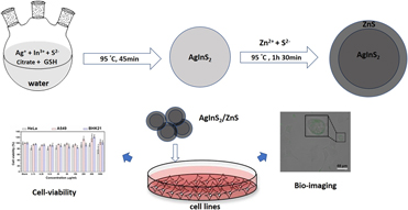
Chalcopyrite quantum dots (QDs) have emerged as a safe alternative to cadmium-based QDs for bio-applications. However, the research on AgInS2 chalcopyrite QDs has not been widely explored in terms of their toxicity. Herein, we report a synthesis of biocompatible AgInS2/ZnS QDs via a greener approach. The emission intensity of the as-synthesized AgInS2 core QDs was enhanced 2-fold after the ZnS shell growth. X-ray diffraction revealed the tetragonal crystal structure of QDs, and high-resolution transmission electron microscope images show that the QDs are spherical in shape and crystalline in nature. Cell viability assays conducted on different cell lines, such as HeLa, A549, and BHK-21 cells, indicated that AgInS2/ZnS QDs are least toxic at a QD concentration range of 100 µg/mL. The fluorescent microscope analysis of A549 cells incubated with AgInS2/ZnS QDs shows that the QDs were accumulated in the cell membranes. The as-synthesized AgInS2/ZnS QDs are less toxic and eco-friendly, and can be used for biolabeling.

Type
Article
Copyright
Copyright © Materials Research Society 2019 

Access options

Get access to the full version of this content by using one of the access options below. (Log in options will check for institutional or personal access. Content may require purchase if you do not have access.)

References

Aboulaich, A., Michalska, M., Schneider, R., Potdevin, A., Deschamps, J., Deloncle, R., Chadeyron, G., and Mahiou, R.: Ce-doped YAG nanophosphor and red emitting CuInS2/ZnS core/shell quantum dots for warm white light-emitting diode with high color rendering index. ACS Appl. Mater. Interfaces 6, 252258 (2013).10.1021/am404108nCrossRefGoogle ScholarPubMed
Carey, G.H., Abdelhady, A.L., Ning, Z., Thon, S.M., Bakr, O.M., and Sargent, E.H.: Colloidal quantum dot solar cells. Chem. Rev. 115, 1273212763 (2015).10.1021/acs.chemrev.5b00063CrossRefGoogle ScholarPubMed
Jing, L., Kershaw, S.V., Li, Y., Huang, X., Li, Y., Rogach, A.L., and Gao, M.: Aqueous based semiconductor nanocrystals. Chem. Rev. 116, 1062310730 (2016).10.1021/acs.chemrev.6b00041CrossRefGoogle ScholarPubMed
Gazouli, M., Lyberopoulou, A., Pericleous, P., Rizos, S., Aravantinos, G., Nikiteas, N., Anagnou, N.P., and Efstathopoulos, E.P.: Development of a quantum-dot-labelled magnetic immunoassay method for circulating colorectal cancer cell detection. World J. Gastroenterol. 18, 4419 (2012).10.3748/wjg.v18.i32.4419CrossRefGoogle ScholarPubMed
Liandris, E., Gazouli, M., Andreadou, M., Sechi, L.A., Rosu, V., and Ikonomopoulos, J.: Detection of pathogenic mycobacteria based on functionalized quantum dots coupled with immunomagnetic separation. PLoS One 6, e20026 (2011).10.1371/journal.pone.0020026CrossRefGoogle ScholarPubMed
Ranjbar-Navazi, Z., Omidi, Y., Eskandani, M., and Davaran, S.: Cadmium-free quantum dot-based theranostics. TrAC. Trends Anal. Chem. 118, 386400 (2019).10.1016/j.trac.2019.05.041CrossRefGoogle Scholar
Petryayeva, E., Algar, W.R., and Medintz, I.L.: Quantum dots in bioanalysis: A review of applications across various platforms for fluorescence spectroscopy and imaging. Appl. Spectrosc. 67, 215252 (2013).10.1366/12-06948CrossRefGoogle Scholar
Chen, Y., Li, S., Huang, L., and Pan, D.: Low-cost and gram-scale synthesis of water-soluble Cu–In–S/ZnS core/shell quantum dots in an electric pressure cooker. Nanoscale 6, 12951298 (2014).10.1039/C3NR05014ACrossRefGoogle Scholar
Li, L., Daou, T.J., Texier, I., Kim Chi, T.T., Liem, N.Q., and Reiss, P.: Highly luminescent CuInS2/ZnS core/shell nanocrystals: Cadmium-free quantum dots for in vivo imaging. Chem. Mater. 21, 24222429 (2009).10.1021/cm900103bCrossRefGoogle Scholar
Lin, Z., Fei, X., Ma, Q., Gao, X., and Su, X.: CuInS2 quantum dots@ silica near-infrared fluorescent nanoprobe for cell imaging. New J. Chem. 38, 9096 (2014).10.1039/C3NJ00957BCrossRefGoogle Scholar
Deng, D., Qu, L., and Gu, Y.: Near-infrared broadly emissive AgInSe2/ZnS quantum dots for biomedical optical imaging. J. Mater. Chem. C 2, 70777085 (2014).10.1039/C4TC01147CCrossRefGoogle Scholar
Fahmi, M.Z. and Chang, J.Y.: Forming double layer-encapsulated quantum dots for bio-imaging and cell targeting. Nanoscale 5, 15171528 (2013).10.1039/c2nr33429aCrossRefGoogle ScholarPubMed
Che, D., Zhu, X., Wang, H., Duan, Y., Zhang, Q., and Li, Y.: Aqueous synthesis of high bright and tunable near-infrared AgInSe2–ZnSe quantum dots for bioimaging. J. Colloid Interface Sci. 463, 17 (2016).10.1016/j.jcis.2015.10.039CrossRefGoogle Scholar
Hu, X., Chen, T., Xu, Y., Wang, M., Jiang, W., and Jiang, W.: Hydrothermal synthesis of bright and stable AgInS2 quantum dots with tunable visible emission. J. Lumin. 200, 189195 (2018).10.1016/j.jlumin.2018.04.025CrossRefGoogle Scholar
Kang, X., Huang, L., Yang, Y., and Pan, D.: Scaling up the aqueous synthesis of visible light emitting multinary AgInS2/ZnS core/shell quantum dots. J. Phys. Chem. C 119, 79337940 (2015).10.1021/acs.jpcc.5b00413CrossRefGoogle Scholar
Bray, F., Ferlay, J., Soerjomataram, I., Siegel, R.L., Torre, L.A., and Jemal, A.: Global cancer statistics 2018: GLOBOCAN estimates of incidence and mortality worldwide for 36 cancers in 185 countries. Ca-Cancer J. Clin. 68, 394424 (2018).10.3322/caac.21492CrossRefGoogle ScholarPubMed
May, B.M., Parani, S., and Oluwafemi, O.S.: Detection of ascorbic acid using green synthesized AgInS2 quantum dots. Mater. Lett. 236, 432435 (2019).10.1016/j.matlet.2018.10.155CrossRefGoogle Scholar
Zi, L., Huang, Y., Yan, Z., and Liao, S.: Thioglycolic acid-capped CuInS2/ZnS quantum dots as fluorescent probe for cobalt ion detection. J. Lumin. 148, 359363 (2014).10.1016/j.jlumin.2013.12.051CrossRefGoogle Scholar
Xiong, W.W., Yang, G.H., Wu, X.C., and Zhu, J.J.: Microwave-assisted synthesis of highly luminescent AgInS2/ZnS nanocrystals for dynamic intracellular Cu(II) detection. J. Mater. Chem. B 1, 41604165 (2013).10.1039/c3tb20638fCrossRefGoogle Scholar
Shang, H., Di, Q., Ji, M., Bai, B., Liu, J., Chen, W., Xu, M., Rong, H., Liu, J., and Zhang, J.: From indium-doped Ag2S to AgInS2 nanocrystals: Low-temperature in situ conversion of colloidal Ag2S nanoparticles and their NIR fluorescence. Chem. - Eur. J. 24, 1367613680 (2018).10.1002/chem.201802973CrossRefGoogle Scholar
Peng, S., Zhang, S., Mhaisalkar, S.G., and Ramakrishna, S.: Synthesis of AgInS2 nanocrystal ink and its photoelectrical application. Phys. Chem. Chem. Phys. 14, 85238529 (2012).10.1039/c2cp40848aCrossRefGoogle ScholarPubMed
Mousavi-Kamazani, M. and Salavati-Niasari, M.: A simple microwave approach for synthesis and characterization of Ag2S–AgInS2 nanocomposites. Composites, Part B 56, 490496 (2014).10.1016/j.compositesb.2013.08.066CrossRefGoogle Scholar
Tan, L., Liu, S., Li, X., Chronakis, I.S., and Shen, Y.: A new strategy for synthesizing AgInS2 quantum dots emitting brightly in near-infrared window for in vivo imaging. Colloids Surf., B 125, 222229 (2015).10.1016/j.colsurfb.2014.11.041CrossRefGoogle ScholarPubMed
Wang, L., Kang, X., and Pan, D.: Gram-scale synthesis of hydrophilic PEI-coated AgInS2 quantum dots and its application in hydrogen peroxide/glucose detection and cell imaging. Inorg. Chem. 56, 61226130 (2017).10.1021/acs.inorgchem.7b00053CrossRefGoogle ScholarPubMed
Shinchi, H., Wakao, M., Nagata, N., Sakamoto, M., Mochizuki, E., Uematsu, T., Kuwabata, S., and Suda, Y.: Cadmium-free sugar-chain-immobilized fluorescent nanoparticles containing low-toxicity ZnS-AgInS2 cores for probing lectin and cells. Bioconjugate Chem. 25, 286295 (2014).10.1021/bc400425wCrossRefGoogle ScholarPubMed
Chang, J.Y., Wang, G.Q., Cheng, C.Y., Lin, W.X., and Hsu, J.C.: Strategies for photoluminescence enhancement of AgInS2 quantum dots and their application as bioimaging probes. J. Mater. Chem. 22, 1060910618 (2012).CrossRefGoogle Scholar
Song, J., Ma, C., Zhang, W., Li, X., Zhang, W., Wu, R., Cheng, X., Ali, A., Yang, M., Zhu, L., and Xia, R.: Bandgap and structure engineering via cation exchange: From binary Ag2S to ternary AgInS2, quaternary AgZnInS alloy and AgZnInS/ZnS core/shell fluorescent nanocrystals for bioimaging. ACS Appl. Mater. Interfaces 8, 2482624836 (2016).10.1021/acsami.6b07768CrossRefGoogle ScholarPubMed
Chen, G., Tian, F., Zhang, Y., Zhang, Y., Li, C., and Wang, Q.: Tracking of transplanted human mesenchymal stem cells in living mice using near-infrared Ag2S quantum dots. Adv. Funct. Mater. 24, 24812488 (2014).CrossRefGoogle Scholar