Hostname: page-component-745bb68f8f-5r2nc Total loading time: 0 Render date: 2025-01-24T13:06:28.729Z Has data issue: false hasContentIssue false

In vitro evaluations of electrospun nanofiber scaffolds composed of poly(ɛ-caprolactone) and polyethylenimine

Published online by Cambridge University Press:  11 May 2015

Xin Jing
Affiliation:
The Key Laboratory of Polymer Processing Engineering of Ministry of Education, South China University of Technology, Guangzhou 510640, China; and Wisconsin Institute for Discovery, University of Wisconsin-Madison, Wisconsin 53715, USA
Hao-Yang Mi
Affiliation:
The Key Laboratory of Polymer Processing Engineering of Ministry of Education, South China University of Technology, Guangzhou 510640, China; and Wisconsin Institute for Discovery, University of Wisconsin-Madison, Wisconsin 53715, USA
Max R. Salick
Affiliation:
Wisconsin Institute for Discovery, University of Wisconsin-Madison, Wisconsin 53715, USA; and Department of Engineering Physics, University of Wisconsin-Madison, Wisconsin 53715, USA
Travis Cordie
Affiliation:
Wisconsin Institute for Discovery, University of Wisconsin-Madison, Wisconsin 53715, USA; and Department of Biomedical Engineering, University of Wisconsin-Madison, Wisconsin 53715, USA
Jason McNulty
Affiliation:
Wisconsin Institute for Discovery, University of Wisconsin-Madison, Wisconsin 53715, USA; and Department of Mechanical Engineering, University of Wisconsin-Madison, Wisconsin 53715, USA
Xiang-Fang Peng*
Affiliation:
The Key Laboratory of Polymer Processing Engineering of Ministry of Education, South China University of Technology, Guangzhou 510640, China
Lih-Sheng Turng*
Affiliation:
Wisconsin Institute for Discovery, University of Wisconsin-Madison, Wisconsin 53715, USA; and Department of Mechanical Engineering, University of Wisconsin-Madison, Wisconsin 53715, USA
*
a)Address all correspondence to these authors. e-mail: [email protected]
Get access

Abstract

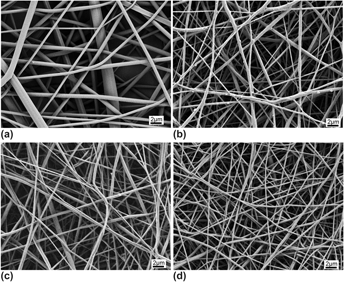
The work was intended to explore the effect of the widely available cationic polymer polyethylenimine (PEI) on small diameter poly(ɛ-caprolactone) (PCL) blood vessel grafts. PEI was blended with PCL and electrospun into nanofibrous vascular scaffolds. The morphologies, wettabilities, mechanical properties, and biological activities of the PCL/PEI electrospun nanofibers were investigated. It was found that by increasing the content of PEI to 5% within the scaffolds, the fiber diameters decreased from 469.7 ± 212.1 to 282.5 ± 107.1 nm, the water contact angle was reduced from 126.6 ± 1.1° to 27.6 ± 3.9°, while the Young's modulus increased from 2.0 ± 0.2 to 4.1 ± 0.1 MPa, the suture retention strength increased from 4.2 ± 0.4 to 6.1 ± 0.7 N, and the burst pressure increased from 801.2 ± 14.1 to 926.2 ± 22.8 mmHg. The in vitro evaluations demonstrated that the nanofibers containing 2% PEI promoted the attachment and proliferation of human umbilical vein endothelial cells (HUVECs).

Type
Articles
Copyright
Copyright © Materials Research Society 2015 

Access options

Get access to the full version of this content by using one of the access options below. (Log in options will check for institutional or personal access. Content may require purchase if you do not have access.)

Footnotes

Contributing Editor: Adrian Mann

References

REFERENCES

Seifu, D.G., Purnama, A., Mequanint, K., and Mantovani, D.: Small-diameter vascular tissue engineering. Nat. Rev. Cardiol. 10(7), 410 (2013).CrossRefGoogle ScholarPubMed
Ma, H., Hu, J., and Ma, P.X.: Polymer scaffolds for small-diameter vascular tissue engineering. Adv. Funct. Mater. 20(17), 2833 (2010).CrossRefGoogle ScholarPubMed
Kannan, R.Y., Salacinski, H.J., Butler, P.E., Hamilton, G., and Seifalian, A.M.: Current status of prosthetic bypass grafts: A review. J. Biomed. Mater. Res., Part B 74(1), 570 (2005).CrossRefGoogle ScholarPubMed
Wang, X., Lin, P., Yao, Q., and Chen, C.: Development of small-diameter vascular grafts. World J. Surg. 31(4), 682 (2007).CrossRefGoogle ScholarPubMed
Keuren, J.F., Wielders, S.J., Driessen, A., Verhoeven, M., Hendriks, M., and Lindhout, T.: Covalently-bound heparin makes collagen thromboresistant. Arterioscler., Thromb., Vasc. Biol. 24(3), 613 (2004).CrossRefGoogle ScholarPubMed
Sayers, R.D., Raptis, S., Berce, M., and Miller, J.H.: Long-term results of femorotibial bypass with vein or polytetrafluoroethylene. Br. J. Surg. 85(7), 934 (1998).CrossRefGoogle ScholarPubMed
Bujan, J., Garcia-Honduvilla, N., and Bellon, J.M.: Engineering conduits to resemble natural vascular tissue. Biotechnol. Appl. Biochem. 39, 17 (2004).CrossRefGoogle ScholarPubMed
Rashid, S.T., Fuller, B., Hamilton, G., and Seifalian, A.M.: Tissue engineering of a hybrid bypass graft for coronary and lower limb bypass surgery. FASEB J. 22(6), 2084 (2008).CrossRefGoogle ScholarPubMed
Nerem, R.M. and Seliktar, D.: Vascular tissue engineering. Annu. Rev. Biomed. Eng. 3, 225 (2001).CrossRefGoogle ScholarPubMed
Edelman, E.R.: Vascular tissue engineering—designer arteries. Circ. Res. 85(12), 1115 (1999).CrossRefGoogle ScholarPubMed
L'Heureux, N., Paquet, S., Labbe, R., Germain, L., and Auger, F.A.: A completely biological tissue-engineered human blood vessel. FASEB J. 12(1), 47 (1998).Google ScholarPubMed
Niklason, L.E., Gao, J., Abbott, W.M., Hirschi, K.K., Houser, S., Marini, R., and Langer, R.: Functional arteries grown in vitro. Science 284(5413), 489 (1999).CrossRefGoogle ScholarPubMed
Hu, J., Sun, X., Ma, H., Xie, C., Chen, Y.E., and Ma, P.X.: Porous nanofibrous PLLA scaffolds for vascular tissue engineering. Biomaterials 31(31), 7971 (2010).CrossRefGoogle ScholarPubMed
Lovett, M., Cannizzaro, C., Daheron, L., Messmer, B., Vunjak-Novakovic, G., and Kaplan, D.L.: Silk fibroin microtubes for blood vessel engineering. Biomaterials 28(35), 5271 (2007).CrossRefGoogle ScholarPubMed
Lovett, M., Eng, G., Kluge, J.A., Cannizzaro, C., Vunjak-Novakovic, G., and Kaplan, D.L.: Tubular silk scaffolds for small diameter vascular grafts. Organogenesis 6(4), 217 (2010).CrossRefGoogle ScholarPubMed
Lindsay, S.W., Konstantinos, T., Eun, S.G., Fiorenzo, G.O., and David, L.K.: Microfabricated porous silk scaffolds for vascularizing engineered tissues. Adv. Funct. Mater. 23(27), 3404 (2013).Google Scholar
Soletti, L., Hong, Y., Guan, J., Stankus, J.J., El-Kurdi, M.S., Wagner, W.R., and Vorp, D.A.: A bilayered elastomeric scaffold for tissue engineering of small diameter vascular grafts. Acta Biomater. 6(1), 110 (2010).CrossRefGoogle ScholarPubMed
McClure, M.J., Wolfe, P.S., Rodriguez, I.A., and Bowlin, G.L.: Bioengineered vascular grafts: Improving vascular tissue engineering through scaffold design. J. Drug Delivery Sci. Technol. 21(3), 211 (2011).CrossRefGoogle Scholar
Cleary, M.A., Geiger, E., Grady, C., Best, C., Naito, Y., and Breuer, C.: Vascular tissue engineering: The next generation. Trends Mol. Med. 18(7), 394 (2012).CrossRefGoogle ScholarPubMed
Naito, Y., Shinoka, T., Duncan, D., Hibino, N., Solomon, D., Cleary, M., Rathore, A., Fein, C., Church, S., and Breuer, C.: Vascular tissue engineering: Towards the next generation vascular grafts. Adv. Drug Delivery Rev. 63(4–5), 312 (2011).CrossRefGoogle ScholarPubMed
Lee, S.J., Liu, J., Oh, S.H., Soker, S., Atala, A., and Yoo, J.J.: Development of a composite vascular scaffolding system that withstands physiological vascular conditions. Biomaterials 29(19), 2891 (2008).CrossRefGoogle ScholarPubMed
Wang, S., Zhang, Y., Wang, H., Yin, G., and Dong, Z.: Fabrication and properties of the electrospun polylactide/silk fibroin-gelatin composite tubular scaffold. Biomacromolecules 10(8), 2240 (2009).CrossRefGoogle ScholarPubMed
Marelli, B., Alessandrino, A., Fare, S., Freddi, G., Mantovani, D., and Tanzi, M.C.: Compliant electrospun silk fibroin tubes for small vessel bypass grafting. Acta Biomater. 6(10), 4019 (2010).CrossRefGoogle ScholarPubMed
Du, F., Wang, H., Zhao, W., Li, D., Kong, D., Yang, J., and Zhang, Y.: Gradient nanofibrous chitosan/poly epsilon-caprolactone scaffolds as extracellular microenvironments for vascular tissue engineering. Biomaterials 33(3), 762 (2012).CrossRefGoogle ScholarPubMed
Han, F., Jia, X., Dai, D., Yang, X., Zhao, J., Zhao, Y., Fan, Y., and Yuan, X.: Performance of a multilayered small-diameter vascular scaffold dual-loaded with VEGF and PDGF. Biomaterials 34(30), 7302 (2013).CrossRefGoogle ScholarPubMed
Prabhakaran, M.P., Venugopal, J.R., Chyan, T.T., Hai, L.B., Chan, C.K., Lim, A.Y., and Ramakrishna, S.: Electrospun biocomposite nanofibrous scaffolds for neural tissue engineering. Tissue Eng., Part A 14(11), 1787 (2008).CrossRefGoogle ScholarPubMed
Prabhakaran, M.P., Venugopal, J., Chan, C.K., and Ramakrishna, S.: Surface modified electrospun nanofibrous scaffolds for nerve tissue engineering. Nanotechnology 19(45), 455102 (2008).CrossRefGoogle ScholarPubMed
Zhu, Y., Gao, C., Liu, X., and Shen, J.: Surface modification of polycaprolactone membrane via aminolysis and biomacromolecule immobilization for promoting cytocompatibility of human endothelial cells. Biomacromolecules 3(6), 1312 (2002).CrossRefGoogle ScholarPubMed
Ogris, M., Brunner, S., Schuller, S., Kircheis, R., and Wagner, E.: PEGylated DNA/transferrin-PEI complexes: Reduced interaction with blood components, extended circulation in blood and potential for systemic gene delivery. Gene Ther. 6(4), 595 (1999).CrossRefGoogle ScholarPubMed
Zhou, X., Laroche, F.J.F., Lamers, G., Torraca, V., Voskamp, P., Lu, T., Chu, F., Spaink, H.P., Abrahams, J.P., and Liu, Z.: Ultra-small graphene oxide functionalized with polyethylenimine (PEI) for very efficient gene delivery in cell and zebrafish embryos. Nano Res. 5(10), 703 (2012).CrossRefGoogle Scholar
Andersson, M.M. and Hatti-Kaul, R.: Protein stabilising effect of polyethyleneimine. J. Biotechnol. 72(1–2), 21 (1999).CrossRefGoogle Scholar
Vancha, A.R., Govindaraju, S., Parsa, K.V., Jasti, M., Gonzalez-Garcia, M., and Ballestero, R.P.: Use of polyethyleneimine polymer in cell culture as attachment factor and lipofection enhancer. BMC Biotechnol. 4, 23 (2004).CrossRefGoogle ScholarPubMed
Boussif, O., Lezoualch, F., Zanta, M.A., Mergny, M.D., Scherman, D., Demeneix, B., and Behr, J.P.: A versatile vector for gene and oligonucleotide transfer into cells in culture and in-vivo: Polyethylenimine. Proc. Natl. Acad. Sci. U. S. A. 92(16), 7297 (1995).CrossRefGoogle ScholarPubMed
Forrest, M.L., Meister, G.E., Koerber, J.T., and Pack, D.W.: Partial acetylation of polyethylenimine enhances in vitro gene delivery. Pharm. Res. 21(2), 365 (2004).CrossRefGoogle ScholarPubMed
Kim, J.H., Choung, P-H., Kim, I.Y., Lim, K.T., Son, H.M., Choung, Y-H., Cho, C-S., and Chung, J.H.: Electrospun nanofibers composed of poly(epsilon-caprolactone) and polyethylenimine for tissue engineering applications. J. Mater. Sci. Eng. B 29(5), 1725 (2009).CrossRefGoogle Scholar
Vaz, C.M., van Tuijl, S., Bouten, C.V., and Baaijens, F.P.: Design of scaffolds for blood vessel tissue engineering using a multi-layering electrospinning technique. Acta Biomater. 1(5), 575 (2005).CrossRefGoogle ScholarPubMed
Ghezzi, C.E., Marelli, B., Muja, N., and Nazhat, S.N.: Immediate production of a tubular dense collagen construct with bioinspired mechanical properties. Acta Biomater. 8(5), 1813 (2012).CrossRefGoogle ScholarPubMed
Courtney, T., Sacks, M.S., Stankus, J., Guan, J., and Wagner, W.R.: Design and analysis of tissue engineering scaffolds that mimic soft tissue mechanical anisotropy. Biomaterials 27(19), 3631 (2006).Google ScholarPubMed
Lee, S.J., Lim, G.J., Lee, J.W., Atala, A., and Yoo, J.J.: In vitro evaluation of a poly(lactide-co-glycolide)-collagen composite scaffold for bone regeneration. Biomaterials 27(18), 3466 (2006).CrossRefGoogle ScholarPubMed
Cui, W., Cheng, L., Li, H., Zhou, Y., Zhang, Y., and Chang, J.: Preparation of hydrophilic poly(l-lactide) electrospun fibrous scaffolds modified with chitosan for enhanced cell biocompatibility. Polymer 53(11), 2298 (2012).CrossRefGoogle Scholar
Mikos, A.G., Lyman, M.D., Freed, L.E., and Langer, R.: Wetting of poly(L-lactic acid) and poly(DL-lactic-co-glycolic acid) foams for tissue-culture. Biomaterials 15(1), 55 (1994).CrossRefGoogle ScholarPubMed
Amiel, G.E., Komura, M., Shapira, O., Yoo, J.J., Yazdani, S., Berry, J., Kaushal, S., Bischoff, J., Atala, A., and Soker, S.: Engineering of blood vessels from acellular collagen matrices coated with human endothelial cells. Tissue Eng. 12(8), 2355 (2006).CrossRefGoogle ScholarPubMed
Stekelenburg, M., Rutten, M.C., Snoeckx, L.H., and Baaijens, F.P.: Dynamic straining combined with fibrin gel cell seeding improves strength of tissue-engineered small-diameter vascular grafts. Tissue Eng., Part A 15(5), 1081 (2009).CrossRefGoogle ScholarPubMed
Fung, Y.C.: Bioviscoelastic solids: Collagen. In Biomechanics; Mechanical Properties of Living Tissues, Springer, New York, NY, 1999; p. 261.Google Scholar
Konig, G., McAllister, T.N., Dusserre, N., Garrido, S.A., Iyican, C., Marini, A., Fiorillo, A., Avila, H., Wystrychowski, W., Zagalski, K., Maruszewski, M., Jones, A.L., Cierpka, L., de la Fuente, L.M., and L'Heureux, N.: Mechanical properties of completely autologous human tissue engineered blood vessels compared to human saphenous vein and mammary artery. Biomaterials 30(8), 1542 (2009).CrossRefGoogle ScholarPubMed
Billiar, K., Murray, J., Laude, D., Abraham, G., and Bachrach, N.: Effects of carbodiimide crosslinking conditions on the physical properties of laminated intestinal submucosa. J. Biomed. Mater. Res. 56(1), 101 (2001).3.0.CO;2-6>CrossRefGoogle ScholarPubMed
Oskoui, P., Stadler, I., and Lanzafame, R.J.: A preliminary study of laser tissue soldering as arterial wall reinforcement in an acute experimental aneurysm model. Laser Surg. Med. 32(5), 346 (2003).CrossRefGoogle Scholar
Sarkar, S., Salacinski, H.J., Hamilton, G., and Seifalian, A.M.: The mechanical properties of infrainguinal vascular bypass grafts: Their role in influencing patency. Eur. J. Vasc. Endovasc. Surg. 31(6), 627 (2006).CrossRefGoogle ScholarPubMed
Seliktar, D., Black, R.A., Vito, R.P., and Nerem, R.M.: Dynamic mechanical conditioning of collagen-gel blood vessel constructs induces remodeling in vitro. Ann. Biomed. Eng. 28(4), 351 (2000).CrossRefGoogle ScholarPubMed
Tryoen-Toth, P., Vautier, D., Haikel, Y., Voegel, J.C., Schaaf, P., Chluba, J., and Ogier, J.: Viability, adhesion, and bone phenotype of osteoblast-like cells on polyelectrolyte multilayer films. J. Biomed. Mater. Res. 60(4), 657 (2002).CrossRefGoogle ScholarPubMed
Kuo, Y.C. and Ku, I.N.: Application of polyethyleneimine-modified scaffolds to the regeneration of cartilaginous tissue. Biotechnol. Prog. 25(5), 1459 (2009).CrossRefGoogle ScholarPubMed
Chen, C.S., Mrksich, M., Huang, S., Whitesides, G.M., and Ingber, D.E.: Geometric control of cell life and death. Science 276(5317), 1425 (1997).CrossRefGoogle ScholarPubMed
Zhu, H., Ji, J., Tan, Q., Barbosa, M.A., and Shen, J.: Surface engineering of poly(DL-lactide) via electrostatic self-assembly of extracellular matrix-like molecules. Biomacromolecules 4(2), 378 (2003).CrossRefGoogle ScholarPubMed
Hersel, U., Dahmen, C., and Kessler, H.: RGD modified polymers: Biomaterials for stimulated cell adhesion and beyond. Biomaterials 24(24), 4385 (2003).CrossRefGoogle ScholarPubMed
Ku, S.H. and Park, C.B.: Human endothelial cell growth on mussel-inspired nanofiber scaffold for vascular tissue engineering. Biomaterials 31(36), 9431 (2010).CrossRefGoogle ScholarPubMed