Hostname: page-component-745bb68f8f-f46jp Total loading time: 0 Render date: 2025-01-11T01:21:51.320Z Has data issue: false hasContentIssue false

Assessment of cell proliferation on 6H–SiC biofunctionalized with self-assembled monolayers

Published online by Cambridge University Press:  31 July 2012

A. Oliveros*
Affiliation:
Electrical Engineering Department, University of South Florida, Tampa, Florida 33620
C.L. Frewin
Affiliation:
Department of Molecular Pharmacology and Physiology, University of South Florida, Tampa, Florida 33612
S.J. Schoell
Affiliation:
Walter Schottky Institut and Physik Department, Technische Universität München, Garching 85748, Germany
M. Hoeb
Affiliation:
Walter Schottky Institut and Physik Department, Technische Universität München, Garching 85748, Germany
M. Stutzmann
Affiliation:
Walter Schottky Institut and Physik Department, Technische Universität München, Garching 85748, Germany
I.D. Sharp
Affiliation:
Walter Schottky Institut and Physik Department, Technische Universität München, Garching 85748, Germany
S.E. Saddow
Affiliation:
Electrical Engineering Department, University of South Florida, Tampa, Florida 33620; and Department of Molecular Pharmacology and Physiology, University of South Florida, Tampa, Florida 33612
*
a)Address all correspondence to this author. e-mail: [email protected]
Get access

Abstract

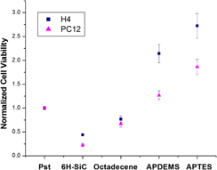
In this article, the biofunctionalization of 6H–SiC (0001) surfaces via self-assembled monolayers (SAMs) has been studied as a means to modify the in vitro biocompatibility of this semiconductor substrate with H4 (human neuroglioma) and PC12 (rat pheochromocytoma) cells. Silanization with aminopropyldiethoxymethylsilane (APDEMS) and aminopropyltriethoxysilane (APTES), which provided moderately hydrophilic surfaces, and alkylation with 1-octadecene that produced hydrophobic surfaces were used to control the 6H–SiC surface chemistry and evaluate changes in cell viability and morphology due to these surface modifications. The morphology of the cells was evaluated with atomic force microscopy. In addition, 3-(4,5-dimethylthiazol-2-yl)-2,5-diphenyltetrazolium bromide (MTT) assays were used to quantitatively evaluate the cell viability on the SAM-modified surfaces. In all cases, the cell proliferation was observed to improve with respect to untreated 6H–SiC surfaces, with up to a 2x increase in viability on the 1-octadecene-modified surfaces, up to 6x increase with APDEMS-modified surfaces, and up to 8x increase with APTES-modified surfaces. This proves the potential of SiC as a substrate for medical devices given the possibility to tailor its surface chemistry for specific applications.

Type
Articles
Copyright
Copyright © Materials Research Society 2012

Access options

Get access to the full version of this content by using one of the access options below. (Log in options will check for institutional or personal access. Content may require purchase if you do not have access.)

References

REFERENCES

Williams, D.F.: On the mechanisms of biocompatibility. Biomaterials 29, 29412953 (2008).Google Scholar
Miyamoto, S., Akiyama, S., and Yamada, K.: Synergistic roles for receptor occupancy and aggregation in integrin transmembrane function. Science 267, 883885 (1995).Google Scholar
Juliano, R.L., Haskill, S., and Carolina, N.: Mini-review signal transduction from the extracellular matrix. Cell 120, 577585 (1993).Google Scholar
Underwood, P., Steele, J.G., and Dalton, B.: Effects of polystyrene surface chemistry on the biological activity of solid phase fibronectin and vitronectin, analysed with monoclonal antibodies. J. Cell Sci. 104, 793803 (1993).Google Scholar
Lee, M.H., Brass, D., Morris, R., Composto, R.J., and Ducheyne, P.: The effect of non-specific interactions on cellular adhesion using model surfaces. Biomaterials 26, 17211730 (2005).Google Scholar
McClary, K.B., Ugarova, T., and Grainger, D.W.: Modulating fibroblast adhesion, spreading, and proliferation using self-assembled monolayer films of alkylthiolates on gold. J. Biomed. Mater. Res. 50, 428439 (2000).Google Scholar
Sundaram, R.S., Gómez-Navarro, C., Balasubramanian, K., Burghard, M., and Kern, K.: Electrochemical modification of graphene. Adv. Mater. 20, 30503053 (2008).Google Scholar
Catellani, A. and Cicero, G.: Modifications of cubic SiC surfaces studied by ab initio simulations: From gas adsorption to organic functionalization. J. Phys. D: Appl. Phys. 40, 62156224 (2007).Google Scholar
Vahlberg, C., Yazdi, G.R., Petoral, R.M., Syvajarvi, M., Uvdal, K., Spetz, A.L., Yakimova, R., and Khranovsky, V.: Surface engineering of functional materials for biosensors. IEEE Sensors J. 2005, 504507 (2005).Google Scholar
Yakimova, R., Steinhoff, G., Petoral, R.M., Vahlberg, C., Khranovskyy, V., Yazdi, G.R., Uvdal, K., and Lloyd Spetz, A.: Novel material concepts of transducers for chemical and biosensors. Biosens. Bioelectron. 22, 27802785 (2007).Google Scholar
Fawcett, T.J., Wolan, J.T., Lloyd Spetz, A., Reyes, M., and Saddow, S.E.: Thermal detection mechanism of SiC based hydrogen resistive gas sensors. Appl. Phys. Lett. 89, 182102 (2006).Google Scholar
Carter, G.E., Casady, J.B., Bonds, J., Okhuysen, M.E., Scofield, J.D., and Saddow, S.E.: Pendeo epitaxy of 3C-SiC on Si substrates. Mater. Sci. Forum 1149, 338342 (2000).Google Scholar
Gabriel, G., Erill, I., Caro, J., Gomez, R., Riera, D., Villa, R., and Godignon, P.: Manufacturing and full characterization of silicon carbide-based multi-sensor micro-probes for biomedical applications. Microelectron. J. 38, 406415 (2007).Google Scholar
Yakimova, R., Petoral, R.M., Yazdi, G.R., Vahlberg, C., Lloyd Spetz, A., and Uvdal, K.: Surface functionalization and biomedical applications based on SiC. J. Phys. D: Appl. Phys. 40, 64356442 (2007).Google Scholar
Saddow, S.E., Frewin, C.L., Coletti, C., Schettini, N., Weeber, E., Oliveros, A., and Jarosezski, M.: Single-crystal silicon carbide: A biocompatible and hemocompatible semiconductor for advanced biomedical applications. Mater. Sci. Forum 679680, 824830 (2011).Google Scholar
Coletti, C., Jaroszeski, M.J., Pallaoro, A., Hoff, M., Iannotta, S., and Saddow, S.E.: Biocompatibility and wettability of crystalline SiC and Si surfaces. In 29th Annual International Conference of the IEEE Engineering in Medicine and Biology Society, Lyon, France, 2007; pp. 58495852.Google Scholar
Frewin, C.L., Jaroszeski, M., Weeber, E., Muffly, K.E., Kumar, A., Peters, M., Oliveros, A., and Saddow, S.E.: Atomic force microscopy analysis of central nervous system cell morphology on silicon carbide and diamond substrates. J. Mol. Recognit. 22, 380388 (2009).Google Scholar
Frewin, C.L.: The Neuron-Silicon Carbide Interface: Biocompatibility Study and BMI Device Development (University of South Florida, Tampa, FL, 2009).Google Scholar
Hehrlein, C.: Stent passivation with silicon carbide as a possible alternative to drug-eluting stents – A comprehensive review of pre-clinical and clinical results. Interventional Cardiol. 4, 6063 (2009).Google Scholar
Rzany, A. and Schaldach, M.: Smart material silicon carbide: Reduced activation of cells and proteins on a-SiC: H-coated stainless steel. Prog. Biomed. Res. 5, 182194 (2001).Google Scholar
Kalnins, U., Erglis, A., Dinne, I., Kumsars, I., and Jegere, S.: Clinical outcomes of silicon carbide coated stents in patients with coronary artery disease. Med. Sci. Monit. 8, PI1620 (2002).Google Scholar
Schettini, N., Jaroszeski, M., West, L., and Saddow, S.: SiC hemacompatibility for cardiovascular applications. In Silicon Carbide Biotechnology; Saddow, S.E. ed. (Elsevier Ltd., Oxford, UK, 2012); pp. 153208.Google Scholar
Santavirta, S., Takagi, M., Nordsletten, L., Anttila, A., Lappalainen, R., and Konttinen, T.: Biocompatibility of silicon carbide in colony formation test in vitro. A promising new ceramic THR implant coating material. J. Biomater. Appl. 118, 8991 (1998).Google Scholar
Faucheux, N., Schweiss, R., Lützow, K., Werner, C., and Groth, T.: Self-assembled monolayers with different terminating groups as model substrates for cell adhesion studies. Biomaterials 25, 27212730 (2004).Google Scholar
Kapur, R. and Rudolph, A.S.: Cellular and cytoskeleton morphology and strength of adhesion of cells on self-assembled monolayers of organosilanes. Exp. Cell. Res. 244, 275285 (1998).Google Scholar
Stenger, D., Pike, C.J., Hickman, J.J., and Cotman, C.W.: Surface determinants of neuronal survival and growth on self-assembled monolayers in culture. Brain Res. 630, 136147 (1993).Google Scholar
Low, S.P., Williams, K.A., Canham, L.T., and Voelcker, N.H.: Evaluation of mammalian cell adhesion on surface-modified porous silicon. Biomaterials 27, 45384546 (2006).Google Scholar
Linford, M.R. and Chidsey, C.E.D.: Alkyl monolayers covalently bonded to silicon surfaces. J. Am. Chem. Soc. 115, 1263112632 (1993).Google Scholar
Bierbaum, K., Kinzler, M., Woell, C., Grunze, M., Haehner, G., Heid, S., and Effenberger, F.: A near edge x-ray absorption fine structure spectroscopy and x-ray photoelectron spectroscopy study of the film properties of self-assembled monolayers of organosilanes on oxidized Si(100). Langmuir 11, 512518 (1995).Google Scholar
Stutzmann, M., Garrido, J.A., Eickhoff, M., and Brandt, M.S.: Direct biofunctionalization of semiconductors: A survey. Phys. Status Solidi A 203, 34243437 (2006).Google Scholar
Rosso, M., Arafat, A., Schroën, K., Giesbers, M., Roper, C.S., Maboudian, R., and Zuilhof, H.: Covalent attachment of organic monolayers to silicon carbide surfaces. Langmuir 24, 40074012 (2008).Google Scholar
Cicero, G. and Catellani, A.: Towards SiC surface functionalization: An ab initio study. J. Chem. Phys. 122, 214716 (2005).Google Scholar
Rosso, M., Giesbers, M., Arafat, A., Schroën, K., and Zuilhof, H.: Covalently attached organic monolayers on SiC and SixN4 surfaces: Formation using UV light at room temperature. Langmuir 25, 21722180 (2009).Google Scholar
Schoell, S.J., Hoeb, M., Sharp, I.D., Steins, W., Eickhoff, M., Stutzmann, M., and Brandt, M.S.: Functionalization of 6H-SiC surfaces with organosilanes. Appl. Phys. Lett. 92, 153301 (2008).Google Scholar
Tsuchida, H., Kamata, I., and Izumi, K.: Infrared attenuated total reflection spectroscopy of 6H–SiC (0001). J. Appl. Phys. 85, 35703574 (1999).Google Scholar
Mossman, T.: Rapid colorimetric assay for cellular growth and survival: Application to proliferation and cytotoxicity assays. J. Immunol. Methods 65, 5563 (1983).Google Scholar
Richards, R.G.: The effect of surface roughness on fibroblast adhesion in vitro. Injury 1996, 27 Suppl 3, SC38SC43.CrossRefGoogle ScholarPubMed
Goodman, C.: Mechanisms and molecules that control growth cone guidance. Annu. Rev. Neurosci. 19, 341347 (1996).Google Scholar
Levitan, I. and Kaczmarek, L.: Adhesion molecules and axon pathfinding. In The Neuron Cell and Molecular Biology; Oxford University Press, New York, 2002; pp. 435446.Google Scholar
Frewin, C.L., Coletti, C., Riedl, C., Starke, U., and Saddow, S.E.: A comprehensive study of hydrogen etching on the major SiC polytypes and crystal orientations. Mater. Sci. Forum 615617, 589592 (2009).CrossRefGoogle Scholar
Coletti, C., Frewin, C.L., Saddow, S.E., Hetzel, M., Virojanadara, C., and Starke, U.: Surface studies of hydrogen etched 3C-SiC(001) on Si(001). Appl. Phys. Lett. 91, 061914 (2007).Google Scholar
Bhowmick, D.K., Linden, S., Devaux, A., De Cola, L., and Zacharias, H.: Functionalization of amorphous SiO2 and 6H-SiC(0001) surfaces with benzo[ghi]perylene-1,2-dicarboxylic anhydride via an APTES linker. Small 8, 592601, 619 (2012).Google Scholar
Sharp, I.D., Schoell, S.J., Hoeb, M., Brandt, M.S., and Stutzmann, M.: Electronic properties of self-assembled alkyl monolayers on Ge surfaces. Appl. Phys. Lett. 92, 223306 (2008).Google Scholar
Mrksich, M. and Whitesides, G.M.: Using self-assembled monolayers to understand the interactions of man-made surfaces with proteins and cells. Annu. Rev. Biophys. 25, 5578 (1996).Google Scholar
Van Damme, M-P., Taglias, J., Nemat, N., and Preston, B.: Determination of cell charge at the surface. Anal. Biochem. 223, 6270 (1994).Google Scholar
Dan, N.: The effect of charge regulation on cell adhesion to substrates: salt-induced repulsion. Colloids Surf., B 27, 4147 (2003).Google Scholar
Arima, Y. and Iwata, H.: Effect of wettability and surface functional groups on protein adsorption and cell adhesion using well-defined mixed self-assembled monolayers. Biomaterials 28, 30743082 (2007).Google Scholar
Robertus, J., Browne, W.R., and Feringa, B.L.: Dynamic control over cell adhesive properties using molecular-based surface engineering strategies. Chem. Soc. Rev. 39, 354378 (2010).Google Scholar
Ostuni, E.: The interaction of proteins and cells with self-assembled monolayers of alkanethiolates on gold and silver. Colloids Surf., B 15, 330 (1999).Google Scholar
Naji, A. and Harmand, M.F.: Cytocompatibility of two coating materials, amorphous alumina and silicon carbide, using human differentiated cell cultures. Biomaterials 12, 690694 (1991).Google Scholar
Webb, K., Hlady, V., and Tresco, P.A.: Relative importance of surface wettability and charged functional groups on NIH 3T3 fibroblast attachment, spreading, and cytoskeletal organization. J. Biomed. Mater. Res. 41, 422430 (1998).Google Scholar
Sorribas, H., Braun, D., Leder, L., Sonderegger, P., and Tiefenauer, L.: Adhesion proteins for a tight neuron-electrode contact. J. Neurosci. Methods 104, 133141 (2001).Google Scholar
Moon, J.H., Shin, J.W., Kim, S.Y., and Park, J.W.: Formation of uniform aminosilane thin layers: An imine formation to measure relative surface density of the amine group. Langmuir 12, 46214624 (1996).Google Scholar
Hernando, J., Pourrostami, T., Garrido, J., Williams, O., Gruen, D., Kromka, A., Steinmuller, D., and Stutzmann, M.: Immobilization of horseradish peroxidase via an amino silane on oxidized ultrananocrystalline diamond. Diamond Relat. Mater. 16, 138143 (2007).Google Scholar