Introduction
Sinonasal malignancies are rare anterior skull base tumours; they account for 0.2–0.8 per cent of all cancers and 3 per cent of upper respiratory tract malignancies.Reference Hoppe, Stegman, Zelefsky, Rosenzweig, Wolden and Patel1 Sinonasal tumours appear to remain asymptomatic until they are locally advanced. The tumours eventually alter the adjacent structures by obstructing sinonasal airways or by infiltrating local tissues, and may therefore imitate respiratory inflammations.Reference Boo and Hogg2 Some of the clinical manifestations are nasal obstruction, nasal discharge, sinusitis, epistaxis, palate ulcers, numb or loose teeth, tooth pain, facial pain or oedema, cranial nerve alterations, proptosis, and even trismus. These signs and symptoms are not tumour-specific, and their chronic nature and resistance to symptomatic treatments eventually necessitate a more precise diagnostic approach. Usually, nasal endoscopy or other appropriate radiological means unveil the existence of a tumour, and a biopsy confirms tissue diagnosis.Reference Boo and Hogg2, Reference Blanch, Ruiz, Alos, Traserra-Coderch and Bernal-Sprekelsen3
Oncological treatment decisions for these tumours are primarily based on tumour histology. For surgical resection with curative intent, the craniofacial approach is considered the ‘gold standard’ for most cases.Reference Batra, Citardi, Worley, Lee and Lanza4–Reference Howard, Lund and Wei6 For early stage (1 and 2) tumours, radical resection is recommended; for more advanced stages of tumour, multimodality treatments may result in better outcomes.Reference Guntinas-Lichius, Kreppel, Stuetzer, Semrau, Eckel and Mueller7 Neo-adjuvant radiotherapy may be considered when wide resection is planned, or when post-surgical tissue hypoxaemia causes tissue resistance to radiation therapy. For those with massive local invasion, perineural invasion, positive surgical margins or infeasibility of complete resection, adjuvant radiotherapy is suggested;Reference Hoppe, Stegman, Zelefsky, Rosenzweig, Wolden and Patel1 for patients with distant metastases, chemotherapy and sometimes palliative surgery are performed.
As an alternative, an endoscopic approach can be used in selected early stage skull base tumour cases as part of a minimally invasive surgical armamentarium.Reference Dave, Bared and Casiano8–Reference Ong, Solares, Carrau and Snyderman10 Although surgeons cannot employ all their hand skills while operating intranasally, this approach is sometimes preferred over the traditional ‘open’ approach. The endoscopic approach is associated with less post-surgical morbidity and better cosmetic outcomes as no facial incisions are required.Reference Batra, Citardi, Worley, Lee and Lanza4, Reference Buchmann, Larsen, Pollack, Tawfik, Sykes and Hoover11 It can yield the same oncological treatment results as the traditional approach, even in advanced cases with skull base involvement.Reference Solares, Grindler, Luong, Kanowitz, Sade and Citardi12, Reference Carrau, Ong, Solares and Snyderman13
Many studies have investigated the efficacy of the endoscopic approach for treatment of sinonasal malignancies. In some reports, it is considered an efficient method.Reference Hoppe, Stegman, Zelefsky, Rosenzweig, Wolden and Patel1, Reference Buchmann, Larsen, Pollack, Tawfik, Sykes and Hoover11–Reference Shipchandler, Batra, Citardi, Bolger and Lanza22 Some consider it to be the treatment of choice for inverted papilloma.Reference Lombardi, Tomenzoli, Butta, Bizzoni, Farina and Sberze23 Others do not consider it as a replacement for the traditional craniofacial approach.Reference Castelnuovo, Belli, Bignami, Battaglia, Sberze and Tomei5, Reference Howard, Lund and Wei6 Finally, there are those that still consider craniofacial surgery to be the gold-standard approach.Reference Batra, Citardi, Worley, Lee and Lanza4–Reference Howard, Lund and Wei6 The low prevalence and various tumour histotypes have limited the number of institutes with enough experience to treat these tumours.Reference Waldron, O'Sullivan, Warde, Gullane, Lui and Payne24
We conducted a large prospective study on referred patients with sinonasal malignancies who received surgery as part of their treatment course. Oncological outcomes were analysed in terms of the surgical approach used, based on whether open or endoscopic surgery was performed.
Materials and methods
Patients
Patients with nasal tumours who were referred to our academic tertiary referral centre (Imam Khomeini Hospital Complex) from 1999 to January 2010 were included in this study. A total of 160 patients were eligible for inclusion; all received surgery as part of their oncological treatment. Patients were allocated to two groups according to the surgical approach employed: open or endoscopic.
All patients who underwent open or endoscopic resection of the malignant sinonasal tumour with curative intent, and had at least one-year follow up, were included in this study. Patients with systemic tumour involvement and revision cases were not included.
In this historical cohort study, every patient was labelled with a number and allocated a data sheet in which their contributing data were recorded. Tumour control and survival was evaluated through this means.
Surgical approach decision
In all patients, tumours were primarily resected using either a conventional open sinus surgery approach or an endoscopic approach. Patients were subsequently allocated to the following two groups: an open surgery group or an endoscopic surgery group.
The endoscopic approach was not considered in patients with: extension to the frontal sinus, involvement of the bony walls of the maxillary sinus (with the exception of the medial one), erosion of the nasal fossa floor and extension to the palate, orbital involvement, massive skull base involvement, skin or extensive subcutaneous involvement, or muscular invasion (pterygoids or infratemporal fossa).
Except for the above-mentioned conditions, the choice of surgical method was based on the expertise of the surgeon.
Evaluated variables
Patients' demographic data were gathered and recorded. In addition, the location of the primary tumour (sinus or nasal cavity involvement, or both) and tumour side(s) were determined by nasal endoscopy or other appropriate radiological means. Clinical manifestations of the disease (mainly patients' chief complaints), and the time period between the onset of symptoms and final diagnosis, were also taken into account. Tissue diagnosis was made via tissue biopsy for all patients, prior to surgical treatment. All pathological evaluations were conducted by the same pathologist. There were reports on tumour pathology, tumour stage and tumour grade for all patients. Invasion of adjacent tissues and/or other organ metastases were evaluated by the same method. Computed tomography and magnetic resonance imaging were both conducted before surgery as part of a routine protocol in our centre for staging and better understanding of tumour boundaries.
Surgical technique
All patients received antibiotics and steroids peri-operatively (regardless of tumour characteristics). Surgical margins were examined with frozen section analysis.
Endoscopic resection
Tumour debulking was performed primarily to reach the tumour origin, in order to resect the entire tumour with sufficient margins. Skull base tumours with a diffuse or ambiguous origin were identified by approaching the sphenoid sinus once the ethmoid roof had been recognised. The pterygopalatine fossa was dissected in cases of tumour extension to this part or if internal maxillary artery ligation was required. Haemostasis was performed at the end of surgery using suction cautery.
Open resection
A lateral rhinotomy incision with or without craniotomy was the primary external incision used. If a craniofacial resection approach was employed, a standard bicoronal craniotomy incision was performed. The tumour was then exposed and completely removed with sufficient margins.
An endoscope or microscope was used in the later stages of surgery for better mapping of the critical area affected by the tumour and complete removal of the tumour margin.
Adjunctive treatment
Standard oncological treatments were delivered to the patients if indicated, based on the pathology of the tumour and according to national guidelines. For patients receiving adjuvant radiotherapy, the radiation dosage was also recorded.
Post-operative evaluation and follow up
Margin involvement was evaluated through pathological analysis of the excised specimens. Immediate surgical complications were dealt with during hospitalisation. After discharge, the patients visited at regular intervals and when they needed medical care. At the follow-up visits, patients were examined for possible recurrence, metastases, late complications (e.g. adhesions and infections) and radiotherapy complications. Surgery dates were used to calculate overall survival.
Ethical approval
All data were extracted from patients' charts and no direct contact was made with the patients. Data sheets were labelled with numbers and the patients' names were kept confidential in all study steps. The study was approved by the ethical board of Tehran University of Medical Sciences.
Statistical methods
Statistical analysis was performed using SPSS software, version 11.5 (Chicago, Illinois, USA). Comparative and inter-variable analyses were carried out using t-tests, chi-square tests and analyses of variance (ANOVA). The calculation of survival was conducted using the Kaplan–Mayer method. A p value of 0.05 or less was considered significant. Data are presented as means ± standard deviations.
Results
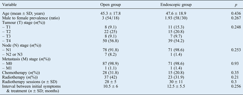
A total of 160 patients with sinonasal tumours were included in this study; 112 patients (70 per cent) were male and the remaining 48 (30 per cent) were female. Their mean age at the time of surgery was 46.56 ± 18.4 years, with an age range of 11 to 86 years. The open surgery group comprised 88 patients (55 per cent) and the endoscopic surgery group consisted of 72 patients (45 per cent). Table I displays patients' baseline information, indicating no significant difference between the groups.

Fig. 1 Overall survival rates for the endoscopic surgery group (blue line) and open surgery group (red line).
Table I Patients' characteristics

SD = standard deviation
Pre-operative tumour characteristics
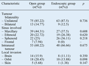
As outlined in Table II, the maxillary sinus was the most susceptible sinus for cancer and was involved in 66 cases (41.25 per cent). The majority of the case series had locally advanced tumours with minimal lymphatic spread, or metastasis and higher tumour grades. Tumour grade was significantly correlated with the patient's age (ANOVA, p = 0.006). Fifty-six patients (35 per cent) had local invasions with orbits more frequently involved in both groups. The groups were not significantly different in terms of tumour characteristics.
Table II Pre-operative tumour characteristics

Clinical manifestations
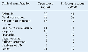
A wide range of signs and symptoms were present in our patient series, among which epistaxis, nasal obstruction and intranasal mass sensation were the most common (in that order) (Table III). Other manifestations were visual acuity problems, proptosis, headache, facial oedema, sensation of sinus fullness and paralysis of a cranial nerve. Of the clinical manifestations, nasal obstruction was significantly correlated with adenocarcinoma and esthesioneuroblastoma (t-test, p = 0.023). The mean duration between the development of signs and symptoms and the time of diagnosis was calculated as 11.17 ± 5.7 months (10.5 ± 6 months for the open surgery group and 12 ± 5.5 months for the endoscopic surgery group).
Table III Clinical presentation of nasal tumours

CN = cranial nerves.
Tumour histopathology
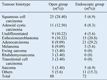
Table IV shows the histopathological diversity of the tumours, with squamous cell carcinoma (SCC) at the top of the list (the most common tumour histotype).
Table IV Tumour histopathology

Oncological treatment
All patients underwent surgical resection of the tumour as part of their oncological treatment. In addition, chemotherapy was delivered to 28 patients (31.81 per cent) in the open surgery group and 15 patients (20.83 per cent) in the endoscopic surgery group. The need for chemotherapy was not significantly different between the groups (t-test, p = 0.352). Radiotherapy was administered with a mean of 28 ± 5 sessions to 37 patients (42.04 per cent) in the open surgery group, and with a mean of 30 ± 11 sessions to 23 patients (31.94 per cent) in the endoscopic surgery group. There were no significant differences between the groups in terms of the need for radiotherapy or the number of radiotherapy sessions received (p = 0.212 and p = 0.305 respectively).
Post-operative tumour outcomes
Follow-up period
The mean period of follow up was 20 ± 14 months for the open surgery group and 22 ± 15 months for the endoscopic surgery group, with no significant difference between the groups (p = 0.252).
Surgical complications
No specific immediate surgical complications were reported after the surgery; however, eight patients (four in each group) experienced post-operative epistaxis, six (four in the open surgery group and two in the endoscopic group) developed epiphora and one in the endoscopic group experienced sinus wall adhesion complications. There was no significant correlation with the type of surgical approach (t-test, p = 0.656).
Recurrence
Fifty-eight patients (65.9 per cent) in the open surgery group and 59 (81.94 per cent) in the endoscopic surgery group experienced tumour recurrence 18 ± 10 months and 20 ± 11 months after the procedure, respectively. Although endoscopic surgery was associated with a higher percentage of recurrence, surgical approach was not significantly correlated with recurrence rate (p = 0.168). Moreover, there was no relationship in terms of the time period between the procedure and recurrence (p = 0.217).
Multivariate logistical regression analysis revealed no significant correlation between the type of surgical approach and recurrence and complication rate (t = −1.33, p = 0.198).
Survival
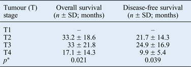
Mean overall survival and mean disease-free survival rates for the patients were 24 ± 20 and 15.2 ± 14.5 months in the endoscopic surgery group, and 28 ± 0.16 and 17.6 ± 14.3 months in the open surgery group respectively, showing a small, insignificant difference (p = 0.125). Figure 1 depicts survival rates for the first five post-surgical years. However, both overall survival and disease-free survival rates were significantly correlated with tumour stage, as summarised in Table V.
Table V Tumour stage and survival rate association

*Analysis of variance. SD = standard deviation
Discussion
In this study, a large series of patients with sinonasal tumours received either conventional open surgery or endoscopic surgery, and the outcomes of survivors were reported. The numbers in both the open surgery group and the endoscopic surgery group, with 88 and 72 patients respectively, were sufficient for the analysis to yield precise results.
The ‘nasal sinus’ is hard to reach surgically. Using conventional approaches, many surrounding structures that are critical have to be dissected but preserved. No external incision is made in the endoscopic approach and there is therefore limited room for surgical manoeuvres. The surgical approach used becomes even more important when tackling a tumour with complicated borders and invasions. Some surgeons consider the conventional approach to be the gold-standard method for nasal sinus and skull base tumour resection.Reference Batra, Citardi, Worley, Lee and Lanza4–Reference Howard, Lund and Wei6, Reference Carrau, Ong, Solares and Snyderman13 Others advocate an endoscopic approach to these tumours in previously selected patients, given sufficient evidence for its feasibility.Reference Dave, Bared and Casiano8, Reference Nicolai, Castelnuovo, Lombardi, Battaglia, Bignami and Pianta9, Reference Roh, Batra, Citardi, Lee, Bolger and Lanza21 The endoscopic approach may be a good alternative to the conventional approach if tumour-free margins are likely (the oncological goal). This is because it involves no facial incision (thereby minimising cosmetic concerns), other surrounding structures are not manipulated and the patients benefit from less morbidity in the post-operative period.
Despite advances in the diagnosis and treatment of sinonasal tumours, multimodality aggressive treatments have been suggested to improve disease conditions in light of the poor prognosis for these patients.Reference Hoppe, Stegman, Zelefsky, Rosenzweig, Wolden and Patel1, Reference Guntinas-Lichius, Kreppel, Stuetzer, Semrau, Eckel and Mueller7, Reference Bhattacharyya25 Therefore, there are still some proponents of conventional surgical treatment in many centres. However, Blanch et al. did not find any significant difference between endoscopic and open surgical approaches in terms of aggressive treatment, and proposed that tumour staging was a more important factor.Reference Blanch, Ruiz, Alos, Traserra-Coderch and Bernal-Sprekelsen3
Sinonasal tumours are more frequent in the fourth to sixth decades of life.Reference Myers, Nussenbaum, Bradford, Teknos, Esclamado and Wolf26, Reference Dulguerov, Jacobsen, Allal, Lehmann and Calcaterra27 The mean age of the patients in this study was 46.56 years, which is similar to that reported in the literature. However, a considerable percentage of patients were in their 30s and 40s; this could be because of different genetic characteristics and the living conditions in Iran as compared with those in the western world. Sexual diversity has been reported in some studies, with a male preponderance in someReference Blanch, Ruiz, Alos, Traserra-Coderch and Bernal-Sprekelsen3 and a female preponderance in others.Reference Dulguerov, Jacobsen, Allal, Lehmann and Calcaterra27 The majority of the patients in our study were male (a male to female ratio of 7:3). However, statistical analysis of these data, as well as reports in the literature, indicate that gender is not necessarily a factor of significance in the formation or development of sinonasal tumours.
An important issue regarding sinonasal tumours is their specific location, which makes them liable to remain undiagnosed until the more advanced stages of disease. Sinonasal tumours clinically manifest when they have invaded adjacent tissues or have physically occluded sinonasal airways; even at this stage, the presentation somewhat resembles an inflammatory upper airway disease.Reference Dulguerov, Jacobsen, Allal, Lehmann and Calcaterra27 Up to 104 (65 per cent) of our patients had stage 3 or 4 tumours upon examination in our centre. Our study revealed that higher survival rates were significantly correlated with earlier stages of the tumour; a delay in diagnosis is likely to cost many patients their lives in Third World countries. The delay in diagnosis may occur because the normal population is not given enough information to take their chronic symptoms seriously, or maybe the first-line healthcare staff who initially deal with these patients are not adequately trained. Nevertheless, there is at present no efficient diagnostic means to detect sinonasal tumours at earlier stages, although better evaluations have been reported to enable earlier diagnosis.Reference Blanch, Ruiz, Alos, Traserra-Coderch and Bernal-Sprekelsen3, Reference Waldron, O'Sullivan, Warde, Gullane, Lui and Payne24, Reference Dulguerov, Jacobsen, Allal, Lehmann and Calcaterra27
Squamous cell carcinoma was the most common tumour histotype in our case series, followed by adenocarcinoma. This finding has been reported by many other studies too,Reference Myers, Nussenbaum, Bradford, Teknos, Esclamado and Wolf26, Reference Dulguerov, Jacobsen, Allal, Lehmann and Calcaterra27 indicating that geographic location and racial factors do not necessarily affect this aspect of tumour development.
Maxillary sinuses were the most common sinuses involved. This may be because these sinuses are the largest paranasal sinuses, containing the majority of the squamous epithelium, and may therefore be more predisposed to tumour formation (especially SCC). In addition, nasal obstruction symptoms were significantly more frequently associated with adenocarcinoma and esthesioneuroblastoma histotypes, which may be due to the fact that these tumours commonly involve the nasal cavity.
In light of advances in the technique, endoscopic sinus surgery is now regarded as the primary tool for treating benign tumours. There have been some promising reports on the treatment of malignant tumours in selected patients. Some surgeons have used endoscopic sinus surgery in larger series of patients with malignant tumours, with promising results.Reference Nicolai, Castelnuovo, Lombardi, Battaglia, Bignami and Pianta9 However, it is worth noting that the outcome of each series is completely dependent upon patients' characteristics such as the pathology or tumour stage.Reference Nicolai, Castelnuovo, Lombardi, Battaglia, Bignami and Pianta9
Nicolai et al. reported that selected patients with sinonasal tumours in the early stages of disease were appropriate for the endoscopic approach.Reference Nicolai, Castelnuovo, Lombardi, Battaglia, Bignami and Pianta9 However, tumour–node–metastasis staging is not always a good predictor of advanced staging or endoscopic resection feasibility.Reference Solares, Grindler, Luong, Kanowitz, Sade and Citardi12, Reference Hanna, DeMonte, Ibrahim, Roberts, Levine and Kupferman16 Hana et al. suggested the use of endoscopic resection for malignant nasal tumours in selected cases, and reported acceptable outcomes.Reference Hanna, DeMonte, Ibrahim, Roberts, Levine and Kupferman16 A combined approach, using conventional techniques with the endoscopic technique, has been shown to have promising results in patients with more advanced tumours.Reference Buchmann, Larsen, Pollack, Tawfik, Sykes and Hoover11
Some authors recommend endoscopic resection for more advanced tumours with skull base involvement.Reference Solares, Grindler, Luong, Kanowitz, Sade and Citardi12, Reference Carrau, Ong, Solares and Snyderman13 For instance, Carrau et al. state that transnasal endoscopic surgery is an outstanding approach for the treatment of sinus and skull base tumours.Reference Carrau, Ong, Solares and Snyderman13 However, all authors using this technique have employed multimodality treatments for adjunctive, oncological control of the disease.Reference Carrau, Ong, Solares and Snyderman13, Reference Lund, Howard and Wei17
In this study, we compared conventional and endoscopic approaches in the treatment of sinus tumours, and reported on the outcomes of survivors. The numbers in both the open surgery group (n = 88) and the endoscopic surgery group (n = 72) were sufficient to yield precise results. The characteristics of the patients and the tumours were not significantly different between the groups. The surgical procedures performed were part of the patients' oncological treatment. The method of surgical approach was likely to have contributed more to the outcome than other adjunctive treatments. Although each treatment has its own strict indications, the surgical approach was the variable that underwent investigation for its specific role in oncological outcomes.
• The current study evaluated the outcome of sinonasal tumours resected using an open or endoscopic approach
• Endoscopic management of sinonasal malignancies can be a feasible and comparable alternative to the conventional approach in the early stages of nasal malignancy
• If the endoscopic approach is considered, patient selection (based on tumour stage and operation feasibility) should be precise
Chemotherapy and radiotherapy, as adjunctive oncological treatments, were respectively delivered to 43 (26.87 per cent) and 60 (37.5 per cent) of the patients after surgical tumour resection. The need for adjunctive therapies was not significantly different between the groups, indicating that both surgical approaches were equally efficient.
One of the shortcomings of this study and other similar investigations was the inability, because of ethical considerations, to carry out a randomised study to compare the two methods. In addition, the rarity of nasal tumours, the different pathological characteristics and the more advanced stages of tumour in the conventional open surgery group (despite no significant difference between the two groups) may have affected the results of this study.
The findings of this study suggest that patients' sinonasal manifestations should be taken more seriously, especially those with a chronic nature, as earlier diagnosis is likely to result in better oncological outcomes. When the diagnosis has been made, and if use of the endoscopic approach is considered, patient selection (based on tumour stage and operation feasibility) should be precise. If given appropriate consideration, the endoscopic approach appears to be as effective as the conventional approach in sinonasal tumour treatment, provided that it is accompanied by suitable adjunctive therapies.
Conclusion
The endoscopic approach for sinonasal malignancy could be an equivalent to the conventional craniofacial approach if the tumour is in the early stages and the patient is precisely selected in terms of operation feasibility.


