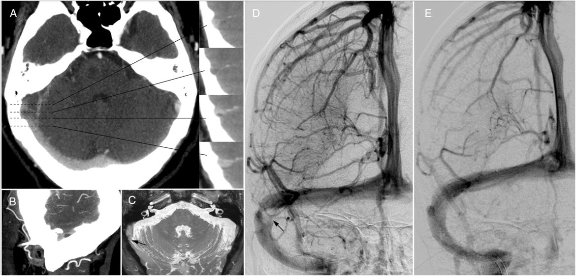
A 61-year-old man was referred to the neurology clinic due to a persistent, disabling, right-sided pulsatile tinnitus (PT), which he noticed 4 years before and had recently worsened. The tinnitus ameliorated with manual pressure over the ipsilateral cervical region and cervical flexion to the right side. With the exception of an audible high-pitch bruit below the right mastoid process, the neurological examination was unremarkable. After careful evaluation, computed tomography angiography and magnetic resonance imaging showed a stenosis in the right lateral venous sinus (Figure 1A–C). Lumbar puncture revealed an intracranial pressure of 12 cmH2O. The presence of a stenosis-associated pressure gradient was confirmed in digital subtraction angiography (9 mmHg), by using a co-axial system consisting of a NeuronMax 0.88 long sheath (placed in the right internal jugular vein) and a 0.021” microcatheter positioned on the superior sagittal sinus (Prowler Select Plus). Following multidisciplinary discussion and patient agreement, recalibration of the venous sinus was successfully performed with a nontapered X-Act stent (9 x 30 mm) (Figure 1D–E), after progression along a microguidewire Synchro 0.014”. Tinnitus resolved immediately after the procedure, and it had not recurred at the 6-month follow-up.

Figure 1: Computed tomography angiography shows a right lateral sinus stenosis in axial, coronal (A) and sagittal (B) reconstructions, caused by a probable Pacchioni’s arachnoid granulation. Magnetic resonance imaging (CISS sequence) also shows the right lateral sinus stenosis (C, arrow). Digital subtraction angiography confirms a stenosis in the right lateral venous sinus (D, arrow), where a pressure gradient of 9 mmHg was quantified, and normal sinus calibre re-establishment after stenting, with no residual pressure gradient (E).
Only 10% of all patients with tinnitus describe it as pulsatile in nature.Reference Hofmann, Behr, Neumann-Haefelin and Schwager 1 There are multiple causes for PT, namely, intracranial or cervical arterial/venous/arteriovenous abnormalities, vascularized tumors in the vicinity of the ear, intracranial hypertension, and semicircular canal dehiscence, which must be carefully searched for.Reference Levine and Oron 2 Venous origin may be suspected when PT decreases or resolves with ipsilateral venous compression, valsalva maneuvers and rotation of the head toward the affected side, and increases with contralateral venous compression and rotation of the head to the contralateral side. Lateral venous sinus stenosis has long been recognized as a cause for isolated PT.Reference Russel, Michaelis, Wiet and Meyer 3 Even though underlying pathophysiological mechanisms are unclear, a significant trans-stenotic pressure gradient is probably required so that when systolic arterial pulses occur, the cerebral veins and sinuses are rhythmically compressed, leading to an increased, turbulent trans-stenotic venous flow. Venous sinus stenting has gained acceptance as treatment option for patients with intracranial hypertension and venous sinus stenosis, with improved papilledema, headache, and PT outcomes.Reference Nicholson, Brinjikji and Radovanovic 4 However, for patients with isolated PT with no intracranial hypertension, the benefit of stenting and the peri- and postprocedural management protocols are not completely established, even though a small patient series show promising results.Reference Baomin, Yongbing and Xiangyu 5 , Reference Levitt, Albuquerque and Gross 6
Venous sinus stenosis needs to be considered in the differential workup of isolated PT, namely, when the characteristics of the tinnitus suggest a venous origin. Careful evaluation of the venous sinuses using angiographic methods may reveal inconspicuous stenosis, and endovascular treatment with stenting may be considered in selected cases.
Ethics Information
This study complies with the Declaration of Helsinki and was conducted in accordance to the local ethics committee requirements. No figures or videos contain information which allows patient identification; therefore, written patient consent for the publication was not obtained.
Authors’ Disclosures
None.
Statement of Authorship
The acquisition of data, clinical and imaging data review, literature review, and final manuscript writing were done by MQN. The clinical data review and literature review were done by EF. The acquisition of data, imaging data review, and literature review were done by JMA. The imaging data review and important intellectual contribution were done by JR. The clinical data review, literature review, and final manuscript writing were done by JP.


