Introduction
Cardiac emboli account for 20% of all ischemic strokes, with atrial fibrillation being the most important cause of cardioembolism.Reference Sandercock, Warlow, Jones and Starkey 1 , Reference Wolf, Abbott and Kannel 2 Previous studies have shown that strokes resulting in cortical deficits (i.e., visual field abnormalities, neglect, aphasia) are strongly associated with a potential cardiac source of embolism.Reference Bogousslavsky, Cachin and Regli 3 , Reference Kittner, Sharkness and Sloan 4 Language deficits are the most common type of higher cortical deficit to occur after acute stroke, and the finding of aphasia appears to be particularly specific for cardioembolism.Reference Kittner, Sharkness and Sloan 4 , Reference Hoffmann, Sacco, Mohr and Tatemichi 5 Indeed, most strokes resulting in aphasia are caused by an embolism of cardiac origin.Reference Hanlon, Lux and Dromerick 6 - Reference Levine, Dulli and Dixit 8 It is unclear whether the same is true for isolated aphasia during transient ischemic attack (TIA).
Strokes of cardioembolic origin also have a high rate of recurrence, as do TIAs, which have a 10% risk of stroke at 90 days, with half of these events occurring within 48 hours.Reference Gladstone, Kapral and Fang 9 - Reference Kolominsky-Rabas, Weber and Gefeller 11 As such, cardioembolism provides an important and time-sensitive opportunity for secondary stroke prevention. At present, the immediate risk of stroke after a TIA characterized by isolated language disturbance is not well known.
Among patients diagnosed in the emergency department with TIA, we hypothesized that patients presenting with isolated aphasia would be more likely to have a cardiac source of embolism. We also hypothesized that such patients with isolated aphasia would have a higher short-term risk of stroke.
Methods
Study design and setting
This study is a secondary analysis of a larger, prospective cohort study conducted at eight Canadian emergency departments as part of the derivation of a clinical decision rule to predict imminent stroke.Reference Perry, Sharma and Sivilotti 12 Ethics board approval was obtained at all participating sites. The combined annual census at all participating sites was 394,000 visits.
Selection of participants
We prospectively enrolled adult patients with a final emergency department diagnosis of TIA between October 2006 and April 2010. Details regarding patient selection and data collection for this population have been previously described.Reference Perry, Sharma and Sivilotti 12 Patients were excluded for 1) neurological deficit>24 hours at presentation, 2) decreased level of consciousness (i.e., Glasgow Coma Scale score of<15), 3) obvious alternate cause for the deficit other than TIA (e.g., hypoglycemia, seizure, electrolyte imbalance, migraine), 4) receiving tissue plasminogen activator, or 5) presentation to the enrolling emergency department>7 days after TIA. The diagnosis of TIA was defined using the World Health Organization definition: a sudden, focal neurological deficit lasting for less than 24 hours, presumed to be of vascular origin, and confined to an area of the brain or eye perfused by a specific artery. A standardized data collection form was completed by the emergency physician at the time of presentation, and the following information was prospectively collected: 1) the history of neurological symptoms, 2) physical findings, 3) electrocardiogram results, and 4) past medical history. For this study, only patients with persistent deficits at the time of presentation were included.
Identification of patients with isolated aphasia
Treating physicians were explicitly asked to identify the presence or absence of language disturbance on the history section of the study data form, and of speech difficulty on the physical examination section. When speech difficulty was identified, physicians were then asked to characterize this as aphasia or dysarthria, and with the explicit definition (aphasia=loss of ability to use written/oral language; dysarthria=difficulty articulating) appearing next to the item on the form. For the current study, we defined isolated aphasia to be those in whom the language deficit was the only neurological deficit identified on physical examination, whether expressive or receptive. The patient complaint of language disturbance on history was insufficient to classify the patient as having aphasia for the purposes of this study. Accordingly, individuals demonstrating normal language comprehension and expression at the time of initial examination by physician were not deemed to be aphasic.
Outcome and outcome assessment
The primary outcome for this analysis was the identification of a potential cardioembolic source for the TIA. Potential cardiac sources for embolism were defined as either a previous diagnosis of atrial fibrillation; atrial fibrillation identified by electrocardiogram, Holter monitor, or echocardiography; or thrombus seen at echocardiography. Secondary outcomes were the occurrence of stroke at 2, 7, 30, and 90 days. All participants were followed using medical record review and a validated, standardized telephone questionnaire.Reference Jones, Williams and Meschia 13 Potential subsequent strokes identified in follow-up were adjudicated by a panel blinded to the initial presentation, unless the subsequent stroke was diagnosed by the treating neurologist, in which case it was automatically deemed an outcome event.
Investigations
As part of routine clinical care, all patients had an electrocardiogram performed at the index visit in the emergency department, and most were referred to a dedicated stroke clinic where they generally received the existing standard of care, the details of which have been described previously.Reference Wasserman, Perry and Dowlatshahi 14 Forty-eight-hour Holter monitoring and echocardiography were arranged at either the index visit or subsequently through the stroke clinic and were performed on an outpatient basis.
Statistical analysis
Data were collected and analysed in SPSS 17 (SPSS Inc., Chicago, IL). Proportions were compared using Fisher’s exact test with confidence intervals (CIs) reported at 95%. Continuous data are presented as mean±standard deviation (SD) where applicable. All tests were two-sided, and results were considered significant if p<0.05.
Results
A total of 2,360 TIA patients were enrolled in the study, and 1,155 patients diagnosed as having a TIA presented with deficits and were included in the final analysis. Of these patients, 41 were classified as having isolated aphasia at the time of presentation (Figure 1). Patients with isolated aphasia were older (73.9±10.0 v. 67.2±14.5 years; p=0.003) and were more likely to have a history of heart failure (9.8% v. 2.6%; p=0.027) (Table 1).

Figure 1 Patient flow.
Table 1 Patient Characteristics of Transient Ischemic Attack Patients with Neurological Deficits at the Time of Emergency Physician Assessment

The investigations used to identify potential cardiac sources of embolism are shown in Table 2. Patients with isolated aphasia were twice as likely to have any cardiac source of embolism (22.0% v. 10.6%; p=0.037). Specifically, patients with isolated aphasia were more likely than patients without isolated aphasia to have atrial fibrillation identified on electrocardiogram (12.2% v. 4.3%; p=0.036) or Holter monitor (4.9% v. 0.7%; p=0.046). The average delay from presentation to investigation was 27.1±21.1 days for Holter monitor and 13.1±14.4 days for echocardiography. All electrocardiograms were performed at the time of presentation.
Table 2 Potential Cardiac Sources of Embolism

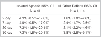
The overall stroke rate in the study cohort at 2, 7, 30, and 90 days was 1.7% (CI 1.1% to 2.7%), 2.5% (CI 1.7% to 3.6%), 3.2% (CI 2.3% to 4.4%), and 3.9% (CI 2.9% to 5.2%), respectively. A comparison of the stroke rate between patients with and without isolated aphasia is shown in Table 3. Patients with isolated aphasia had a higher rate of stroke than patients without isolated aphasia at 2 days, although the differences between the two groups were not significant.
Table 3 Stroke Rate Among Patients With And Without Transient Isolated Aphasia

Discussion
In this prospective cohort study, we demonstrated that TIA patients presenting with isolated aphasia have twice the rate of potential cardiac sources of embolism than patients with other forms of TIA. Patients with transient isolated aphasia were also more likely to have a stroke in the next 90 days, although this difference did not reach statistical significance. Accordingly, the finding of isolated aphasia on physical examination may allow the physician to rapidly identify patients with a potential cardiac source of embolism who are also at increased risk of stroke.
Language disturbance can result from dysfunction of any of the following structures in the dominant hemisphere: the superior temporal gyrus, the inferior frontal gyrus and adjacent subcortical white matter, the pre- or post-central gyrus, or the thalamus. Aphasia commonly occurs after occlusion of the middle cerebral artery or one of its branches, but it has also been observed after occlusion of the posterior cerebral artery.Reference Maulaz, Bezerra and Bogousslavsky 15 Regardless of the territory involved, cardioembolism is the most common cause of language disturbance,Reference Maulaz, Bezerra and Bogousslavsky 15 , Reference Ferro 16 including isolated Broca’s or Wernicke’s aphasia,Reference Knepper, Biller and Tranel 7 , Reference Levine, Dulli and Dixit 8 and the relatively uncommon syndrome of global aphasia without hemiparesis.Reference Hanlon, Lux and Dromerick 6 We found that, in patients with persistent deficits at time of initial emergency presentation, nearly 1 in 4 patients with transient isolated aphasia had a potential cardiac source of embolism compared to 1 in 10 patients with other forms of TIA. Accordingly, isolated aphasia appears to be a strong predictor of a potential cardiac source of embolism.
Atrial fibrillation is an increasingly common cardiac arrhythmia and is the leading cause of cardioembolism to the brain.Reference Wolf, Abbott and Kannel 2 Oral anticoagulation therapy markedly reduces the risk of stroke, yet many patients with paroxysmal atrial fibrillation go undetected and untreated for years.Reference Wann, Curtis and January 17 A recent systematic review of noninvasive cardiac monitoring after stroke or TIA found that 5% of patients had previously undiagnosed atrial fibrillation.Reference Liao, Khalid and Scallan 18 More recently, Sposato et al. used continuous cardiac monitoring initiated immediately after stroke or TIA to show that 13.5% of patients had previously undiagnosed atrial fibrillation.Reference Sposato, Klein and Jauregui 19 Similarly, Fennis et al. recently analysed the results of two large clinical trials and found that patients diagnosed with TIA and presenting with isolated aphasia were more likely to have a history of atrial fibrillation.Reference Fennis, Compter and van den Broek 20 The results of the present study confirm the strong association between isolated aphasia and a history of atrial fibrillation. However, we also now show that one-third of patients with isolated aphasia and a potential cardiac source of embolism did not have a known history of atrial fibrillation. The high rate of undiagnosed atrial fibrillation in patients presenting with isolated aphasia suggests that urgent screening for a potential cardiac source of embolism is warranted.
Historically, TIA has been found to have a 2-day stroke risk of 5% and a 90-day stroke risk of approximately 10%, making this presentation an important but time-sensitive opportunity for stroke prevention.Reference Gladstone, Kapral and Fang 9 , Reference Johnston, Gress, Browner and Sidney 10 The rapid initiation of antiplatelet agents and early assessment in a dedicated stroke prevention clinic can cut this risk dramatically, and both interventions were used in our cohort,Reference Wasserman, Perry and Dowlatshahi 14 , Reference Lavallee, Meseguer and Abboud 21 , Reference Rothwell, Giles and Chandratheva 22 as evidenced by a relatively low overall rate of stroke. Despite these interventions, patients with isolated aphasia in this study had double the rate of stroke at all time points assessed and a 7.3% rate of stroke at 90 days. Although our study lacked sufficient power to demonstrate whether this clinically important difference was due to chance alone, it would not be surprising that antiplatelet agents and urgent investigation, including carotid imaging, Holter monitoring, and echocardiography would be less likely to reduce the immediate risk of stroke after TIA due to cardioembolism. Antiplatelet agents, such as aspirin, are less effective at preventing stroke caused by cardioembolism.Reference Aguilar, Hart and Pearce 23 , Reference Hart, Pearce and Koudstaal 24 Furthermore, ischemic strokes of cardioembolic origin have the highest rate of recurrence,Reference Kolominsky-Rabas, Weber and Gefeller 11 and the same may be true for patients with TIA. Speech disturbance is a component of the ABCDReference Wolf, Abbott and Kannel 2 score, a tool developed to predict the risk of stroke after TIA,Reference Johnston, Rothwell and Nguyen-Huynh 25 even if an independent verification analysis has found this score to be inaccurate and nonspecific.Reference Perry, Sharma and Sivilotti 12 It is possible that the accuracy of scoring tools may be improved by including aphasia, only when it occurs in the absence of other deficits or studying other interactions between components of the score.
The diagnosis of TIA in the present study was based upon the treating physician’s discharge diagnosis in the emergency department. Some investigators have reported a false diagnosis rate of up to 60% in one U.S. study of inpatients initially assessed by an emergency physician, including those with gradual onset and nonspecific neurological symptoms.Reference Prabhakaran, Silver and Warrior 26 These results are not unexpected given the transient nature of the symptoms and the fact that many patients present at a time when their symptoms have already started to resolve. We believe that, given the specific nature of isolated aphasia, the subsequent stroke risk, and the explicit, prospective identification of neurological signs and symptoms as well as other elements of our study, the large majority of patients in our cohort had a TIA.
Limitations
This study has several limitations. First, for many patients, there was a substantial delay of at least 1 week between the index event and subsequent echocardiography and Holter monitoring. Thus, the diagnostic yield and benefits of earlier investigation for potential cardiac source of embolism immediately after the index event cannot be estimated. Moreover, only 25% and 70% of all patients received Holter monitoring or echocardiography, respectively. Accordingly, we likely underestimated the true number of patients with a potential cardiac source of embolism. Second, the definition of TIA in this study was time-based, whereas the recently revised definition is tissue-based.Reference Albers, Caplan and Easton 27 Because most patients in this study did not receive an MRI within the first 24 hours, we could not assess for acute infarction. Thus, some patients diagnosed with a TIA may be better classified as transient symptoms with infarction.Reference Ay, Koroshetz and Benner 28
Conclusions
Cardioembolism is a leading cause of stroke, and identifying patients requiring urgent evaluation is critical.Reference Sandercock, Warlow, Jones and Starkey 1 In this study, nearly one-quarter of all patients presenting with transient isolated aphasia had a potential cardiac source of embolism, double the rate found in patients with other forms of TIA, and persistent neurological findings at initial evaluation. Most of these patients had atrial fibrillation, and in one-third this diagnosis was new. Importantly, patients with isolated aphasia were also more likely to have a stroke in the next 90 days. Taken together, these results support the rapid evaluation and treatment of all patients with transient isolated aphasia for potential cardiac sources of embolism in order to prevent subsequent stroke.
Acknowledgements
We would like to thank the following authors for their contributions: Jason Wasserman and Jeffrey Perry conceived the idea. Jason Wasserman prepared the manuscript. Jane Sutherland coordinated the study and collected data. Mukul Sharma and Ian Stiell provided much input into the study design and revision of the manuscript. Marco Sivilotti, Marcel Émond, Andrew Worster, and Dar Dowlatshahi assisted with study design, oversaw the collection of data at their respective sites, and critically revised the manuscript.
Competing interests: The authors would like to supply their funding source: Canadian Institutes of Health Research (Grant 172524).


