Skip to main content Accessibility help
×
Hostname: page-component-78c5997874-j824f Total loading time: 0 Render date: 2024-11-05T15:40:00.567Z Has data issue: false hasContentIssue false

Chapter 17 - The Use of EEG and aEEG in Assessing the Term and Preterm Brain

from Section 3 - Diagnosis of the Infant with Brain Injury

Published online by Cambridge University Press:  13 December 2017

David K. Stevenson
Affiliation:
Stanford University, California
William E. Benitz
Affiliation:
Stanford University, California
Philip Sunshine
Affiliation:
Stanford University, California
Susan R. Hintz
Affiliation:
Stanford University, California
Maurice L. Druzin
Affiliation:
Stanford University, California
Get access

Summary

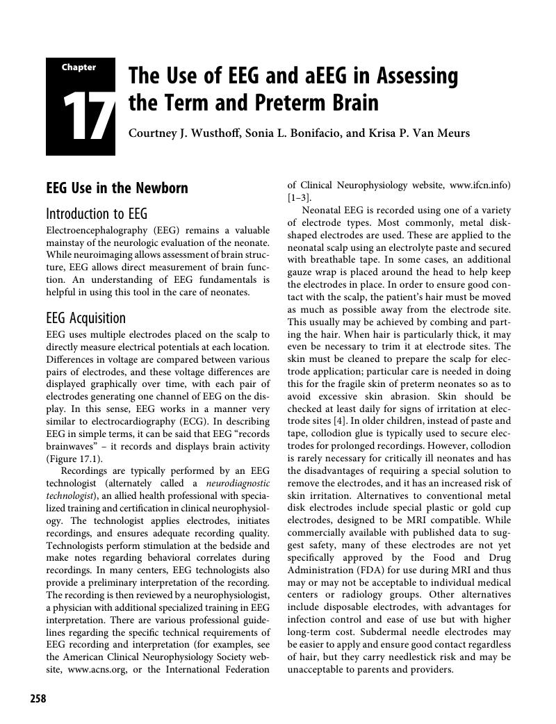
Image of the first page of this content. For PDF version, please use the ‘Save PDF’ preceeding this image.'
Type
Chapter
Information
Publisher: Cambridge University Press
Print publication year: 2017

Access options

Get access to the full version of this content by using one of the access options below. (Log in options will check for institutional or personal access. Content may require purchase if you do not have access.)

References

Guideline II: Minimum technical standards for pediatric electroencephalography, April 2016. Available at www.acns.org/practice/guidelines.Google Scholar
Shellhaas, RA, Chang, T, Tsuchida, T, et al. The American Clinical Neurophysiology Society’s guideline on continuous electroencephalography monitoring in neonates. J Clin Neurophysiol 2011; 28(6): 611–7.Google Scholar
Tsuchida, TN, Wusthoff, CJ, Shellhaas, RA, et al. American Clinical Neurophysiology Society standardized EEG terminology and categorization for the description of continuous EEG monitoring in neonates: report of the American Clinical Neurophysiology Society critical care monitoring committee. J Clin Neurophysiol 2013; 30(2): 161–73.Google Scholar
Neurodiagnostic Society. Skin safety during EEG procedures, April 2016. Available at www.aset.org/i4a/pages/index.cfm?pageid=4134.Google Scholar
Klem, GH, Lüders, HO, Jasper, HH, Elger, C. The ten-twenty electrode system of the International Federation. The International Federation of Clinical Neurophysiology. Electroencephalogr Clin Neurophysiol Suppl 1999; 52: 36.Google Scholar
World Health Organization (WHO). Guidelines on neonatal seizures, WHO Guidelines Approved by the Guidelines Review Committee, 2011. Available at www.ncbi.nlm.nih.gov/books/NBK304092/.Google Scholar
Murray, DM, Boylan, GB, Ali, I, et al. Defining the gap between electrographic seizure burden, clinical expression and staff recognition of neonatal seizures. Arch Dis Child Fetal Neonatal Ed 2008; 93(3): F187–91.Google Scholar
Clancy, RR, Legido, A, Lewis, D. Occult neonatal seizures. Epilepsia 1988; 29(3): 256–61.Google Scholar
Glass, HC, Wusthoff, CJ, Shellhaas, RA, et al. Risk factors for EEG seizures in neonates treated with hypothermia: a multicenter cohort study. Neurology 2014; 82(14): 1239–44.Google Scholar
Wusthoff, CJ, Dlugos, DJ, Gutierrez-Colina, A, et al. Electrographic seizures during therapeutic hypothermia for neonatal hypoxic-ischemic encephalopathy. J Child Neurol. 2011; 26(6): 724–8.Google Scholar
Committee on Fetus and Newborn, Papile, L-A, Baley, JE, et al. Hypothermia and neonatal encephalopathy. Pediatrics 2014; 133(6): 1146–50.Google Scholar
Scher, MS, Alvin, J, Gaus, L, et al. Uncoupling of EEG-clinical neonatal seizures after antiepileptic drug use. Pediatr Neurol 2003; 28(4): 277–80.Google Scholar
Tsuchida, TN. EEG background patterns and prognostication of neonatal encephalopathy in the era of hypothermia. J Clin Neurophysiol 2013; 30(2): 122–5.Google Scholar
Bye, AM, Lee, D, Naidoo, D, Flanagan, D. The effects of morphine and midazolam on EEGs in neonates. J Clin Neurosci 1997; 4(2): 173–5.Google Scholar
Young, GB, da Silva, OP. Effects of morphine on the electroencephalograms of neonates: a prospective, observational study. Clin Neurophysiol 2000; 111(11): 1955–60.Google Scholar
Le Bihannic, A, Beauvais, K, Busnel, A, et al. Prognostic value of EEG in very premature newborns. Arch Dis Child Fetal Neonatal Ed 2012; 97(2): F106–9.Google Scholar
Abend, NS, Wusthoff, CJ. Neonatal seizures and status epilepticus. J Clin Neurophysiol 2012; 29(5): 441–8.Google Scholar
Hrachovy, RA, Mizrahi, EM. Atlas of Neonatal Electroencephalography, 4th edn. New York: Demos Medical, 2015.Google Scholar
Clancy, R, Wusthoff, C. Brain Monitoring: Normal Neonatal EEG. Ambler, PA: Moberg Multimedia, 2011.Google Scholar
Hellström-Westas, L, Rosén, I, Vries, LS, Greisen, G. Amplitude-integrated EEG classification and interpretation in preterm and term infants. NeoReviews 2006; 7(2): e7687.Google Scholar
al Naqeeb, N, Edwards, AD, Cowan, FM, Azzopardi, D. Assessment of neonatal encephalopathy by amplitude-integrated electroencephalography. Pediatrics 1999; 103(6 Pt 1): 1263–71.Google Scholar
Bjerre, I, Hellström-Westas, L, Rosén, I, Svenningsen, N. Monitoring of cerebral function after severe asphyxia in infancy. Arch Dis Child 1983; 58(12): 9971002.Google Scholar
Hellström-Westas, L, Rosén, I, Svenningsen, NW. Predictive value of early continuous amplitude integrated EEG recordings on outcome after severe birth asphyxia in full term infants. Arch Dis Child Fetal Neonatal Ed 1995; 72(1): F34–8.Google Scholar
Hellström-Westas, L, Rosén, I, Swenningsen, NW. Silent seizures in sick infants in early life: diagnosis by continuous cerebral function monitoring. Acta Paediatr Scand 1985; 74(5): 741–8.Google Scholar
Bonifacio, SL, Glass, HC, Peloquin, S, Ferriero, DM. A new neurological focus in neonatal intensive care. Nat Rev Neurol 2011; 7(9): 485–94.Google Scholar
Toet, MC, van der Meij, W, de Vries, LS, et al. Comparison between simultaneously recorded amplitude integrated electroencephalogram (cerebral function monitor) and standard electroencephalogram in neonates. Pediatrics. 2002; 109(5): 772–9.Google Scholar
Shellhaas, RA, Soaita, AI, Clancy, RR. Sensitivity of amplitude-integrated electroencephalography for neonatal seizure detection. Pediatrics 2007; 120(4): 770–7.Google Scholar
Hellström-Westas, L, Rosén, I. Continuous brain-function monitoring: state of the art in clinical practice. Semin Fetal Neonatal Med 2006; 11(6): 503–11.Google Scholar
Norman, E, Wikström, S, Rosén, I, et al. Premedication for intubation with morphine causes prolonged depression of electrocortical background activity in preterm infants. Pediatr Res 2013; 73(1): 8794.Google Scholar
Hellström-Westas, L, Bell, AH, Skov, L, et al. Cerebroelectrical depression following surfactant treatment in preterm neonates. Pediatrics 1992; 89(4 Pt 1): 643–7.Google Scholar
Merchant, N, Azzopardi, D. Early predictors of outcome in infants treated with hypothermia for hypoxic-ischaemic encephalopathy. Dev Med Child Neurol 2015; 57: 816.Google Scholar
Thoresen, M, Hellström-Westas, L, Liu, X, de Vries, LS. Effect of hypothermia on amplitude-integrated electroencephalogram in infants with asphyxia. Pediatrics 2010; 126(1): e131–9.Google Scholar
Gluckman, PD, Wyatt, JS, Azzopardi, D, et al. Selective head cooling with mild systemic hypothermia after neonatal encephalopathy: multicentre randomised trial. Lancet 2005; 365(9460): 663–70.Google Scholar
Azzopardi, DV, Strohm, B, Edwards, AD, et al. Moderate hypothermia to treat perinatal asphyxial encephalopathy. N Engl J Med 2009; 361(14): 1349–58.Google Scholar
Toet, MC, Hellström-Westas, L, Groenendaal, F, et al. Amplitude integrated EEG 3 and 6 hours after birth in full term neonates with hypoxic-ischaemic encephalopathy. Arch Dis Child Fetal Neonatal Ed 1999; 81(1): F1923.Google Scholar
Bonifacio, SL, deVries, LS, Groenendaal, F. Impact of hypothermia on predictors of poor outcome: how do we decide to redirect care? Semin Fetal Neonatal Med 2015; 20(2): 122–7.Google Scholar
Abend, NS, Wusthoff, CJ, Goldberg, EM, Dlugos, DJ. Electrographic seizures and status epilepticus in critically ill children and neonates with encephalopathy. Lancet Neurol. 2013; 12(12): 1170–9.Google Scholar
Nash, KB, Bonifacio, SL, Glass, HC, et al. Video-EEG monitoring in newborns with hypoxic-ischemic encephalopathy treated with hypothermia. Neurology 2011; 76(6): 556–62.Google Scholar
Shah, DK, Wusthoff, CJ, Clarke, P, et al. Electrographic seizures are associated with brain injury in newborns undergoing therapeutic hypothermia. Arch Dis Child Fetal Neonatal Ed 2014; 99(3): F219–24.Google Scholar
Rakshasbhuvankar, A, Paul, S, Nagarajan, L, et al. EEG for detection of neonatal seizures: a systematic review. Seizure. 2015; 33: 90–8.Google Scholar
Olischar, M, Shany, E, Aygün, C, et al. Amplitude-integrated electroencephalography in newborns with inborn errors of metabolism. Neonatology 2012; 102(3): 203–11.Google Scholar
Tekgunduz, , Caner, I, Eras, Z, et al. Prognostic value of amplitude-integrated electroencephalography in neonates with hypernatremic dehydration. J Matern-Fetal Neonatal Med 2014; 27(7): 658–63.Google Scholar
ter Horst, HJ, van Olffen, M, Remmelts, HJ, et al. The prognostic value of amplitude integrated EEG in neonatal sepsis and/or meningitis. Acta Paediatr Oslo Nor 2010; 99(2): 194200.Google Scholar
Verboon-Maciolek, MA, Groenendaal, F, Hahn, CD, et al. Human parechovirus causes encephalitis with white matter injury in neonates. Ann Neurol 2008; 64(3): 266–73.Google Scholar
Kidokoro, H, Kubota, T, Hayashi, N, et al. Absent cyclicity on aEEG within the first 24 h is associated with brain damage in preterm infants. Neuropediatrics 2010; 41(6): 241–5.Google Scholar
Wood, NS, Costeloe, K, Gibson, AT, et al. The EPICure study: associations and antecedents of neurological and developmental disability at 30 months of age following extremely preterm birth. Arch Dis Child Fetal Neonatal Ed 2005; 90(2): F134–40.Google Scholar
Hintz, SR, Kendrick, DE, Wilson-Costello, DE, et al. Early-childhood neurodevelopmental outcomes are not improving for infants born at <25 weeks’ gestational age. Pediatrics 2011; 127(1): 6270.Google Scholar
Hellström-Westas, L. Comparison between tape-recorded and amplitude-integrated EEG monitoring in sick newborn infants. Acta Paediatr Oslo Nor 1992; 81(10): 812–9.Google Scholar
Olischar, M, Klebermass, K, Kuhle, S, et al. Reference values for amplitude-integrated electroencephalographic activity in preterm infants younger than 30 weeks’ gestational age. Pediatrics 2004; 113(1 Pt 1):e61–6.Google Scholar
Selton, D, Andre, M, Hascoët, JM. Normal EEG in very premature infants: reference criteria. Clin Neurophysiol 2000; 111(12): 2116–24.Google Scholar
Hayakawa, M, Okumura, A, Hayakawa, F, et al. Background electroencephalographic (EEG) activities of very preterm infants born at less than 27 weeks gestation: a study on the degree of continuity. Arch Dis Child Fetal Neonatal Ed 2001; 84(3): F163–7.Google Scholar
Sisman, J, Campbell, DE, Brion, LP. Amplitude-integrated EEG in preterm infants: maturation of background pattern and amplitude voltage with postmenstrual age and gestational age. J Perinatol 2005; 25(6): 391–6.Google Scholar
Klebermass, K, Kuhle, S, Olischar, M, et al. Intra- and extrauterine maturation of amplitude-integrated electroencephalographic activity in preterm infants younger than 30 weeks of gestation. Biol Neonate 2006; 89(2): 120–5.Google Scholar
Burdjalov, VF, Baumgart, S, Spitzer, AR. Cerebral function monitoring: a new scoring system for the evaluation of brain maturation in neonates. Pediatrics 2003; 112(4): 855–61.Google Scholar
Kuhle, S, Klebermass, K, Olischar, M, et al. Sleep-wake cycles in preterm infants below 30 weeks of gestational age: preliminary results of a prospective amplitude-integrated EEG study. Wien Klin Wochenschr. 2001; 113(7–8): 219–23.Google Scholar
Hellström-Westas, L, Klette, H, Thorngren-Jerneck, K, Rosén, I. Early prediction of outcome with aEEG in preterm infants with large intraventricular hemorrhages. Neuropediatrics 2001; 32(6): 319–24.Google Scholar
Aso, K, Abdab-Barmada, M, Scher, MS. EEG and the neuropathology in premature neonates with intraventricular hemorrhage. J Clin Neurophysiol 1993; 10(3): 304–13.Google Scholar
Olischar, M, Klebermass, K, Waldhoer, T, et al. Background patterns and sleep-wake cycles on amplitude-integrated electroencephalography in preterms younger than 30 weeks gestational age with peri-/intraventricular haemorrhage. Acta Paediatr Oslo Nor 2007; 96(12): 1743–50.Google Scholar
Chalak, LF, Sikes, NC, Mason, MJ, Kaiser, JR. Low-voltage aEEG as predictor of intracranial hemorrhage in preterm infants. Pediatr Neurol 2011; 44(5): 364–9.Google Scholar
Olischar, M, Klebermass, K, Kuhle, S, et al. Progressive posthemorrhagic hydrocephalus leads to changes of amplitude-integrated EEG activity in preterm infants. Childs Nerv Syst 2004; 20(1): 41–5.Google Scholar
Olischar, M, Klebermass, K, Hengl, B, et al. Cerebrospinal fluid drainage in posthaemorrhagic ventricular dilatation leads to improvement in amplitude-integrated electroencephalographic activity. Acta Paediatr Oslo Nor 2009; 98(6): 1002–9.Google Scholar
Klebermass-Schrehof, K, Rona, Z, Waldhör, T, et al. Can neurophysiological assessment improve timing of intervention in posthaemorrhagic ventricular dilatation? Arch Dis Child Fetal Neonatal Ed 2013; 98(4): F291–7.Google Scholar
de Vries, LS, Liem, KD, van Dijk, K, et al. Early versus late treatment of posthaemorrhagic ventricular dilatation: results of a retrospective study from five neonatal intensive care units in the Netherlands. Acta Paediatr Oslo Nor 2002; 91(2): 212–7.Google Scholar
Song, J, Zhu, C, Xu, F, et al. Predictive value of early amplitude-integrated electroencephalography for later diagnosed cerebral white matter damage in preterm infants. Neuropediatrics 2014; 45(5): 314–20.Google Scholar
Song, J, Xu, F, Wang, L, et al. Early amplitude-integrated electroencephalography predicts brain injury and neurological outcome in very preterm infants. Sci Rep 2015; 5: 13810.Google Scholar
Wikström, S, Pupp, IH, Rosén, I, et al. Early single-channel aEEG/EEG predicts outcome in very preterm infants. Acta Paediatr Oslo Nor 2012; 101(7): 719–26.Google Scholar
Klebermass, K, Olischar, M, Waldhoer, T, et al. Amplitude-integrated EEG pattern predicts further outcome in preterm infants. Pediatr Res 2011; 70(1): 102–8.Google Scholar
Welch, C, Helderman, J, Williamson, E, O’Shea, TM. Brain wave maturation and neurodevelopmental outcome in extremely low gestational age neonates. J Perinatol 2013; 33(11): 867–71.Google Scholar
Natalucci, G, Leuchter, RH-V, Bucher, HU, et al. Functional brain maturation assessed during early life correlates with anatomical brain maturation at term-equivalent age in preterm infants. Pediatr Res 2013; 74(1): 6874.Google Scholar
Greisen, G, Pryds, O. Low CBF, discontinuous EEG activity, and periventricular brain injury in ill, preterm neonates. Brain Dev 1989; 11(3): 164–8.Google Scholar
Van Os, S, Klaessens, J, Hopman, J, et al. Preservation of electrocortical brain activity during hypoxemia in preterm lambs. Exp Brain Res 2003; 151(1): 54–9.Google Scholar
West, CR, Groves, AM, Williams, CE, et al. Early low cardiac output is associated with compromised electroencephalographic activity in very preterm infants. Pediatr Res 2006; 59(4 Pt 1): 610–5.Google Scholar
Shah, D, Paradisis, M, Bowen, JR. Relationship between systemic blood flow, blood pressure, inotropes, and aEEG in the first 48 h of life in extremely preterm infants. Pediatr Res 2013; 74(3): 314–20.Google Scholar
Lemmers, PMA, Toet, MC, van Bel, F. Impact of patent ductus arteriosus and subsequent therapy with indomethacin on cerebral oxygenation in preterm infants. Pediatrics 2008; 121(1): 142–7.Google Scholar
Evans, N, Kluckow, M. Early ductal shunting and intraventricular haemorrhage in ventilated preterm infants. Arch Dis Child Fetal Neonatal Ed 1996; 75(3): F183–6.Google Scholar
Miller, SP, Mayer, EE, Clyman, RI, et al. Prolonged indomethacin exposure is associated with decreased white matter injury detected with magnetic resonance imaging in premature newborns at 24 to 28 weeks’ gestation at birth. Pediatrics 2006; 117(5): 1626–31.Google Scholar
Kabra, NS, Schmidt, B, Roberts, RS, et al. Neurosensory impairment after surgical closure of patent ductus arteriosus in extremely low birth weight infants: results from the Trial of Indomethacin Prophylaxis in Preterms. J Pediatr 2007; 150(3): 229–34, 234.e1.Google Scholar
Lemmers, PMA, Molenschot, MC, Evens, J, et al. Is cerebral oxygen supply compromised in preterm infants undergoing surgical closure for patent ductus arteriosus? Arch Dis Child Fetal Neonatal Ed 2010; 95(6): F429–34.Google Scholar
Leslie, ATFS, Jain, A, El-Khuffash, A, et al. Evaluation of cerebral electrical activity and cardiac output after patent ductus arteriosus ligation in preterm infants. J Perinatol 2013; 33(11): 861–6.Google Scholar
Helderman, JB, Welch, CD, Leng, X, O’Shea, TM. Sepsis-associated electroencephalographic changes in extremely low gestational age neonates. Early Hum Dev 2010; 86(8): 509–13.Google Scholar

Save book to Kindle

To save this book to your Kindle, first ensure [email protected] is added to your Approved Personal Document E-mail List under your Personal Document Settings on the Manage Your Content and Devices page of your Amazon account. Then enter the ‘name’ part of your Kindle email address below. Find out more about saving to your Kindle.

Note you can select to save to either the @free.kindle.com or @kindle.com variations. ‘@free.kindle.com’ emails are free but can only be saved to your device when it is connected to wi-fi. ‘@kindle.com’ emails can be delivered even when you are not connected to wi-fi, but note that service fees apply.

Find out more about the Kindle Personal Document Service.

Available formats
×

Save book to Dropbox

To save content items to your account, please confirm that you agree to abide by our usage policies. If this is the first time you use this feature, you will be asked to authorise Cambridge Core to connect with your account. Find out more about saving content to Dropbox.

Available formats
×

Save book to Google Drive

To save content items to your account, please confirm that you agree to abide by our usage policies. If this is the first time you use this feature, you will be asked to authorise Cambridge Core to connect with your account. Find out more about saving content to Google Drive.

Available formats
×