Skip to main content Accessibility help
×
Hostname: page-component-78c5997874-lj6df Total loading time: 0 Render date: 2024-11-09T05:47:46.675Z Has data issue: false hasContentIssue false

7 - Breast applications of diffusion-weighted MRI

Published online by Cambridge University Press:  10 November 2010

Bachir Taouli
Affiliation:
Mount Sinai School of Medicine, New York
Get access

Summary

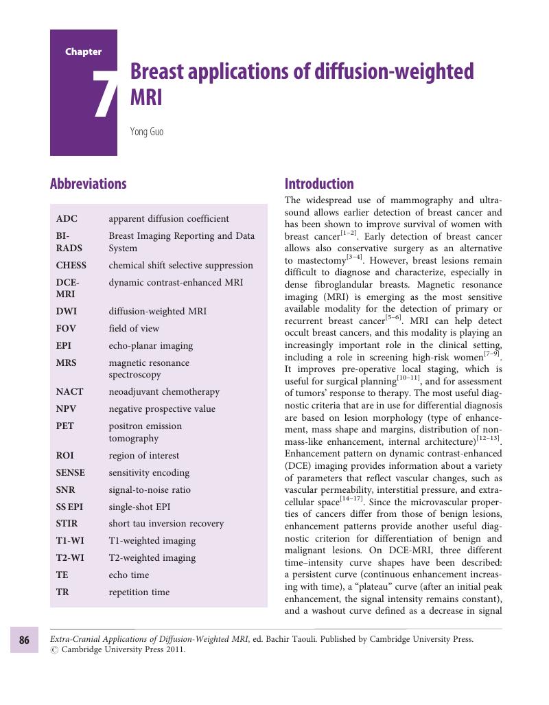
Image of the first page of this content. For PDF version, please use the ‘Save PDF’ preceeding this image.'
Type
Chapter
Information
Publisher: Cambridge University Press
Print publication year: 2010

Access options

Get access to the full version of this content by using one of the access options below. (Log in options will check for institutional or personal access. Content may require purchase if you do not have access.)

References

Senie, RT, Lesser, M, Kinne, DW, Rosen, PP. Method of tumor detection influences disease-free survival of women with breast carcinoma. Cancer 1994;73 (6):1666–72.3.0.CO;2-E>CrossRefGoogle ScholarPubMed
Margolin, FR.Detecting early breast cancer: experience in a community hospital. Cancer 1989;64 (12 Suppl):2702–5.3.0.CO;2-D>CrossRefGoogle Scholar
Thibault, F, Nos, C, Meunier, M, et al. MRI for surgical planning in patients with breast cancer who undergo preoperative chemotherapy. Am J Roentgenol 2004;183 (4):1159–68.CrossRefGoogle ScholarPubMed
Esserman, L, Hylton, N, Yassa, L, et al. Utility of magnetic resonance imaging in the management of breast cancer: evidence for improved preoperative staging. J Clin Oncol 1999;17 (1):110–19.CrossRefGoogle ScholarPubMed
Kuhl, C.The current status of breast MR imaging. I. Choice of technique, image interpretation, diagnostic accuracy, and transfer to clinical practice. Radiology 2007;244 (2):356–78.CrossRefGoogle ScholarPubMed
Kuhl, CK.Current status of breast MR imaging. II. Clinical applications. Radiology 2007;244 (3): 672–91.CrossRefGoogle Scholar
Liberman, L. Breast cancer screening with MRI: what are the data for patients at high risk?N Engl J Med 2004;351 (5):497–500.CrossRefGoogle ScholarPubMed
Robson, ME, Offit, K. Breast MRI for women with hereditary cancer risk. JAMA 2004;292 (11): 1368–70.CrossRefGoogle ScholarPubMed
Morris, EA, Liberman, L, Ballon, DJ, et al. MRI of occult breast carcinoma in a high-risk population. Am J Roentgenol 2003;181 (3):619–26.CrossRefGoogle Scholar
Bedrosian, I, Mick, R, Orel, SG, et al. Changes in the surgical management of patients with breast carcinoma based on preoperative magnetic resonance imaging. Cancer 2003;98 (3):468–73.CrossRefGoogle ScholarPubMed
Fischer, U, Kopka, L, Grabbe, E. Breast carcinoma: effect of preoperative contrast-enhanced MR imaging on the therapeutic approach. Radiology 1999;213 (3):881–8.CrossRefGoogle ScholarPubMed
Hochman, MG, Orel, SG, Powell, CM, et al. Fibroadenomas: MR imaging appearances with radiologic-histopathologic correlation. Radiology 1997; 204 (1):123–9.CrossRefGoogle ScholarPubMed
Nunes, LW, Schnall, MD, Orel, SG. Update of breast MR imaging architectural interpretation model. Radiology 2001;219 (2):484–94.CrossRefGoogle ScholarPubMed
Kaiser, WA, Zeitler, E. MR imaging of the breast: fast imaging sequences with and without Gd-DTPA – preliminary observations. Radiology 1989;170 (3 Pt 1):681–6.CrossRefGoogle ScholarPubMed
Buadu, LD, Murakami, J, Murayama, S, et al. Patterns of peripheral enhancement in breast masses: correlation of findings on contrast medium enhanced MRI with histologic features and tumor angiogenesis. J Comput Assist Tomogr 1997;21 (3):421–30.CrossRefGoogle ScholarPubMed
Su, MY, Jao, JC, Nalcioglu, O. Measurement of vascular volume fraction and blood-tissue permeability constants with a pharmacokinetic model: studies in rat muscle tumors with dynamic Gd-DTPA enhanced MRI. Magn Reson Med 1994;32 (6):714–24.CrossRefGoogle ScholarPubMed
Stack, JP, Redmond, OM, Codd, MB, Dervan, PA, Ennis, JT. Breast disease: tissue characterization with Gd-DTPA enhancement profiles. Radiology 1990;174 (2):491–4.CrossRefGoogle ScholarPubMed
Kuhl, CK, Mielcareck, P, Klaschik, S, et al. Dynamic breast MR imaging: are signal intensity time course data useful for differential diagnosis of enhancing lesions?Radiology 1999;211 (1):101–10.CrossRefGoogle ScholarPubMed
Bluemke, DA, Gatsonis, CA, Chen, MH, et al. Magnetic resonance imaging of the breast prior to biopsy. JAMA 2004;292 (22):2735–42.CrossRefGoogle ScholarPubMed
Wiener, JI, Schilling, KJ, Adami, C, Obuchowski, NA. Assessment of suspected breast cancer by MRI: a prospective clinical trial using a combined kinetic and morphologic analysis. Am J Roentgenol 2005;184 (3):878–86.CrossRefGoogle ScholarPubMed
Macura, KJ, Ouwerkerk, R, Jacobs, MA, Bluemke, DA. Patterns of enhancement on breast MR images: interpretation and imaging pitfalls. Radiographics 2006;26 (6):1719–34.CrossRefGoogle ScholarPubMed
Schnall, MD, Blume, J, Bluemke, DA, et al. Diagnostic architectural and dynamic features at breast MR imaging: multicenter study. Radiology 2006;238 (1):42–53.CrossRefGoogle ScholarPubMed
Szabo, BK, Aspelin, P, Wiberg, MK, Bone, B. Dynamic MR imaging of the breast: analysis of kinetic and morphologic diagnostic criteria. Acta Radiol 2003;44 (4):379–86.Google ScholarPubMed
Heywang-Kobrunner, SH, Bick, U, Bradley, WG, et al. International investigation of breast MRI: results of a multicentre study (11 sites) concerning diagnostic parameters for contrast-enhanced MRI based on 519 histopathologically correlated lesions. Eur Radiol 2001;11 (4):531–46.CrossRefGoogle ScholarPubMed
Kinkel, K, Helbich, TH, Esserman, LJ, et al. Dynamic high-spatial-resolution MR imaging of suspicious breast lesions: diagnostic criteria and interobserver variability. Am J Roentgenol 2000;175 (1):35–43.CrossRefGoogle ScholarPubMed
,ACR breast imaging reporting and data system (BIRADS). Breast Imaging Atlas. Reston, VA:American College of Radiology; 2003.Google Scholar
Kuhl, CK, Schmutzler, RK, Leutner, CC, et al. Breast MR imaging screening in 192 women proved or suspected to be carriers of a breast cancer susceptibility gene: preliminary results. Radiology 2000;215 (1):267–79.CrossRefGoogle ScholarPubMed
Smyczek-Gargya, B, Fersis, N, Dittmann, H, et al. PET with [18F]fluorothymidine for imaging of primary breast cancer: a pilot study. Eur J Nucl Med Mol Imag 2004;31 (5):720–4.CrossRefGoogle Scholar
Samson, DJ, Flamm, CR, Pisano, ED, Aronson, N. Should FDG PET be used to decide whether a patient with an abnormal mammogram or breast finding at physical examination should undergo biopsy?Acad Radiol 2002;9 (7):773–83.CrossRefGoogle ScholarPubMed
Avril, N, Rose, CA, Schelling, M, et al. Breast imaging with positron emission tomography and fluorine-18 fluorodeoxyglucose: use and limitations. J Clin Oncol 2000;18 (20):3495–502.CrossRefGoogle ScholarPubMed
Eliat, PA, Dedieu, V, Bertino, C, et al. Magnetic resonance imaging contrast-enhanced relaxometry of breast tumors: an MRI multicenter investigation concerning 100 patients. Magn Reson Imag 2004;22 (4):475–81.CrossRefGoogle ScholarPubMed
Jacobs, MA, Barker, PB, Bluemke, DA, et al. Benign and malignant breast lesions: diagnosis with multiparametric MR imaging. Radiology 2003;229 (1):225–32.CrossRefGoogle ScholarPubMed
Englander, SA, Ulug, AM, Brem, R, Glickson, JD, Zijl, PC. Diffusion imaging of human breast. NMR Biomed 1997;10 (7):348–52.3.0.CO;2-R>CrossRefGoogle ScholarPubMed
Lucas-Quesada, FA, Sinha, S, DeBruhl, N, Sinha, U, Bassett, LW. Estimation of diffusion coefficients for benign and malignant breast lesions using echo planar MRI. (Abstract.) Radiology 1998;209;468.Google Scholar
Partridge, SC, McKinnon, GC, Henry, RG, Hylton, NM. Menstrual cycle variation of apparent diffusion coefficients measured in the normal breast using MRI. J Magn Reson Imag 2001;14 (4):433–8.CrossRefGoogle ScholarPubMed
Guo, Y, Cai, YQ, Cai, ZL, et al. Differentiation of clinically benign and malignant breast lesions using diffusion-weighted imaging. J Magn Reson Imag 2002;16 (2):172–8.CrossRefGoogle ScholarPubMed
Sinha, S, Lucas-Quesada, FA, Sinha, U, DeBruhl, N, Bassett, LW. In vivo diffusion-weighted MRI of the breast: potential for lesion characterization. J Magn Reson Imag 2002;15 (6):693–704.CrossRefGoogle ScholarPubMed
Schmithorst, VJ, Dardzinski, BJ, Holland, SK. Simultaneous correction of ghost and geometric distortion artifacts in EPI using a multiecho reference scan. IEEE Trans Med Imag 2001;20 (6):535–9.CrossRefGoogle ScholarPubMed
Kurihara, Y, Yakushiji, YK, Tani, I, Nakajima, Y, Cauteren, M. Coil sensitivity encoding in MR imaging: advantages and disadvantages in clinical practice. Am J Roentgenol 2002;178 (5):1087–91.CrossRefGoogle ScholarPubMed
Lee, RF, Westgate, CR, Weiss, RG, Bottomley, PA. An analytical SMASH procedure (ASP) for sensitivity-encoded MRI. Magn Reson Med 2000;43 (5):716–25.3.0.CO;2-K>CrossRefGoogle ScholarPubMed
Bammer, R, Keeling, SL, Augustin, M, et al. Improved diffusion-weighted single-shot echo-planar imaging (EPI) in stroke using sensitivity encoding (SENSE). Magn Reson Med 2001;46 (3):548–54.CrossRefGoogle Scholar
Kuroki, Y, Nasu, K. Advances in breast MRI: diffusion-weighted imaging of the breast. Breast Cancer 2008;15 (3):212–17.CrossRefGoogle ScholarPubMed
Bihan, D, Breton, E, Lallemand, D, et al. Separation of diffusion and perfusion in intravoxel incoherent motion MR imaging. Radiology 1988;168 (2):497–505.CrossRefGoogle ScholarPubMed
Kuroki, Y, Nasu, K, Kuroki, S, et al. Diffusion-weighted imaging of breast cancer with the sensitivity encoding technique: analysis of the apparent diffusion coefficient value. Magn Reson Med Sci 2004;3 (2):79–85.CrossRefGoogle ScholarPubMed
Wenkel, E, Geppert, C, Schulz-Wendtland, R, et al. Diffusion weighted imaging in breast MRI: comparison of two different pulse sequences. Acad Radiol 2007;14 (9):1077–83.CrossRefGoogle ScholarPubMed
Rubesova, E, Grell, AS, Maertelaer, V, et al. Quantitative diffusion imaging in breast cancer: a clinical prospective study. J Magn Reson Imag 2006;24 (2):319–24.CrossRefGoogle ScholarPubMed
Woodhams, R, Matsunaga, K, Iwabuchi, K, et al. Diffusion-weighted imaging of malignant breast tumors: the usefulness of apparent diffusion coefficient (ADC) value and ADC map for the detection of malignant breast tumors and evaluation of cancer extension. J Comput Assist Tomogr 2005;29 (5):644–9.CrossRefGoogle ScholarPubMed
Marini, C, Iacconi, C, Giannelli, M, et al. Quantitative diffusion-weighted MR imaging in the differential diagnosis of breast lesion. Eur Radiol 2007;17 (10):2646–55.CrossRefGoogle ScholarPubMed
Park, MJ, Cha, ES, Kang, BJ, Ihn, YK, Baik, JH. The role of diffusion-weighted imaging and the apparent diffusion coefficient (ADC) values for breast tumors. Korean J Radiol 2007;8 (5):390–6.CrossRefGoogle ScholarPubMed
Yoshikawa, MI, Ohsumi, S, Sugata, S, et al. Comparison of breast cancer detection by diffusion-weighted magnetic resonance imaging and mammography. Radiat Med 2007;25 (5):218–23.CrossRefGoogle ScholarPubMed
Kinoshita, T, Yashiro, N, Ihara, N, et al. Diffusion-weighted half-Fourier single-shot turbo spin echo imaging in breast tumors: differentiation of invasive ductal carcinoma from fibroadenoma. J Comput Assist Tomogr 2002;26 (6):1042–6.CrossRefGoogle ScholarPubMed
Langer, SA, Horst, KC, Ikeda, DM, et al. Pathologic correlates of false positive breast magnetic resonance imaging findings: which lesions warrant biopsy?Am J Surg 2005;190 (4):633–40.CrossRefGoogle ScholarPubMed
Kim, T, Murakami, T, Takahashi, S, et al. Diffusion-weighted single-shot echoplanar MR imaging for liver disease. Am J Roentgenol 1999;173 (2):393–8.CrossRefGoogle ScholarPubMed
Bammer, R. Basic principles of diffusion-weighted imaging. Eur J Radiol 2003;45 (3):169–84.CrossRefGoogle ScholarPubMed
Woodhams, R, Matsunaga, K, Kan, S, et al. ADC mapping of benign and malignant breast tumors. Magn Reson Med Sci 2005;4 (1):35–42.CrossRefGoogle ScholarPubMed
Bihan, D, Breton, E, Lallemand, D, et al. MR imaging of intravoxel incoherent motions: application to diffusion and perfusion in neurologic disorders. Radiology 1986;161 (2):401–7.CrossRefGoogle ScholarPubMed
Buadu, LD, Murakami, J, Murayama, S, et al. Breast lesions: correlation of contrast medium enhancement patterns on MR images with histopathologic findings and tumor angiogenesis. Radiology 1996;200 (3):639–49.CrossRefGoogle ScholarPubMed
Hatakenaka, M, Soeda, H, Yabuuchi, H, et al. Apparent diffusion coefficients of breast tumors: clinical application. Magn Reson Med Sci 2008;7 (1):23–9.CrossRefGoogle ScholarPubMed
Yamashita, Y, Tang, Y, Takahashi, M. Ultrafast MR imaging of the abdomen: echo planar imaging and diffusion-weighted imaging. J Magn Reson Imag 1998;8 (2):367–74.CrossRefGoogle ScholarPubMed
Kang, BK, Na, DG, Ryoo, JW, et al. Diffusion-weighted MR imaging of intracerebral hemorrhage. Korean J Radiol 2001;2 (4):183–91.CrossRefGoogle ScholarPubMed
Buzdar, AU, Singletary, SE, Booser, DJ, et al. Combined modality treatment of stage III and inflammatory breast cancer: M.D. Anderson Cancer Center experience. Surg Oncol Clin N Am 1995;4 (4):715–34.Google Scholar
Partridge, SC, Gibbs, JE, Lu, Y, et al. MRI measurements of breast tumor volume predict response to neoadjuvant chemotherapy and recurrence-free survival. Am J Roentgenol 2005;184 (6):1774–81.CrossRefGoogle ScholarPubMed
Wahl, RL, Zasadny, K, Helvie, M, et al. Metabolic monitoring of breast cancer chemohormonotherapy using positron emission tomography: initial evaluation. J Clin Oncol 1993;11 (11):2101–11.CrossRefGoogle ScholarPubMed
Bassa, P, Kim, EE, Inoue, T, et al. Evaluation of preoperative chemotherapy using PET with fluorine-18-fluorodeoxyglucose in breast cancer. J Nucl Med 1996;37 (6):931–8.Google ScholarPubMed
Smith, IC, Welch, AE, Hutcheon, AW, et al. Positron emission tomography using [(18)F]-fluorodeoxy-D-glucose to predict the pathologic response of breast cancer to primary chemotherapy. J Clin Oncol 2000;18 (8):1676–88.CrossRefGoogle Scholar
Schelling, M, Avril, N, Nahrig, J, et al. Positron emission tomography using [(18)F]fluorodeoxyglucose for monitoring primary chemotherapy in breast cancer. J Clin Oncol 2000;18 (8):1689–95.CrossRefGoogle Scholar
Chen, X, Moore, MO, Lehman, CD, et al. Combined use of MRI and PET to monitor response and assess residual disease for locally advanced breast cancer treated with neoadjuvant chemotherapy. Acad Radiol 2004;11 (10):1115–24.CrossRefGoogle ScholarPubMed
Galons, JP, Altbach, MI, Paine-Murrieta, GD, Taylor, CW, Gillies, RJ. Early increases in breast tumor xenograft water mobility in response to paclitaxel therapy detected by non-invasive diffusion magnetic resonance imaging. Neoplasia 1999;1 (2):113–17.CrossRefGoogle ScholarPubMed
Sharma, U, Danishad, KK, Seenu, V, Jagannathan, NR. Longitudinal study of the assessment by MRI and diffusion-weighted imaging of tumor response in patients with locally advanced breast cancer undergoing neoadjuvant chemotherapy. NMR Biomed 2009;22 (1):104–13.CrossRefGoogle ScholarPubMed
Rajan, R, Poniecka, A, Smith, TL, et al. Change in tumor cellularity of breast carcinoma after neoadjuvant chemotherapy as a variable in the pathologic assessment of response. Cancer 2004;100 (7):1365–73.CrossRefGoogle Scholar
Archer, CD, Parton, M, Smith, IE, et al. Early changes in apoptosis and proliferation following primary chemotherapy for breast cancer. Br J Cancer 2003;89 (6):1035–41.CrossRefGoogle ScholarPubMed

Save book to Kindle

To save this book to your Kindle, first ensure [email protected] is added to your Approved Personal Document E-mail List under your Personal Document Settings on the Manage Your Content and Devices page of your Amazon account. Then enter the ‘name’ part of your Kindle email address below. Find out more about saving to your Kindle.

Note you can select to save to either the @free.kindle.com or @kindle.com variations. ‘@free.kindle.com’ emails are free but can only be saved to your device when it is connected to wi-fi. ‘@kindle.com’ emails can be delivered even when you are not connected to wi-fi, but note that service fees apply.

Find out more about the Kindle Personal Document Service.

Available formats
×

Save book to Dropbox

To save content items to your account, please confirm that you agree to abide by our usage policies. If this is the first time you use this feature, you will be asked to authorise Cambridge Core to connect with your account. Find out more about saving content to Dropbox.

Available formats
×

Save book to Google Drive

To save content items to your account, please confirm that you agree to abide by our usage policies. If this is the first time you use this feature, you will be asked to authorise Cambridge Core to connect with your account. Find out more about saving content to Google Drive.

Available formats
×