Skip to main content Accessibility help
×
Hostname: page-component-78c5997874-v9fdk Total loading time: 0 Render date: 2024-11-19T14:54:30.533Z Has data issue: false hasContentIssue false

15 - MR perfusion imaging in cardiac diseases

from Section 2 - Clinical applications

Published online by Cambridge University Press:  05 May 2013
Peter B. Barker
Affiliation:
The Johns Hopkins University School of Medicine
Xavier Golay
Affiliation:
National Hospital for Neurology and Neurosurgery, London
Gregory Zaharchuk
Affiliation:
Stanford University Medical Center
Get access

Summary

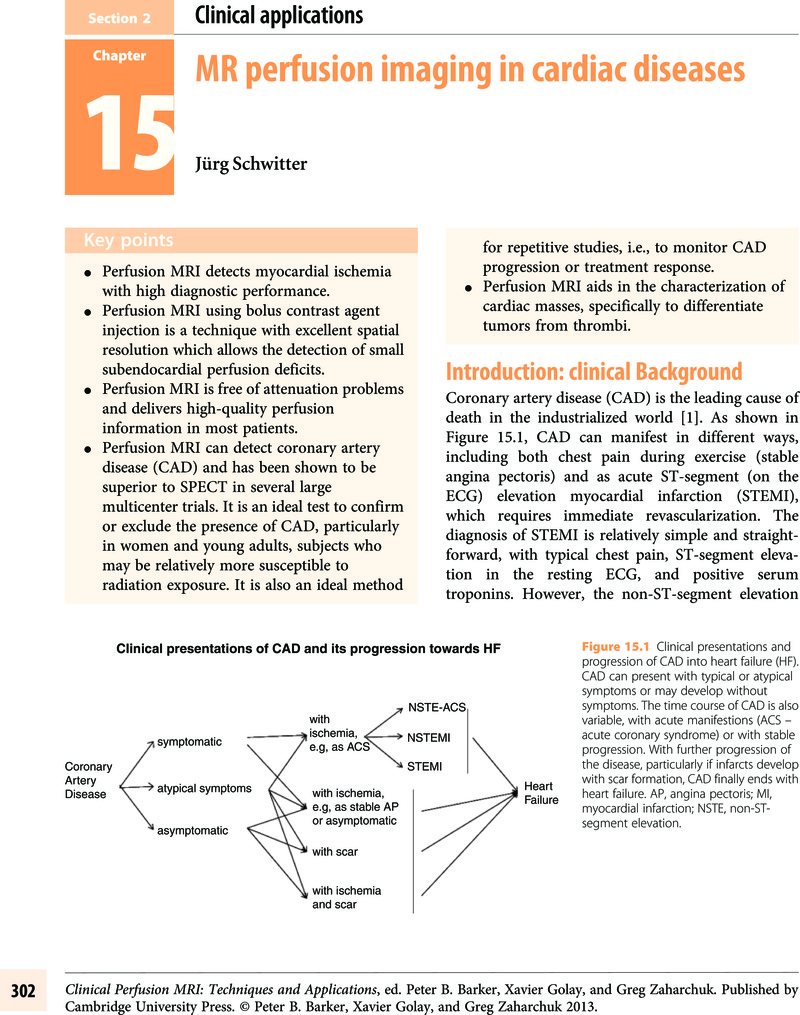
Image of the first page of this content. For PDF version, please use the ‘Save PDF’ preceeding this image.'
Type
Chapter
Information
Clinical Perfusion MRI
Techniques and Applications
, pp. 302 - 325
Publisher: Cambridge University Press
Print publication year: 2013

Access options

Get access to the full version of this content by using one of the access options below. (Log in options will check for institutional or personal access. Content may require purchase if you do not have access.)

References

Lloyd-Jones, D, Adams, R, Carnethon, M, et al. American Heart Association Statistics Committee and Stroke Statistics Subcommittee. Heart disease and stroke statistics – 2009 update: report from the American Heart Association Statistics Committee and Stroke Statistics Subcommittee. Circulation 2009;119.
Volmink, J, Newton, J, Hicks, N, et al. Coronary event and case fatality rates in an English population: results of the Oxford myocardial infarction incidence study. The Oxford Myocardial Infarction Incidence Study Group. Heart 1998;80:40–4.CrossRefGoogle Scholar
Savonitto, S, Ardissino, D, Granger, C, et al. Prognostic value of the admission electrocardiogram in acute coronary syndromes. JAMA 1999;281:707–13.CrossRefGoogle ScholarPubMed
Hachamovitch, R, Rozanski, A, Shaw, L, et al. Impact of ischaemia and scar on the therapeutic benefit derived from myocardial revascularization vs. medical therapy among patients undergoing stress-rest myocardial perfusion scintigraphy. Eur Heart J 2011;32:1012–24.CrossRefGoogle ScholarPubMed
Schwitter, J.Future strategies in the management of coronary artery disease. Fut Cardiol 2006;2(5):555–65.CrossRefGoogle ScholarPubMed
Kwong, RY, Chan, AK, Brown, KA, et al. Impact of unrecognized myocardial scar detected by cardiac magnetic resonance imaging on event-free survival in patients presenting with signs or symptoms of coronary artery disease. Circulation 2006;113(23):2733–43.CrossRefGoogle ScholarPubMed
Allman, K, Shaw, L, Hachamovitch, R, Udelson, J.Myocardial viability testing and impact of revascularization on prognosis in patients with coronary artery disease and left ventricular dysfunction: a meta-analysis. J Am Coll Cardiol 2002;39:1151–8.CrossRefGoogle ScholarPubMed
Beanlands, RS, Ruddy, TD, deKemp, RA, et al. Positron emission tomography and recovery following revascularization (PARR-1): the importance of scar and the development of a prediction rule for the degree of recovery of left ventricular function. J Am Coll Cardiol 2002;40(10):1735–43.CrossRefGoogle ScholarPubMed
Bonow, RO, Maurer, G, Lee, KL, et al. Myocardial viability and survival in ischemic left ventricular dysfunction. N Engl J Med 2011;364(17):1617–25.CrossRefGoogle ScholarPubMed
Wagner, A, Mahrholdt, H, Holly, TA, et al. Contrast-enhanced MRI and routine single photon emission computed tomography (SPECT) perfusion imaging for detection of subendocardial myocardial infarcts: an imaging study. Lancet 2003;361(9355):374–9.CrossRefGoogle ScholarPubMed
Iskander, S, Iskandrian, AE.Risk assessment using single-photon emission computed tomographic technetium-99m sestamibi imaging. J Am Coll Cardiol 1998;32(1):57–62.CrossRefGoogle ScholarPubMed
Jahnke, C, Nagel, E, Gebker, R, et al. Prognostic value of cardiac magnetic resonance stress tests: adenosine stress perfusion and dobutamine stress wall motion imaging. Circulation 2007;115:1769–76.CrossRefGoogle ScholarPubMed
Steel, K, Broderick, R, Gandla, V, et al. Complementary prognostic values of stress myocardial perfusion and late gadolinium enhancement imaging by cardiac magnetic resonance in patients with known or suspected coronary artery disease. Circulation 2009;120:1390–400.CrossRefGoogle ScholarPubMed
Hundley, WG, Morgan, TM, Neagle, CM, et al. Magnetic resonance imaging determination of cardiac prognosis. Circulation 2002;106(18):2328–33.CrossRefGoogle ScholarPubMed
Hoffman, JIE, Spaan, JAE.Pressure-flow relations in coronary circulation. Physiol Rev 1990;70:331–90.CrossRefGoogle ScholarPubMed
Mosher, P, Ross, JJ, McFate, P, Shaw, R.Control of coronary blood flow by an autoregulatory mechanism. Circ Res 1964;14:250–9.CrossRefGoogle ScholarPubMed
Schwitter, J, DeMarco, T, Kneifel, S, et al. Magnetic resonance-based assessment of global coronary flow and flow reserve and its relation to left ventricular functional parameters: a comparison with positron emission tomography. Circulation 2000;101(23):2696–702.CrossRefGoogle ScholarPubMed
Tomanek, R, Palmer, P, Pfeiffer, G, et al. Morphometry of canine coronary arteries, arterioles and capillaries during hypertension and left ventricular hypertrophy. Circ Res 1986;58:38–46.CrossRefGoogle ScholarPubMed
Uren, N, Melin, J, De Bruyne, B, et al. Relation between myocardial blood flow and the severity of coronary artery stenosis. N Engl J Med 1994;330:1782–8.CrossRefGoogle ScholarPubMed
Picano, E, Parodi, O, Lattanzi, F, et al. Assessment of anatomic and physiological severity of single-vessel coronary artery lesions by dipyridamole echocardiography. Comparison with positron emission tomography and quantitative arteriography. Circulation 1994;89:753–61.CrossRefGoogle ScholarPubMed
Di Carli, MF, Czernin, J, Hoh, C, et al. Relation among stenosis severity, myocardial blood flow, and flow reserve in patients with coronary artery disease. Circulation 1995;91:1944–51.CrossRefGoogle ScholarPubMed
Sambuceti, G, Parodi, O, Marcassa, C, et al. Alterations in regulation of myocardial blood flow in one-vessel coronary artery disease determined by positron emission tomography. Am J Cardiol 1993;72:538–43.CrossRefGoogle Scholar
Schwitter, J, Nanz, D, Kneifel, S, et al. Assessment of myocardial perfusion in coronary artery disease by magnetic resonance: a comparison with positron emission tomography and coronary angiography. Circulation 2001;103(18):2230–5.CrossRefGoogle ScholarPubMed
Bertschinger, KM, Nanz, D, Buechi, M, et al. Magnetic resonance myocardial first-pass perfusion imaging: parameter optimization for signal response and cardiac coverage. J Magn Reson Imaging 2001;14(5):556–62.CrossRefGoogle ScholarPubMed
Giang, T, Nanz, D, Coulden, R, et al. Detection of coronary artery disease by magnetic resonance myocardial perfusion imaging with various contrast medium doses: first European multicenter experience. Eur Heart J 2004;25:1657–65.CrossRefGoogle Scholar
Schwitter, J, Wacker, C, van Rossum, A, et al. MR-IMPACT: comparison of perfusion-cardiac magnetic resonance with single-photon emission computed tomography for the detection of coronary artery disease in a multicentre, multivendor, randomized trial. Eur Heart J 2008;29:480–9.CrossRefGoogle Scholar
Plein, S, Kozerke, S, Suerder, D, et al. High spatial resolution myocardial perfusion cardiac magnetic resonance for the detection of coronary artery disease. Eur Heart J 2008;29:2148–55.CrossRefGoogle ScholarPubMed
Schwitter, J. CMR Update, 2nd edn. Lausanne: Schwitter, J. 2012; Available from (accessed November 19, 2012).
Schwitter, J, Arai, A.Imaging: assessment of cardiac ischaemia and viability: role of cardiovascular magnetic resonance. Eur Heart J 2011;32:799–809.CrossRefGoogle Scholar
Bruder, O, Schneider, S, Nothnagel, D, et al. EuroCMR (European Cardiovascular Magnetic Resonance) Registry. J Am Coll Cardiol 2009;54:1457–66.CrossRefGoogle ScholarPubMed
Schwitter, J, Wacker, CM, Wilke, N, et al. Superior diagnostic performance of perfusion-cardiovascular magnetic resonance versus SPECT to detect coronary artery disease: the secondary endpoints of the multicenter multivendor MR-IMPACT II (Magnetic Resonance Imaging for Myocardial Perfusion Assessment in Coronary Artery Disease Trial). J. Cardiovasc Magn Reson 2012;14:61–71.CrossRefGoogle Scholar
Nagel, E, Klein, C, Paetsch, I, et al. Magnetic resonance perfusion measurements for the noninvasive detection of coronary artery disease. Circulation 2003;108(4):432–7.CrossRefGoogle ScholarPubMed
Plein, S, Radjenovic, A, Ridgway, JP, et al. Coronary artery disease: myocardial perfusion MR imaging with sensitivity encoding versus conventional angiography. Radiology 2005;235(2):423–30.CrossRefGoogle ScholarPubMed
Paetsch, I, Jahnke, C, Wahl, A, et al. Comparison of dobutamine stress magnetic resonance, adenosine stress magnetic resonance, and adenosine stress magnetic resonance perfusion. Circulation 2004;110(7):835–42.CrossRefGoogle ScholarPubMed
Kwong, R, Schussheim, A, Rekhraj, S, et al. Detecting acute coronary syndrome in the emergency department with cardiac magnetic resonance imaging. Circulation 2003;107:531–7.CrossRefGoogle ScholarPubMed
Ingkanisorn, WP, Kwong, RY, Bohme, NS, et al. Prognosis of negative adenosine stress magnetic resonance in patients presenting to an emergency department with chest pain. J Am Coll Cardiol 2006;47(7):1427–32.CrossRefGoogle Scholar
Plein, S, Greenwood, J, Ridgeway, J, et al. Assessment of non-ST-segment elevation acute coronary syndromes with cardiac magnetic resonance imaging. J Am Coll Cardiol 2004;44:2173–81.CrossRefGoogle ScholarPubMed
Hoffmann, U, Globits, S, Schima, W, et al. Usefulness of magnetic resonance imaging of cardiac and paracardiac masses. Am J Cardiol 2003;92(7):890–5.CrossRefGoogle ScholarPubMed
Schuler, P, Weber, A, Bode, P, et al. MRI of intimal sarcoma of the pulmonary arteries. Circ Cardiovasc Imaging 2009;2:e37–9.CrossRefGoogle ScholarPubMed
Fussen, S, De Boeck, BWL, Zellweger, MJ, et al. Cardiovascular magnetic resonance imaging for diagnosis and clinical management of suspected cardiac masses and tumours. Eur Heart J 2011;32(12):1551–60.CrossRefGoogle ScholarPubMed
Srichai, MB, Junor, C, Rodriguez, LL, et al. Clinical, imaging, and pathological characteristics of left ventricular thrombus: a comparison of contrast-enhanced magnetic resonance imaging, transthoracic echocardiography, and transesophageal echocardiography with surgical or pathological validation. Am Heart J 2006;152(1):75–84.CrossRefGoogle ScholarPubMed
Knuesel, PR, Nanz, D, Wyss, C, et al. Characterization of dysfunctional myocardium by positron emission tomography and magnetic resonance: relation to functional outcome after revascularization. Circulation 2003;108(9):1095–100.CrossRefGoogle ScholarPubMed
Christian, TF, Rettmann, DW, Aletras, AH, et al. Absolute myocardial perfusion in canines measured by using dual-bolus first-pass MR imaging. Radiology 2004;232(3):677–84.CrossRefGoogle ScholarPubMed
Gatehouse, PD, Elkington, AG, Ablitt, NA, et al. Accurate assessment of the arterial input function during high-dose myocardial perfusion cardiovascular magnetic resonance. J Magn Reson Imaging 2004;20(1):39–45.CrossRefGoogle ScholarPubMed
Biological effects of ionizing radiation (BEIR) reports VII-Phase 2. National Research Council. Available from: (accessed November 19, 2012).
Eisenberg, M, Afilalo, J, Lawler, P, et al. Cancer risk related to low-dose ionizing radiation from cardiac imaging in patients after acute myocardial infarction. CMAJ 2011;183:430–6.CrossRefGoogle ScholarPubMed
Cardis, E, Vrijheid, M, Blettner, M, et al. Risk of cancer after low doses of ionising radiation: retrospective cohort study in 15 countries. Br Med J 2005;331:77–82.CrossRefGoogle ScholarPubMed
Buechel, ER, Balmer, C, Bauersfeld, U, Kellenberger, CJ, Schwitter, J.Feasibility of perfusion cardiovascular magnetic resonance in paediatric patients. J Cardiovasc Magn Reson 2009;11:51.CrossRefGoogle ScholarPubMed
Burke, A, Kolodgie, F, Farb, A, et al. Healed plaque ruptures and sudden coronary death: evidence that subclinical rupture has a role in plaque progression. Circulation 2001;103:934–40.CrossRefGoogle Scholar
Schwitter, J, Oelhafen, M, Wyss, BM, et al. 2D-spatially-selective real-time magnetic resonance imaging for the assessment of microvascular function and its relation to the cardiovascular risk profile. J Cardiovasc Magn Reson 2006;8(5):759–69.CrossRefGoogle ScholarPubMed
Schwitter, J, Kozerke, S, Bremerich, J, et al. Oral administration of 17beta-estradiol over 3 months without progestin co-administration does not improve coronary flow reserve in post-menopausal women: a randomized placebo-controlled cross-over CMR study. J Cardiovasc Magn Reson 2007;9:665–72.CrossRefGoogle Scholar
Koskenvuo, J, Hartiala, J, Knuuti, J, et al. Assessing coronary sinus blood flow in patients with coronary artery diseaseAJR Am J Roentgenol 2001;177:1161–6.CrossRefGoogle ScholarPubMed
Kawada, N, Sakuma, H, Yamakado, T, et al. Hypertrophic cardiomyopathy: MR measurement of coronary blood flow and vasodilator flow reserve in patients and healthy subjects. Radiology 1999;211:129–35.CrossRefGoogle ScholarPubMed
Hays, A, Hirsch, G, Kelle, S, et al. Noninvasive visualization of coronary artery endothelial function in healthy subjects and in patients with coronary artery disease. J Am Coll Cardiol 2010;56:1657–65.CrossRefGoogle ScholarPubMed
Niemi, P, Poncelet, BP, Kwong, KK, et al. Myocardial intensity changes associated with flow stimulation in blood oxygenation sensitive magnetic resonance imaging. Magn Reson Med 1996;36:78–82.CrossRefGoogle ScholarPubMed
Fieno, DS, Shea, SM, Li, Y, et al. Myocardial perfusion imaging based on the blood oxygen level-dependent effect using T2-prepared steady-state free-precession magnetic resonance imaging. Circulation 2004;110:1284–90.CrossRefGoogle ScholarPubMed
Wright, K, Klocke, F, Deshpande, V, et al. Assessment of regional differences in myocardial blood flow using T2-weighted 3D BOLD imaging. Magn Reson Med 2001;46:573–8.CrossRefGoogle ScholarPubMed
Reeder, S, Atalay, M, McVeigh, E, Zerhouni, E, Forder, J.Quantitative cardiac perfusion: a noninvasive spin-labeling method that exploits coronary vessel geometry. Radiology 1996;200:177–84.CrossRefGoogle ScholarPubMed
Belle, V, Kahler, E, Waller, C, et al. In vivo quantitative mapping of cardiac perfusion in rats using a noninvasive MR spin-labeling method. J Magn Reson Imaging 1998;8:1240–5.CrossRefGoogle ScholarPubMed
McCommis, KS, Zhang, H, Herrero, P, Grapler, RJ, Zheng, J.Feasibility study of myocardial perfusion and oxygenation by noncontrast MRI: comparison with PET study in a canine model. Magn Reson Imaging 2008;26:11–19.CrossRefGoogle Scholar
Golman, K, Ardenkjaer-Larsen, JH, Petersson, JS, Mansson, S, Leunbach, I.Molecular imaging with endogenous substances. Proc Natl Acad Sci U S A 2003;100(18):10435–9.CrossRefGoogle ScholarPubMed
Schwitter, J.Myocardial perfusion imaging by cardiac magnetic resonance. J Nucl Cardiol 2006;13(6):841–54.CrossRefGoogle ScholarPubMed
Schwitter, J.Extending the frontiers of cardiac magnetic resonance. Circulation 2008;118:109–12.CrossRefGoogle ScholarPubMed
Wahl, A, Paetsch, I, Gollesch, A, et al. Safety and feasibility of high-dose dobutamine-atropine stress cardiovascular magnetic resonance for diagnosis of myocardial ischaemia: experience in 1000 consecutive cases. Eur Heart J 2004;25(14):1230–6.CrossRefGoogle ScholarPubMed
Hundley, WG, Hamilton, CA, Thomas, MS, et al. Utility of fast cine magnetic resonance imaging and display for the detection of myocardial ischemia in patients not well suited for second harmonic stress echocardiography. Circulation 1999;100(16):1697–702.CrossRefGoogle Scholar
Wilkoff, BL, Bello, D, Taborsky, M, et al. Magnetic resonance imaging in patients with a pacemaker system designed for the magnetic resonance environment. Heart Rhythm 2011;8(1):65–73.CrossRefGoogle ScholarPubMed
Syed, MA, Carlson, K, Murphy, M.Long-term safety of cardiac magnetic resonance imaging performed in the first few days after bare-metal stent implantation. J Magn Reson Imaging 2006;24(5):1056–61.CrossRefGoogle ScholarPubMed
Kellman, P, Derbyshire, J, Agyeman, K, McVeigh, E, Arai, A.Extended coverage first-pass perfusion imaging using slice-interleaved TSENSE. Magn Reson Med 2004;51:200–4.CrossRefGoogle ScholarPubMed
Zaret, BL, Rigo, P, Wackers, FJ, et al. Myocardial perfusion imaging with 99mTc tetrofosmin. Comparison to 201TI imaging and coronary angiography in a phase III multicenter trial. Tetrofosmin International Trial Study Group. Circulation 1995;91:313–19.CrossRefGoogle Scholar
Van Train, KF, Garcia, EV, Maddahi, J, et al. Multicenter trial validation for quantitative analysis of same-day, rest-stress technetium-99m-sestamibi myocardial tomograms. J Nucl Med 1994;35:609–18.Google ScholarPubMed
Hendel, RC, Berman, DS, Cullon, SJ, et al. Multicenter clinical trial to evaluate the efficacy of correction for photon attenuation and scatter in SPECT myocardial perfusion imaging. Circulation 1999;99:2742–9.CrossRefGoogle ScholarPubMed

Save book to Kindle

To save this book to your Kindle, first ensure [email protected] is added to your Approved Personal Document E-mail List under your Personal Document Settings on the Manage Your Content and Devices page of your Amazon account. Then enter the ‘name’ part of your Kindle email address below. Find out more about saving to your Kindle.

Note you can select to save to either the @free.kindle.com or @kindle.com variations. ‘@free.kindle.com’ emails are free but can only be saved to your device when it is connected to wi-fi. ‘@kindle.com’ emails can be delivered even when you are not connected to wi-fi, but note that service fees apply.

Find out more about the Kindle Personal Document Service.

Available formats
×

Save book to Dropbox

To save content items to your account, please confirm that you agree to abide by our usage policies. If this is the first time you use this feature, you will be asked to authorise Cambridge Core to connect with your account. Find out more about saving content to Dropbox.

Available formats
×

Save book to Google Drive

To save content items to your account, please confirm that you agree to abide by our usage policies. If this is the first time you use this feature, you will be asked to authorise Cambridge Core to connect with your account. Find out more about saving content to Google Drive.

Available formats
×