Skip to main content Accessibility help
×
Hostname: page-component-848d4c4894-tn8tq Total loading time: 0 Render date: 2024-06-25T05:19:22.011Z Has data issue: false hasContentIssue false

Chapter 67 - Cerebral Amyloid Angiopathy

from Section 7 - Non-Inflammatory Disorders of the Arterial Wall

Published online by Cambridge University Press:  15 June 2018

Louis Caplan
Affiliation:
Beth Israel-Deaconess Medical Center, Boston
José Biller
Affiliation:
Loyola University Stritch School of Medicine, Chicago
Get access

Summary

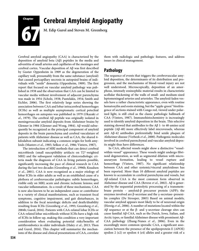
Image of the first page of this content. For PDF version, please use the ‘Save PDF’ preceeding this image.'
Type
Chapter
Information
Publisher: Cambridge University Press
Print publication year: 2018

Access options

Get access to the full version of this content by using one of the access options below. (Log in options will check for institutional or personal access. Content may require purchase if you do not have access.)

References

Akoudad, S., Portegies, M. L., Koudstaal, P. J., et al. (2015). Cerebral microbleeds are associated with an increased risk of stroke: The Rotterdam study. Circulation 132: 509–16.Google Scholar
Auriel, E., Charidimou, A., Gurol, M. E., et al. (2016). Validation of clinicoradiological criteria for the diagnosis of cerebral amyloid angiopathy-related inflammation. JAMA Neurol 73: 197202.Google Scholar
Auriel, E., Gurol, M. E., Ayres, A., et al. (2012). Characteristic distributions of intracerebral hemorrhage-associated diffusion-weighted lesions. Neurology 79: 2335–41.Google Scholar
Black, S., Gao, F., and Bilbao, J. (2009). Understanding white matter disease: Imaging–pathological correlations in vascular cognitive impairment. Stroke 40: S4852.Google Scholar
Blitstein, M. K. and Tung, G. A. (2007). MRI of cerebral microhemorrhages. AJR Am J Roentgenol 189: 720–5.Google Scholar
Boulouis, G., Charidimou, A., Auriel, E., et al. (2016). Intracranial atherosclerosis and cerebral small vessel disease in intracerebral hemorrhage patients. J Neurol Sci 369: 324–9.Google Scholar
Boyle, P. A., Yu, L., Nag, S., et al. (2015). Cerebral amyloid angiopathy and cognitive outcomes in community-based older persons. Neurology 85: 1930–6.Google Scholar
Charidimou, A., Meegahage, R., Fox, Z., et al. (2013a). Enlarged perivascular spaces as a marker of underlying arteriopathy in intracerebral haemorrhage: A multicentre MRI cohort study. J Neurol Neurosurg Psychiatry 84: 624–9.Google Scholar
Charidimou, A., Peeters, A. P., Jager, R., et al. (2013b). Cortical superficial siderosis and intracerebral hemorrhage risk in cerebral amyloid angiopathy. Neurology 81: 1666–73.Google Scholar
Charidimou, A., Linn, J., Vernooij, M. W., et al. (2015). Cortical superficial siderosis: detection and clinical significance in cerebral amyloid angiopathy and related conditions. Brain 138: 2126–39.Google Scholar
Charidimou, A., Boulouis, G., Haley, K., et al. (2016). White matter hyperintensity patterns in cerebral amyloid angiopathy and hypertensive arteriopathy. Neurology 86: 505–11.Google Scholar
De Strooper, B. (2003). Aph-1, Pen-2, and nicastrin with presenilin generate an active gamma-secretase complex. Neuron 38: 912.Google Scholar
Dierksen, G. A., Skehan, M. E., Khan, M. A., et al. (2010). Spatial relation between microbleeds and amyloid deposits in amyloid angiopathy. Ann Neurol 68: 545–8.Google Scholar
Dumas, A., Dierksen, G. A., Gurol, M. E., et al. (2012). Functional magnetic resonance imaging detection of vascular reactivity in cerebral amyloid angiopathy. Ann Neurol 72: 7681.Google Scholar
Eckman, M. H., Rosand, J., Knudsen, K. A., Singer, D. E., and Greenberg, S. M. (2003). Can patients be anticoagulated after intracerebral hemorrhage? A decision analysis. Stroke 34: 1710–16.Google Scholar
Eckman, M. H., Wong, L. K., Soo, Y. O., et al. (2008). Patient-specific decision-making for warfarin therapy in nonvalvular atrial fibrillation: How will screening with genetics and imaging help? Stroke 39: 3308–15.Google Scholar
Eng, J. A., Frosch, M. P., Choi, K., Rebeck, G. W., and Greenberg, S. M. (2004). Clinical manifestations of cerebral amyloid angiopathy-related inflammation. Ann Neurol 55: 250–6.Google Scholar
Fotiadis, P., van Rooden, S., van der Grond, J., et al. (2016). Cortical atrophy in patients with cerebral amyloid angiopathy: A case–control study. Lancet Neurol 15: 811–19.Google Scholar
Glenner, G. G. and Wong, C. W. (1984). Alzheimer’s disease: Initial report of the purification and characterization of a novel cerebrovascular amyloid protein. Biochem Biophys Res Commun 120: 885–90.Google Scholar
Goldstein, L. B., Amarenco, P., Szarek, M., et al. (2008). Hemorrhagic stroke in the Stroke Prevention by Aggressive Reduction in Cholesterol Levels study. Neurology 70: 2364–70.Google Scholar
Greenberg, S. M., Vonsattel, J. P., Stakes, J. W., Gruber, M., and Finklestein, S. P. (1993). The clinical spectrum of cerebral amyloid angiopathy: Presentations without lobar hemorrhage. Neurology 43: 2073–9.Google Scholar
Greenberg, S. M., Briggs, M. E., Hyman, B. T., et al. (1996a). Apolipoprotein E epsilon 4 is associated with the presence and earlier onset of hemorrhage in cerebral amyloid angiopathy. Stroke 27: 1333–7.Google Scholar
Greenberg, S. M., Finklestein, S. P., and Schaefer, P. W. (1996b). Petechial hemorrhages accompanying lobar hemorrhage: Detection by gradient-echo MRI. Neurology 46: 1751–4.Google Scholar
Greenberg, S. M., Vonsattel, J. P., Segal, A. Z., et al. (1998). Association of apolipoprotein E epsilon2 and vasculopathy in cerebral amyloid angiopathy. Neurology 50: 961–5.Google Scholar
Greenberg, S. M., Eng, J. A., Ning, M., Smith, E. E., and Rosand, J. (2004a). Hemorrhage burden predicts recurrent intracerebral hemorrhage after lobar hemorrhage. Stroke 35: 1415–20.Google Scholar
Greenberg, S. M., Gurol, M. E., Rosand, J., and Smith, E. E. (2004b). Amyloid angiopathy-related vascular cognitive impairment. Stroke 35(Suppl 1): 2616–19.Google Scholar
Greenberg, S. M., Rosand, J., Schneider, A. T., et al. (2006). A phase 2 study of tramiprosate for cerebral amyloid angiopathy. Alzheimer Dis Assoc Disord 20: 269–74.Google Scholar
Greenberg, S. M., Grabowski, T., Gurol, M. E., et al. (2008). Detection of isolated cerebrovascular beta-amyloid with Pittsburgh compound B. Ann Neurol 64: 587–91.Google Scholar
Greenberg, S. M., Vernooij, M. W., Cordonnier, C., et al. (2009). Cerebral microbleeds: A guide to detection and interpretation. Lancet Neurology 8: 165–74.Google Scholar
Gurol, M. E. (2016). Molecular neuroimaging in vascular cognitive impairment. Stroke 47: 1146–52.Google Scholar
Gurol, M. E. and Greenberg, S. M. (2008). Management of intracerebral hemorrhage. Curr Atheroscler Rep 10: 324–31.Google Scholar
Gurol, M. E., Irizarry, M. C., Smith, E. E., et al. (2006). Plasma beta-amyloid and white matter lesions in AD, MCI, and cerebral amyloid angiopathy. Neurology 66: 23–9.Google Scholar
Gurol, M. E., Dierksen, G., Betensky, R., et al. (2012). Predicting sites of new hemorrhage with amyloid imaging in cerebral amyloid angiopathy. Neurology 79: 320–6.Google Scholar
Gurol, M. E., Viswanathan, A., Gidicsin, C., et al. (2013). Cerebral amyloid angiopathy burden associated with leukoaraiosis: A positron emission tomography/magnetic resonance imaging study. Ann Neurol 73: 529–36.Google Scholar
Gurol, M. E., Becker, J. A., Fotiadis, P., et al. (2016). Florbetapir-PET to diagnose cerebral amyloid angiopathy: A prospective study. Neurology 87: 2043–9.Google Scholar
Hackam, D. G. and Mrkobrada, M. (2012). Selective serotonin reuptake inhibitors and brain hemorrhage: A meta-analysis. Neurology 79: 1862–5.Google Scholar
Haley, K. E., Greenberg, S. M., and Gurol, M. E. (2013). Cerebral microbleeds and macrobleeds: Should they influence our recommendations for antithrombotic therapies? Curr Cardiol Rep 15: 425–32.Google Scholar
Hemphill, J. C. III, Greenberg, S. M., Anderson, C. S., et al. (2015). Guidelines for the management of spontaneous intracerebral hemorrhage: A Guideline for Healthcare Professionals from the American Heart Association/American Stroke Association. Stroke 46: 2032–60.Google Scholar
Hermann, D. M. and Bassetti, C. L. (2016). Role of sleep-disordered breathing and sleep–wake disturbances for stroke and stroke recovery. Neurology 87: 1407–16.Google Scholar
Herzig, M. C., Van Nostrand, W. E., and Jucker, M. (2006). Mechanism of cerebral beta-amyloid angiopathy: Murine and cellular models. Brain Pathol 16: 4054.Google Scholar
January, C. T., Wann, L. S., Alpert, J. S., et al. (2014). 2014 AHA/ACC/HRS guideline for the management of patients with atrial fibrillation: A report of the American College of Cardiology/American Heart Association Task Force on practice guidelines and the Heart Rhythm Society. Circulation 130: e199267.Google Scholar
Johnson, K. A., Gregas, M., Becker, J. A., et al. (2007). Imaging of amyloid burden and distribution in cerebral amyloid angiopathy. Ann Neurol 62: 229–34.Google Scholar
Kimberly, W. T., Gilson, A., Rost, N. S., et al. (2009). Silent ischemic infarcts are associated with hemorrhage burden in cerebral amyloid angiopathy. Neurology 72: 1230–5.Google Scholar
Klunk, W. E., Engler, H., Nordberg, A., et al. (2004). Imaging brain amyloid in Alzheimer’s disease with Pittsburgh compound-B. Ann Neurol 55: 306–19.Google Scholar
Knudsen, K. A., Rosand, J., Karluk, D., and Greenberg, S. M. (2001). Clinical diagnosis of cerebral amyloid angiopathy: Validation of the Boston criteria. Neurology 56: 537–9.Google Scholar
Lauer, A., Greenberg, S. M., and Gurol, M. E. (2015). Statins in intracerebral hemorrhage. Curr Atheroscler Rep 17: 46.Google Scholar
Linn, J., Halpin, A., Demaerel, P., et al. (2010). Prevalence of superficial siderosis in patients with cerebral amyloid angiopathy. Neurology 74: 1346–50.Google Scholar
Lovelock, C. E., Molyneux, A. J., and Rothwell, P. M. (2007). Change in incidence and aetiology of intracerebral haemorrhage in Oxfordshire, UK, between 1981 and 2006: A population-based study. Lancet Neurol 6: 487–93.Google Scholar
Ly, J. V., Donnan, G. A., Villemagne, V. L., et al. (2010). 11 C-PIB binding is increased in patients with cerebral amyloid angiopathy-related hemorrhage. Neurology 74: 487–93.Google Scholar
Martinez-Ramirez, S., Pontes-Neto, O. M., Dumas, A. P., et al. (2013). Topography of dilated perivascular spaces in subjects from a memory clinic cohort. Neurology 80: 1551–6.Google Scholar
Martinez-Ramirez, S., Romero, J. R., Shoamanesh, A., et al. (2015). Diagnostic value of lobar microbleeds in individuals without intracerebral hemorrhage. Alzheimers Dement 11: 1480–8.Google Scholar
Masoudi, F. A., Calkins, H., Kavinsky, C. J., et al. (2015). 2015 ACC/HRS/SCAI Left atrial appendage occlusion device societal overview. J Am Coll Cardiol 66: 1497–513.Google Scholar
Masters, C. L., Simms, G., Weinman, N. A., et al. (1985). Amyloid plaque core protein in Alzheimer disease and Down syndrome. Proc Natl Acad Sci USA 82: 4245–9.Google Scholar
Masuda, J., Tanaka, K., Ueda, K., and Omae, T. (1988). Autopsy study of incidence and distribution of cerebral amyloid angiopathy in Hisayama, Japan. Stroke 19: 205–10.Google Scholar
Nicoll, J. A., Burnett, C., Love, S., et al. (1996). High frequency of apolipoprotein E epsilon 2 in patients with cerebral hemorrhage due to cerebral amyloid angiopathy. Ann Neurol 39: 682–3.Google Scholar
O’Donnell, H. C., Rosand, J., Knudsen, K. A., et al. (2000). Apolipoprotein E genotype and the risk of recurrent lobar intracerebral hemorrhage. N Engl J Med 342: 240–5.Google Scholar
Okazaki, H., Reagan, T. J., and Campbell, R. J. (1979). Clinicopathologic studies of primary cerebral amyloid angiopathy. Mayo Clin Proc 54: 2231.Google Scholar
Oppenheim, G. (1909). Uber “drusige Nekrosen” in der Großhirnrinde. Neurol Centralbl 28: 410–13.Google Scholar
Pantelakis, S. (1954). A particular type of senile angiopathy of the central nervous system: Congophilic angiopathy, topography and frequency [in French]. Monatsschr Psychiatr Neurol 128: 219–56.Google Scholar
Peca, S., McCreary, C. R., Donaldson, E., et al. (2013). Neurovascular decoupling is associated with severity of cerebral amyloid angiopathy. Neurology 81: 1659–65.Google Scholar
Poels, M. M., Vernooij, M. W., Ikram, M. A., et al. (2010). Prevalence and risk factors of cerebral microbleeds: An update of the Rotterdam scan study. Stroke 41(Suppl): S103–6.Google Scholar
Purrucker, J. C., Haas, K., Rizos, T., et al. (2016). Early clinical and radiological course, management, and outcome of intracerebral hemorrhage related to new oral anticoagulants. JAMA Neurol 73: 169–77.Google Scholar
Reijmer, Y. D., Fotiadis, P., Martinez-Ramirez, S., et al. (2015). Structural network alterations and neurological dysfunction in cerebral amyloid angiopathy. Brain 138(Pt 1): 179–88.Google Scholar
Reijmer, Y. D., van Veluw, S. J., and Greenberg, S. M. (2016). Ischemic brain injury in cerebral amyloid angiopathy. J Cereb Blood Flow Metab 36: 4054.Google Scholar
Revesz, T., Holton, J. L., Lashley, T., et al. (2002). Sporadic and familial cerebral amyloid angiopathies. Brain Pathol 12: 343–57.Google Scholar
Rosand, J., Hylek, E. M., O’Donnell, H. C., and Greenberg, S. M. (2000). Warfarin-associated hemorrhage and cerebral amyloid angiopathy: A genetic and pathologic study. Neurology 55: 947–51.Google Scholar
Rosand, J., Muzikansky, A., Kumar, A., et al. (2005). Spatial clustering of hemorrhages in probable cerebral amyloid angiopathy. Ann Neurol 58: 459–62.Google Scholar
Schmechel, D. E., Saunders, A. M., Strittmatter, W. J., et al. (1993). Increased amyloid beta-peptide deposition in cerebral cortex as a consequence of apolipoprotein E genotype in late-onset Alzheimer disease. Proc Natl Acad Sci USA 90: 9649–53.Google Scholar
Scholz, W. (1938). Studien zur pathologie der hirngefabe II: Die drusige entartung der hirnarterien und Capillaren. Zeit. Gesamte Neurol Psychiatr 162: 694715.Google Scholar
Scolding, N. J., Joseph, F., Kirby, P. A., et al. (2005). Abeta-related angiitis: Primary angiitis of the central nervous system associated with cerebral amyloid angiopathy. Brain 128: 500–15.Google Scholar
Selkoe, D. J., Abraham, C. R., Podlisny, M. B., and Duffy, L. K. (1986). Isolation of low-molecular-weight proteins from amyloid plaque fibers in Alzheimer’s disease. J Neurochem 46: 1820–34.Google Scholar
Smith, E. E. and Eichler, F. (2006). Cerebral amyloid angiopathy and lobar intracerebral hemorrhage. Arch Neurol 63: 148–51.Google Scholar
Smith, E. E., Gurol, M. E., Eng, J. A., et al. (2004). White matter lesions, cognition, and recurrent hemorrhage in lobar intracerebral hemorrhage. Neurology 63: 1606–12.Google Scholar
Smith, E. E., Vijayappa, M., Lima, F., et al. (2008). Impaired visual evoked flow velocity response in cerebral amyloid angiopathy. Neurology 71: 1424–30.Google Scholar
Smith, E. E., Schneider, J. A., Wardlaw, J. M., and Greenberg, S. M. (2012). Cerebral microinfarcts: The invisible lesions. Lancet Neurol 11: 272–82.Google Scholar
Stone, N. J., Robinson, J. G., Lichtenstein, A. H., et al. (2014). 2013 ACC/AHA guideline on the treatment of blood cholesterol to reduce atherosclerotic cardiovascular risk in adults: A report of the American College of Cardiology/American Heart Association Task Force on Practice Guidelines. Circulation 129(Suppl 2): S145.Google Scholar
Thanprasertsuk, S., Martinez-Ramirez, S., Pontes-Neto, O. M., et al. (2014). Posterior white matter disease distribution as a predictor of amyloid angiopathy. Neurology 83: 794800.Google Scholar
Thon, J. M. and Gurol, M. E. (2016). Intracranial hemorrhage risk in the era of antithrombotic therapies for ischemic stroke. Curr Treat Options Cardiovasc Med 18: 29.Google Scholar
van Asch, C. J., Luitse, M. J., Rinkel, G. J., van der Tweel, I., Algra, A., and Klijn, C. J. (2010). Incidence, case fatality, and functional outcome of intracerebral haemorrhage over time, according to age, sex, and ethnic origin: A systematic review and meta-analysis. Lancet Neurol 9: 167–76.Google Scholar
van Etten, E. S., Auriel, E., Haley, K. E., et al. (2014). Incidence of symptomatic hemorrhage in patients with lobar microbleeds. Stroke 45: 2280–5.Google Scholar
van Veluw, S. J., Biessels, G. J., Bouvy, W. H., et al. (2016). Cerebral amyloid angiopathy severity is linked to dilation of juxtacortical perivascular spaces. J Cereb Blood Flow Metab 36: 576–80.Google Scholar
Verbeek, M. M., De Waal, R. M. W., and Vinters, H. V. (2000). Cerebral Amyloid Angiopathy in Alzheimer’s Disease and Related Disorders. Dordrecht: Kluwer Academic.Google Scholar
Verbeek, M. M., Kremer, B. P., Rikkert, M. O., et al. (2009). Cerebrospinal fluid amyloid beta(40) is decreased in cerebral amyloid angiopathy. Ann Neurol 66: 245–49.Google Scholar
Vinters, H. V. (1987). Cerebral amyloid angiopathy: A critical review. Stroke 18: 311–24.Google Scholar
Zhang-Nunes, S. X., Maat-Schieman, M. L., van Duinen, S. G., et al. (2006). The cerebral beta-amyloid angiopathies: Hereditary and sporadic. Brain Pathol 16: 30–9.Google Scholar

Save book to Kindle

To save this book to your Kindle, first ensure [email protected] is added to your Approved Personal Document E-mail List under your Personal Document Settings on the Manage Your Content and Devices page of your Amazon account. Then enter the ‘name’ part of your Kindle email address below. Find out more about saving to your Kindle.

Note you can select to save to either the @free.kindle.com or @kindle.com variations. ‘@free.kindle.com’ emails are free but can only be saved to your device when it is connected to wi-fi. ‘@kindle.com’ emails can be delivered even when you are not connected to wi-fi, but note that service fees apply.

Find out more about the Kindle Personal Document Service.

Available formats
×

Save book to Dropbox

To save content items to your account, please confirm that you agree to abide by our usage policies. If this is the first time you use this feature, you will be asked to authorise Cambridge Core to connect with your account. Find out more about saving content to Dropbox.

Available formats
×

Save book to Google Drive

To save content items to your account, please confirm that you agree to abide by our usage policies. If this is the first time you use this feature, you will be asked to authorise Cambridge Core to connect with your account. Find out more about saving content to Google Drive.

Available formats
×