Hostname: page-component-5c6d5d7d68-wpx84 Total loading time: 0 Render date: 2024-08-20T23:58:52.791Z Has data issue: false hasContentIssue false

Tips and Tricks for Volume Electron Microscopy Workflows in Plants

Published online by Cambridge University Press:  22 July 2022

Kirk J Czymmek*
Affiliation:
Advanced Bioimaging Laboratory, Donald Danforth Plant Science Center, Saint Louis, MO, United States

Abstract

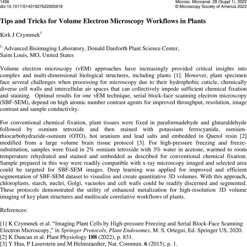
Image of the first page of this content. For PDF version, please use the ‘Save PDF’ preceeding this image.'
Type
3D Volume Electron Microscopy in Biology Research
Copyright
Copyright © Microscopy Society of America 2022

References

Czymmek, K et al. “Imaging Plant Cells by High-pressure Freezing and Serial Block-Face Scanning Electron Microscopy,” in Springer Protocols, Plant Endosomes, Ortegui, M. S., Ed. Springer US, 2020.Google Scholar
Duncan, K et al. Plant Physiology 188 (2022), p. 831.10.1093/plphys/kiab405CrossRefGoogle Scholar
Hua, Y, Laserstein, P and Helmstaedter, M, Nat. Commun. 6 (2015), p. 1.Google Scholar