Hostname: page-component-78c5997874-fbnjt Total loading time: 0 Render date: 2024-11-05T17:14:06.357Z Has data issue: false hasContentIssue false

Revealing the Structure of Graphitic Carbon Nitride through Low-Dose TEM using a Direct Electron Detector

Published online by Cambridge University Press:  04 August 2017

Diane M. Haiber
Affiliation:
School for Engineering of Matter, Transport & Energy, Arizona State University, Tempe, Arizona, USA
Peter A. Crozier
Affiliation:
School for Engineering of Matter, Transport & Energy, Arizona State University, Tempe, Arizona, USA

Abstract

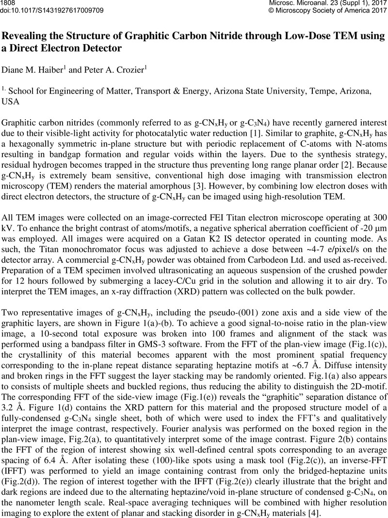
Image of the first page of this content. For PDF version, please use the ‘Save PDF’ preceeding this image.'
Type
Abstract
Copyright
© Microscopy Society of America 2017 

References

[1] Wang, X., et al, Nat. Mater. 8 2009). p. 76.CrossRefGoogle Scholar
[2] Lotsch, B.V., et al, Chem. Eur. J 13 2007). p. 4969.CrossRefGoogle Scholar
[3] Haiber, D.M., et al, Microsc. Microanal. 22(Suppl 3 2016). p. 986.CrossRefGoogle Scholar
[4] Support from the U.S. DOE (DE-SC0004954), use of AC-TEM at the John M. Cowley Center for High Resolution Electron Microscopy, use of the K2 IS camera courtesy of Gatan, and use of XRD equipment within the LeRoy Eyring Center for Solid State Science at ASU are gratefully acknowledged.Google Scholar