Hostname: page-component-77c89778f8-rkxrd Total loading time: 0 Render date: 2024-07-19T06:29:23.081Z Has data issue: false hasContentIssue false

Quick 3D Elemental Mapping of Biological Tissues using Super High Solid Angle EDS

Published online by Cambridge University Press:  05 August 2019

Yuuki Yamaguchi
Affiliation:
JEOL Ltd. 3-1-2 Musashino, Akishima, Tokyo 196-8558JAPAN
Hideo Nishioka
Affiliation:
JEOL Ltd. 3-1-2 Musashino, Akishima, Tokyo 196-8558JAPAN
Yukari Moriya
Affiliation:
JEOL Ltd. 3-1-2 Musashino, Akishima, Tokyo 196-8558JAPAN
Chikako Nakayama
Affiliation:
JEOL Ltd. 3-1-2 Musashino, Akishima, Tokyo 196-8558JAPAN
Tomohiro Haruta
Affiliation:
JEOL Ltd. 3-1-2 Musashino, Akishima, Tokyo 196-8558JAPAN
John Gilbert
Affiliation:
Bruker Nano GmbH. Am Studio 2D, 12489 Berlin, GERMANY
Shunsuke Asahina
Affiliation:
JEOL Ltd. 3-1-2 Musashino, Akishima, Tokyo 196-8558JAPAN

Abstract

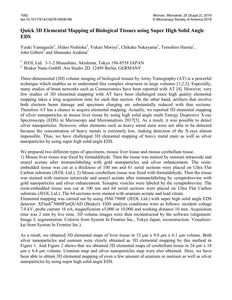
Image of the first page of this content. For PDF version, please use the ‘Save PDF’ preceeding this image.'
Type
Element Analysis of Biological Materials
Copyright
Copyright © Microscopy Society of America 2019 

References

[1]Micheva, K.D. and Smith, S.J., Neuron 55(1) (2007), p.25Google Scholar
[2]Horstmann, H. et al. , PLoS One. 7(4) (2012), e35172.Google Scholar
[3]Koga, D. et al. , Microscopy 65(2)(2016), p.145Google Scholar
[4]Kasthuri, et al. , Cell 162 (2015), p.648Google Scholar
[5]Yamaguchi, Y. et al. , Microsc. Microanal. 23 (Suppl 1) (2017), p.1180Google Scholar