Skip to main content Accessibility help
×
Hostname: page-component-78c5997874-4rdpn Total loading time: 0 Render date: 2024-11-10T02:33:51.976Z Has data issue: false hasContentIssue false

4 - Neuroradiology

Published online by Cambridge University Press:  05 March 2012
Kiat Tsong Tan
Affiliation:
Queen Elizabeth Hospital, Charlottetown
John Curtis
Affiliation:
University Hospital Aintree
Jessie Aw
Affiliation:
University of Chicago
Get access

Summary

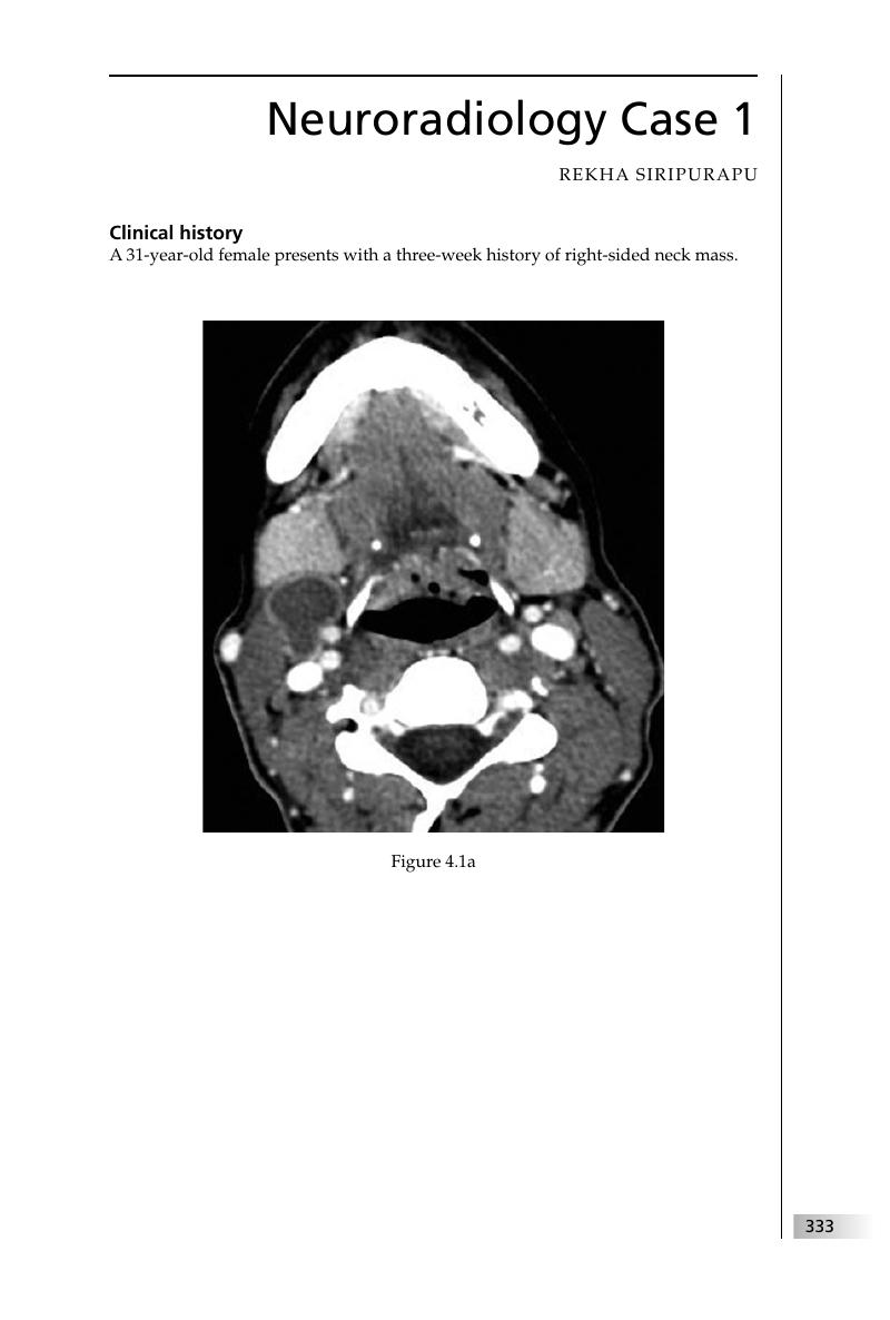
Image of the first page of this content. For PDF version, please use the ‘Save PDF’ preceeding this image.'
Type
Chapter
Information
Final FRCR 2B Viva
A Survival Guide
, pp. 333 - 452
Publisher: Cambridge University Press
Print publication year: 2011

Access options

Get access to the full version of this content by using one of the access options below. (Log in options will check for institutional or personal access. Content may require purchase if you do not have access.)

References

Hudgins, PA, Gillison, M. Second branchial cleft cyst: not!!AJNR Am J Neuroradiol 2009; 30: 1628–9.CrossRefGoogle ScholarPubMed
Koeller, KK, Alamo, L, Adair, CF, Smirniotopoulos, JG. Congenital cystic masses of the neck: radiologic–pathologic correlation. Radiographics 1999; 19: 121–46.CrossRefGoogle ScholarPubMed
Forghani, R, Farb, RI. Diagnosis and temporal evolution of signs of intracranial hypotension on MRI of the brain. Neuroradiology 2008; 50: 1025–34.CrossRefGoogle Scholar
Moyeri, NN, Henson, JW, Schaefer, PW, Zervas, NT. Spinal dural enhancement on magnetic resonance imaging associated with spontaneous intracranial hypotension. J Neurosurg 1998; 88; 912–18.CrossRefGoogle Scholar
Grossman, RI, Yousem, DM. Neuroradiology: the Requisites, 2nd edn. St Louis, MO: Mosby, 2003; 98–105.Google Scholar
Newton, HB, Jolesz, FA, eds. Handbook of Neuro-oncology Neuroimaging. New York, NY: Academic Press, 2008; 3–4, 13–14, 31.
Grossman, RI, Yousem, DM. Neuroradiology: the Requisites, 2nd edn. St Louis, MO: Mosby, 2003; 447–9.Google Scholar
Osborn, AG. Diagnostic Imaging: Brain. Salt Lake City, UT: Amirsys, 2004; I (10): 50–3.Google Scholar
Grossman, RI, Yousem, DM. Neuroradiology: the Requisites, 2nd edn. St Louis, MO: Mosby, 2003; 231–4.Google Scholar
Porter, PJ, Willinsky, RA, Harper, W, Wallace, MC. Cerebral cavernous malformation: natural history and prognosis after clinical deterioration with or without haemorrhage. J Neurosurg 1997; 87: 190–7.CrossRefGoogle ScholarPubMed
Grossman, RI, Yousem, DM. Neuroradiology: the Requisites, 2nd edn. St Louis, MO: Mosby, 2003; 288–90.Google Scholar
Grossman, RI, Yousem, DM. Neuroradiology: the Requisites, 2nd edn. St Louis, MO: Mosby, 2003; 248–52.Google Scholar
Barkovich, AJ.Morphologic characteristics of subcortical heterotopias: MR imaging study. AJNR Am J Neuroradiol 2000; 21: 290–5.Google ScholarPubMed
Kornienko, VN, Pronin, IN.Diagnostic Neuroradiology. Berlin: Springer, 2009; 39, 49, 415, 585.Google Scholar
Mafee, MF, Valvassori, GE, Becker, M. Sinonasal pathology. In Imaging of the Head and Neck, 2nd edn. Stuttgart: Thieme, 2005; 434–6.Google Scholar
Raghavan, P, Phillips, DC. Magnetic resonance imaging of sinonasal malignancies. Top Magn Reson Imaging 2007; 18: 259–67.CrossRefGoogle ScholarPubMed
Som, PM, Curtin, HD. Head and Neck Imaging, 4th edn. St Louis, MO: Mosby, 2002; 285–90.Google Scholar
Grossman, RI, Yousem, DM. Neuroradiology: the Requisites, 2nd edn. St Louis, MO: Mosby, 2003; 128–30.Google Scholar
Bonneville, F, Cattin, F, Marsot-Dupuch, K, et al. T1 signal hyperintensity in the sellar region: spectrum of findings. Radiographics 2006; 26: 93–113.CrossRefGoogle Scholar
Chan, YL, Tse, HY. Imaging in thalassemia. J HK Coll Radiol 2002; 5: 155–61.Google Scholar
Tunaci, M, Tunaci, A, Engin, G, et al. Imaging features of thalassemia. Eur Radiol 1999; 9: 1804–9.Google ScholarPubMed
Leach, JL, Fortuna, RB, Jones, BV, Gaskill-Shipley, MF. Imaging of cerebral venous thrombosis: current techniques, spectrum of findings, and diagnostic pitfalls. Radiographics 2006; 26: S19–43.CrossRefGoogle ScholarPubMed
Rodallec, MH, Krainik, A, Feydy, A, et al. Cerebral venous thrombosis and multidetector CT angiography: tips and tricks. Radiographics 2006; 26: S5–18.CrossRefGoogle ScholarPubMed
Fernandez, C, Girard, N, Paredes, AP, et al. The usefulness of MR imaging in the diagnosis of dysembryoplastic neuroepithelial tumour in children: a study of 14 cases. AJNR Am J Neuroradiol 2003; 24: 829–34.Google Scholar
Grossman, RI, Yousem, DM. Neuroradiology: the Requisites, 2nd edn. St Louis, MO: Mosby, 2003; 139.Google Scholar
Grossman, RI, Yousem, DM. Neuroradiology: the Requisites, 2nd edn. St Louis, MO: Mosby, 2003; 375–7.Google Scholar
Relkin, N, Marmarou, A, Klinge, P, Bergsneider, M, Black, PM. Diagnosing idiopathic normal pressure hydrocephalus. Neurosurgery 2005; 57: S4–16.CrossRefGoogle ScholarPubMed
Grossman, RI, Yousem, DM. Neuroradiology: the Requisites, 2nd edn. St Louis, MO: Mosby, 2003; 653–9.Google Scholar
Given, CA, Burdette, JH, Elster, AD, Williams, DW. Psuedo-subarachnoid haemorrhage: a potential imaging pitfall associated with diffuse cerebral oedema. AJNR Am J Neuroradiol 2003; 24: 254–6.Google Scholar
Gutierrez, LG, Rovira, A, Portela, , Leite C da, C, Lucato, LT. CT and MR in non-neonatal hypoxic–ischemic encephalopathy: radiological findings with pathophysiological correlations. Neuroradiology 2010; 52: 949–76.CrossRefGoogle Scholar
Grossman, RI, Yousem, DM. Neuroradiology: the Requisites, 2nd edn. St Louis, MO: Mosby, 2003; 105–8.Google Scholar
Stone, JA, Cooper, H, Castillo, M, Mukherji, SK. Malignant schwannoma of the trigeminal nerve. AJNR Am J Neuroradiol 2001; 22: 505–7.Google ScholarPubMed
Wippold, FJ, Lubner, M, Perrin, RJ, Lämmle, M, Perry, A. Neuropathology for the neuroradiologist: Antoni A and Antoni B tissue patterns. AJNR Am J Neuroradiol 2007; 28: 1633–8.CrossRefGoogle Scholar
Harnsberger, RH. Handbook of Head and Neck Imaging, 2nd edn. St Louis, MO: Mosby, 1995; 60–74.Google Scholar
Witt, RL.Major salivary gland cancer. Surg Oncol Clin N Am 2004; 13: 113–27.CrossRefGoogle ScholarPubMed
DeAngelis, LM. Brain tumors. N Eng J Med 2001; 344: 114–23.CrossRefGoogle ScholarPubMed
Ferner, RE. Neurofibromatosis 1 and neurofibromatosis 2: a twenty first century perspective. Lancet Neurol 2007; 6: 340–51.CrossRefGoogle ScholarPubMed
Rodriguez, D, Poussaint, TY. Neuroimaging findings in neurofibromatosis type 1 and 2. Neuroimaging Clin N Am 2004; 14: 149–70.CrossRefGoogle ScholarPubMed
Goldberg, AN, Oroszlan, G, Anderson, TD. Complications of frontal sinusitis and their management. Otolaryngol Clin N Am 2001; 34: 211–25.CrossRefGoogle ScholarPubMed
Greenlee, JE. Subdural empyema. Curr Treat Options Neurol 2003; 5: 13–22.CrossRefGoogle ScholarPubMed
Osborn, MK, Steinberg, JP. Subdural empyema and other suppurative complications of paranasal sinusitis. Lancet Infect Dis 2007; 7: 62–7.CrossRefGoogle ScholarPubMed
Thurnher, MM, Donovan Post, MJ. Neuroimaging in the brain in HIV-1-infected patients. Neuroimaging Clin N Am 2008; 18: 93–117.CrossRefGoogle ScholarPubMed
Gavrilovic, IT, Abrey, LE.Primary central nervous system lymphoma. Curr Oncol Rep 2004; 6: 388–95.CrossRefGoogle ScholarPubMed
Biondi, A. Intracranial aneurysms associated with other lesions, disorders or anatomic variations. Neuroimaging Clin N Am 2006; 16: 467–82.CrossRefGoogle ScholarPubMed
Grossman, RI, Yousem, DM. Neuroradiology: the Requisites, 2nd edn. St Louis, MO: Mosby, 2003; 500–1.Google Scholar
Ringer, AJ, Salud, L, Tomsich, TA.Carotid cavernous fistulas: anatomy, classification and treatment. Neurosurg Clin N Am 2005; 16: 279–95.CrossRefGoogle ScholarPubMed
Grossman, RI, Yousem, DM. Neuroradiology: the Requisites, 2nd edn. St Louis, MO: Mosby, 2003; 174–95.Google Scholar
Krapf, H, Widder, B, Skalej, M. Small rosarylike infarctions in the centrum ovale suggest hemodynamic failure. AJNR Am J Neuroradiol 1998; 19: 1479–84.Google ScholarPubMed
Momjian-Mayor, I, Baron, JC. The pathophysiology of watershed infarction in internal carotid artery disease: review of cerebral perfusion studies. Stroke 2005; 36: 567–77.CrossRefGoogle ScholarPubMed
Barbagallo, JS, Kolodzieh, MS, Silverberg, NB, Weinberg, JM. Neurocutaneous disorders. Dermatol Clin 2002; 20: 547–60.CrossRefGoogle ScholarPubMed
Koeller, KK, Rushing, EF. Pilocytic astrocytoma: radiologic–pathologic correlation. Radiographics 2004; 24: 1693–708.CrossRefGoogle ScholarPubMed
Rodriguez, D, Poussaint, TY. Neuroimaging findings in neurofibromatosis type 1 and 2. Neuroimaging Clin N Am 2004; 14: 149–70.CrossRefGoogle ScholarPubMed
Kornienko, VN, Pronin, IN.Diagnostic Neuroradiology. Berlin: Springer, 2009; 151.Google Scholar
Osborn, A, Tong, K. Handbook of Neuroradiology: Brain and Skull. St Louis, MO: Mosby, 1996; 52–3, 568–9.Google Scholar
Tisch, S, Silberstein, P, Limousin-Dowsey, P, Jahanshahi, M. The basal ganglia: anatomy, physiology and pharmacology. Psychiatr Clin N Am 2004; 27: 757–99.CrossRefGoogle ScholarPubMed
Kummer, R, Bourquain, H, Bastianello, S, et al. Early prediction of irreversible brain damage after ischemic stroke at CT. Radiology 2001; 219: 95–100.CrossRefGoogle Scholar
Speer, MC, Enterline, DS, Mehltretter, L, et al. Chiari type I malformation with or without syringomyelia: prevalence and genetics. J Genet Couns 2003; 12: 297–311.CrossRefGoogle ScholarPubMed
Tubbs, RS, Lyerly, MJ, Loukas, M, Shoja, MM, Oakes, WJ. The pediatric Chiari I malformation: a review. Childs Nerv Syst 2007; 23: 1239–50.CrossRefGoogle ScholarPubMed
Bonneville, F, Sourour, N, Biondi, A. Intracranial aneurysms: an overview. Neuroimaging Clin N Am 2006; 16: 371–82.CrossRefGoogle ScholarPubMed
Schievink, WI. Intracranial aneurysms. N Engl J Med 1997; 336: 28–40.CrossRefGoogle ScholarPubMed
Yanaka, K, Nagase, S, Asakawa, H, et al. Management of unruptured cerebral aneurysms in patients with polycystic kidney disease. Surg Neurol 2004; 62: 538–45.CrossRefGoogle ScholarPubMed
Maeder, PP, Holtås, SL, Basibüyük, LN, et al. Colloid cysts of the third ventricle: correlation of MR and CT findings with histology and chemical analysis. AJNR Am J Neuroradiol 1990; 11: 575–81.Google ScholarPubMed
Osborn, AG. Diagnostic Imaging: Brain. Salt Lake City, UT: Amirsys, 2004; I (7): 9–10.Google Scholar
Purdy, RA, Kirby, S. Headaches and brain tumors. Neurol Clin N Am 2004; 22: 39–53.CrossRefGoogle ScholarPubMed
Wilms, G, Marchal, G, Hecke, P, et al. Colloid cysts of the third ventricle: MR findings. J Comput Assist Tomogr 1990; 14: 527–31.CrossRefGoogle ScholarPubMed
Barkovich, AJ.Pediatric Neuroimaging, 4th edn. Philadelphia, PA: Lippincott Williams & Wilkins, 2005; 744–52.Google Scholar
Naidich, TP, Harwood-Nash, DC. Diastematomyelia: hemicord and meningeal sheaths; single and double arachnoid and dural tubes. AJNR Am J Neuroradiol 1983; 4: 633–6.Google ScholarPubMed
Gamba, JL, Woodruff, WW, Djang, WT, Yeates, AE.Craniofacial mucormycosis: assessment with CT. Radiology 1986; 160: 207–12.CrossRefGoogle ScholarPubMed
Manning, SC, Merkel, M, Kriesel, K, Vuitch, F, Marple, B. Computed tomography and magnetic resonance diagnosis of allergic fungal sinusitis. Laryngoscope 1997; 107: 170–6.CrossRefGoogle ScholarPubMed
Mukherji, SK, Figueroa RE, Ginsberg LE, et al. Allergic fungal sinusitis: CT findings. Radiology 1998; 207: 417–22.CrossRefGoogle ScholarPubMed
Suarez, JI, Tarr, RW, Selman, WR. Aneurysmal subarachnoid hemorrhage. N Engl J Med 2006; 354: 387–96.CrossRefGoogle ScholarPubMed
Gijn, J, Kerr, RS, Rinkel, GJ. Subarachnoid haemorrhage. Lancet 2007; 369: 306–18.CrossRefGoogle ScholarPubMed
Ganeshan, D, Narlawar, R, McCann, C, Lewis-Jones, H, Curtis, J. Cerebral venous thrombosis: a pictorial review. Eur J Radiol 2010; 74: 110–16.CrossRefGoogle ScholarPubMed
Linn, J, Pfefferkorn, T, Ivanicova, K, et al. Noncontrast CT in deep cerebral venous thrombosis and sinus thrombosis: comparison of its diagnostic value for both entities. AJNR Am J Neuroradiol 2009; 30: 728–35.CrossRefGoogle ScholarPubMed
Selim, M, Caplan, LR. Radiological diagnosis of cerebral venous thrombosis. Front Neurol Neurosci 2008; 23: 96–111.Google ScholarPubMed
Barkovich, AJ.Pediatric Neuroimaging, 4th edn. Philadelphia, PA: Lippincott Williams & Wilkins, 2005; 710–31.Google Scholar
Raghavan, N, Barkovich, AJ, Edwards, M, Norman, D. MR imaging in the tethered spinal cord syndrome. AJR Am J Roentgenol 1989; 152: 843–52.CrossRefGoogle ScholarPubMed
Go, JL, Vu, VN, Lee, KJ, Becker, TS. Orbital trauma. Neuroimaging Clin N Am 2002; 12: 311–24.CrossRefGoogle ScholarPubMed
Reppucci VS, Movshovich A. Current concepts in the treatment of traumatic injury to the posterior segment. Ophthalmol Clin N Am 1999; 12: 465–74.
Chavhan, GB, Babyn, PS, Thomas, B, Shroff, MM, Haacke, EM. Principles, techniques, and applications of T2*-based MR imaging and its special applications. Radiographics 2009; 29: 1433–49.CrossRef
Hammound, DA, Wasserman, BA. Diffuse axonal injury: pathophysiology and imaging. Neuroimaging Clin N Am 2002; 12: 205–16.CrossRefGoogle Scholar
Poussaint, TY, Moeller, KK. Imaging of pediatric head trauma. Neuroimaging Clin N Am 2002; 12: 271–94.CrossRefGoogle ScholarPubMed
Rhea, JT, Novelline, RA.How to simplify the CT diagnosis of Le Fort fractures. AJR Am J Roentgenol 2005; 184: 1700–5.CrossRefGoogle ScholarPubMed
Sun, JK, LeMay, DR. Imaging of facial trauma. Neuroimaging Clin N Am 2002; 12: 295–309.CrossRefGoogle ScholarPubMed
Aikele, P, Kittner, T, Offergeld, C, et al. Diffusion-weighted MR imaging of cholesteatoma in pediatric and adult patients who have undergone middle ear surgery. AJR Am J Roentgenol 2003; 181: 261–5.CrossRefGoogle ScholarPubMed
Semaan, MT, Megerian, CA. The pathophysiology of cholesteatoma. Otolaryngol Clin N Am 2006: 39; 1143–59.CrossRefGoogle ScholarPubMed
Shohet, JA, Jong, AL. The management of pediatric cholesteatoma. Otolaryngol Clin N Am 2002; 35: 841–51.CrossRefGoogle ScholarPubMed
Smith, JA, Danner, CJ. Complications of chronic otitis media and cholesteatoma. Otolaryngol Clin N Am 2006; 39: 1237–55.CrossRefGoogle ScholarPubMed
Jagannathan, J, Kanter, AS, Sheehan, JP, Jane, JA, Laws, ER. Benign brain tumors: sellar/parasellar tumors. Neurol Clin 2007; 25: 1231–49.CrossRefGoogle ScholarPubMed
Molitch, ME. Nonfunctioning pituitary tumors and pituitary incidentalomas. Endocrinol Metab Clin N Am 2008; 37: 151–71.CrossRefGoogle ScholarPubMed
Aygun, N, Zinreich, SJ. Imaging for functional endoscopic sinus surgery. Otolaryngol Clin N Am 2006; 39: 403–16.CrossRefGoogle ScholarPubMed
Becker, SS. Surgical management of polyps in the treatment of nasal airway obstruction. Otolaryngol Clin N Am 2009; 42: 377–85.CrossRefGoogle ScholarPubMed
Bikhazi, NB. Contemporary management of nasal polyps. Otolaryngol Clin N Am 2004; 37: 327–37.CrossRefGoogle ScholarPubMed
Crozier, E, Sumer, BD. Head and neck cancer. Med Clin North Am 2010; 94: 1031–46.CrossRefGoogle ScholarPubMed
Stambuk, HE, Karimi, S, Lee, N, Patel, SG. Oral cavity and oropharynx tumors. Radiol Clin North Am 2007; 45: 1–20.CrossRefGoogle ScholarPubMed
Apushkin, MA, Apushkin, MA, Shapiro, MJ, Mafee, MF. Retinoblastoma and simulating lesions: role of imaging. Neuroimaging Clin N Am 2005; 15: 49–67.CrossRefGoogle ScholarPubMed
Aygun, N, Zinreich, SJ.Imaging for functional endoscopic sinus surgery. Otolaryngol Clin N Am 2006; 39: 403–16.CrossRefGoogle ScholarPubMed
Har-El, G.Transnasal endoscopic management of frontal mucoceles. Otolaryngol Clin N Am 2001; 34: 243–51.CrossRefGoogle ScholarPubMed
McMains, KC, Kountakis, SE.Fronto-orbital-ethmoid mucoceles. Oper Tech Otolaryngol 2006; 17: 19–23.CrossRefGoogle Scholar
Hamilton, BE, Weissman, JL. Imaging of chemosensory loss. Otolaryngol Clin N Am 2004; 37: 1255–80.CrossRefGoogle ScholarPubMed
Potter, BO, Sturgis, EM. Sarcomas of the head and neck. Surg Oncol Clin N Am 2003; 12: 379–417.CrossRefGoogle ScholarPubMed
Weber, AL, Arayedh, SA, Rashid, A. Nasopharynx: clinical, pathologic, and radiologic assessment. Neuroimaging Clin N Am 2003; 13: 465–83.CrossRefGoogle ScholarPubMed
Altman, DA, Atkinson, DS, Brat, DJ. Best cases from the AFIP: glioblastoma multiforme. Radiographics 2007; 27: 883–8.CrossRefGoogle ScholarPubMed
Bot, JC, Barkhof, F. Spinal-cord MRI in multiple sclerosis: conventional and nonconventional MR techniques. Neuroimaging Clin N Am 2009; 19: 81–99.CrossRefGoogle ScholarPubMed
Courtney, AM, Treadaway, K, Remington, G, Frohman, E. Multiple sclerosis. Med Clin North Am 2009; 93: 451–76.CrossRefGoogle ScholarPubMed
Lubin, FD, Reingold, SC. Defining the clinical course of multiple sclerosis: results of an international survey. Neurology 1996; 46: 907–11.CrossRefGoogle Scholar
Simon, JH, Kleinschmidt-DeMasters, BK. Variants of multiple sclerosis. Neuroimaging Clin N Am 2008; 18: 703–16.CrossRefGoogle ScholarPubMed
Barkovich, AJ.Pediatric Neuroimaging, 4th edn. Philadelphia, PA: Lippincott Williams & Wilkins, 2005; 205–6.Google Scholar
Yuang, BY, Castillo, M. Hypoxic–ischemic brain injury: imaging findings from birth to adulthood. Radiographics 2008; 28: 417–39.Google Scholar
Umeoka, S, Koyama, T, Miki, Y, et al. Pictorial review of tuberous sclerosis in various organs. Radiographics 2008; 28: e32.CrossRefGoogle ScholarPubMed

Save book to Kindle

To save this book to your Kindle, first ensure [email protected] is added to your Approved Personal Document E-mail List under your Personal Document Settings on the Manage Your Content and Devices page of your Amazon account. Then enter the ‘name’ part of your Kindle email address below. Find out more about saving to your Kindle.

Note you can select to save to either the @free.kindle.com or @kindle.com variations. ‘@free.kindle.com’ emails are free but can only be saved to your device when it is connected to wi-fi. ‘@kindle.com’ emails can be delivered even when you are not connected to wi-fi, but note that service fees apply.

Find out more about the Kindle Personal Document Service.

Available formats
×

Save book to Dropbox

To save content items to your account, please confirm that you agree to abide by our usage policies. If this is the first time you use this feature, you will be asked to authorise Cambridge Core to connect with your account. Find out more about saving content to Dropbox.

Available formats
×

Save book to Google Drive

To save content items to your account, please confirm that you agree to abide by our usage policies. If this is the first time you use this feature, you will be asked to authorise Cambridge Core to connect with your account. Find out more about saving content to Google Drive.

Available formats
×