Skip to main content Accessibility help
×
Hostname: page-component-78c5997874-t5tsf Total loading time: 0 Render date: 2024-11-08T18:14:49.355Z Has data issue: false hasContentIssue false

1 - The normal child: growth and development of the infant and child; frequent and important normal variants

Published online by Cambridge University Press:  06 December 2010

Heike E. Daldrup-Link
Affiliation:
University of California, San Francisco
Charles A. Gooding
Affiliation:
University of California, San Francisco
Get access

Summary

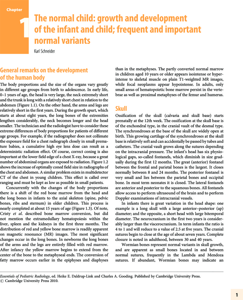
Image of the first page of this content. For PDF version, please use the ‘Save PDF’ preceeding this image.'
Type
Chapter
Information
Essentials of Pediatric Radiology
A Multimodality Approach
, pp. 1 - 18
Publisher: Cambridge University Press
Print publication year: 2010

Access options

Get access to the full version of this content by using one of the access options below. (Log in options will check for institutional or personal access. Content may require purchase if you do not have access.)

References

Allison, JW, Faddis, , Kinder, DLet al. (2000) Intracranial resistive index (RI) values in normal term infants during the first day of life. Pediatr Radiol 30, 618–20.CrossRefGoogle ScholarPubMed
Argyropoulou, M, Perignon, F, Brunelle, Fet al. (1991) Height of normal pituitary gland as a function of age evaluated by magnetic resonance imaging in children. Pediatr Radiol 21, 247–9.CrossRefGoogle ScholarPubMed
Barghouth, G, Prior, JO, Lepori, Det al. (2002) Paranasal sinuses in children: size evaluation of maxillary, sphenoid, and frontal sinuses by percentile curves. Eur Radiol 12, 1451–9.CrossRefGoogle ScholarPubMed
Barkovich, AJPediatric Neuroimaging, 4th edn. (Philadelphia: Lippincott Williams and Wilkins, 2005).Google Scholar
Bar-Ziv, J, Barki, Y, Itzchak, Yet al. (1984) Posterior mediastinal accessory thymus. Pediatr Radiol 14, 165–7.CrossRefGoogle ScholarPubMed
Berdon, WE, Baker, DH, James, LS (1965) The ductus bump. A transient physiologic mass in the chest roentgenograms in newborn infants. AJR Am J Roentgenol 95, 91–8.CrossRefGoogle Scholar
Blane, CE, Bockstein, F, DiPietro, Met al. (1985) Sonographic standards of normal infant kidney length. AJR Am J Roentgenol 145, 1289–92.CrossRefGoogle ScholarPubMed
Brodeur, AE, Silberstein, MJ, Graviss, ERRadiology of the Pediatric Elbow (Boston: Hall, 1981).Google Scholar
Budorick, NE, Pretouius, DH, Grafe, MRet al. (1991) Ossification of the fetal spine, Radiology 181, 561–5.CrossRefGoogle ScholarPubMed
Byrd, SE, Darling, CF, Wilczynski, MA (1993) White matter of the brain. NeuroimagingClin N Am 3, 247–66.Google Scholar
Chiara, A, Chirico, G, Vecchi, E, Rondini, G (1989) Ultrasonic evaluation of kidney length in term and preterm infants. Eur J Pediat 149, 94–5.CrossRefGoogle ScholarPubMed
Cohen, HL, Shapiro, MA, Mandel, FS, Shapiro, ML (1993) Normal ovaries in neonates and infants: sonographic study of 77 patients 1 day to 24 months old. AJR Am J Roentgenol 160, 583–6.CrossRefGoogle ScholarPubMed
Cohen, MM, McLean, RE (eds.) Craniosynostosis (New York: Oxford University Press, 2000).Google Scholar
Cristy, M (1981) Active bone marrow distribution as a function of age in humans. Phys Med Biol 26, 389–400.CrossRefGoogle ScholarPubMed
Deane, MGM, Burton, EM, Harlow, SAet al. (2001) Swallowing dysfunction in infants less than 1 year of age. Pediatr Radiol 31, 423–42.CrossRefGoogle Scholar
Deeg, KH, Rupprecht, T (1989) Pulsed Doppler sonographic measurement of normal values for the flow velocities in the intracranial arteries of healthy newborn. Pediatr Radiol 19, 71–8.CrossRefGoogle Scholar
Dinkel, E, Ertl, M, Dittrich, Met al. (1985) Kidney size in childhood sonographical growth charts for kidney length and volume. Pediatr Radiol 15, 38–43.CrossRefGoogle ScholarPubMed
Dittrich, M, Milde, S, Dinkel, Eet al. (1983) Sonographic biometry of liver and spleen size in childhood. Pediatr Radiol 13, 206.CrossRefGoogle ScholarPubMed
Edwards, DK, Higgins, CB, Gilpin, EA (1981) The cardiothoracic ratio in newborn infants. AJR Am J Roentgenol 136, 907–13.CrossRefGoogle ScholarPubMed
Engelbrecht, V, Scherer, A, Rassek, Met al. (2002) Diffusion-weighted MR imaging in the brain in children: findings in the normal brain and in the brain with white matter disease. Radiology 222, 410–18.CrossRefGoogle Scholar
Enriquez, G, Correa, F, Lucaya, Jet al. (2003) Potential pitfalls in cranial sonography. Pediatr Radiol 33, 110–17.CrossRefGoogle ScholarPubMed
Enriquez, G, Correa, F, Aso, Cet al. (2006) Mastoid fontanel approach for sonographic imaging of the neonatal brain. Pediatr Radiol 36, 532–40.CrossRefGoogle Scholar
Fischer, JK (1978) Technical notes: skin fold vs. pneumothorax. AJR Am J Roentgenol 130, 791–2.CrossRefGoogle Scholar
Fraser-Hill, MA, Atri, M, Bret, PMet al. (1990) Intrahepatic portal venous system: variation demonstrated with duplex and color Doppler US. Radiology 177, 523–6.CrossRefGoogle Scholar
Friedman, DM, Schacht, RG (1991) Doppler waveforms in the renal arteries of normal children. J Clin Ultrasound 19, 387–92.CrossRefGoogle ScholarPubMed
Galanski, M, Nagel, H-D, Stamm, G (2007) (in German) Pädiatrische CT Expositionspraxis in Deutschland. ROFO 179, 1110–11. www.drg-apt.de.Google Scholar
Gallego, C, Miralles, M, Marin, Cet al. (2004) Congenital hepatic shunts. Radiographics 24, 755–72.CrossRefGoogle ScholarPubMed
Girodias, JB, Azouz, EM, Maarton, D (1991) Intervertebral disk space calcification. A report of 51 children with a review of the literature. Pediatr Radiol 21, 541–6.CrossRefGoogle ScholarPubMed
Gollogly, S, Smith, JT, White, SKet al. (2004) The volume of lung parenchyma as a function of age: a review of 1050 normal CT scans of the chest with three-dimensional volumetric reconstruction of the pulmonary system. Spine 29, 2061.CrossRefGoogle ScholarPubMed
Gooding, CACranial sutures and fontanelles. In Radiology of the Skull and Brain, vol. 1 The Skull, ed. Newton, TH, Potts, DJ (St. Louis: Mosby, 1971) pp. 216–40.Google Scholar
Gooding, CA, Neuhauser, EBD (1965) Growth and development of the vertebral bodies in the presence and absence of normal stress. AJR Am J Roentgenol 93, 388–93.Google Scholar
Graf, R (1984) Classification of hip joint dysplasia by means of sonography. Arch Orthop Trauma Surg 102, 248–55.CrossRefGoogle ScholarPubMed
Graf, R, Tschauner, C, Klapsch, W (1993) Process in prevention of late developmental dislocation of the hip by sonographic newborn hip “screening”: results of a comparative follow-up study. J Pediatr Orthop 2, 115–21.CrossRefGoogle Scholar
Grattan-Smith, JD, Jones, RA (2006) MR urography in children. Pediatr Radiol 36, 1119–32.CrossRefGoogle ScholarPubMed
Gressens, P, Hüppi, PSNormal and abnormal brain development. In Fanaroff and Martin's Neonatal-Perinatal Medicine: Diseases of the Fetus and Infant, 8th edn., ed. Martin, RJ, Fanaroff, AA, Walsh, MC (Philadelphia: Mosby, 2006) pp. 883–908.Google Scholar
Greulich, WW, Pyle, SIRadiographic Atlas of Skeletal Development of the Hand and the Wrist, 2nd edn. (Stanford: Stanford University Press, 1959).Google Scholar
Griscom, NT (1982) Computed tomographic determination of tracheal dimensions in children and adolescents. Radiology 145, 361.CrossRefGoogle ScholarPubMed
Haber, HP, Mayer, EI (1994) Ultrasound evaluation of uterine and ovarian size from birth to puberty. Pediatr Radiol 24, 11–13.CrossRefGoogle ScholarPubMed
Han, BK, Babcock, DS (1985) Sonographic measurements and appearance of normal kidneys in children. AJR Am J Roentgenol 145, 611.CrossRefGoogle ScholarPubMed
Harcke, HT (1995) The role of ultrasound in diagnosis and management of developmental dysplasia of the hip. Pediatr Radiol 25, 225–7.CrossRefGoogle ScholarPubMed
Harcke, HT, Grissom, (1990) Performing dynamic sonography of the infant hip. AJR Am J Roentgenol 155, 837–44.CrossRefGoogle ScholarPubMed
Hernanz-Schulman, M, Ambrosino, MM, Freeman, PCet al. (1995) Common bile duct in children: sonographic dimensions. Radiology 195, 193–5.CrossRefGoogle ScholarPubMed
Hoffer, FA, Hanabergh, AM, Teele, RL (1985) The interreniculare junction: a mimic of renal scarring on normal pediatric sonograms. AJR Am J Roentgenol 145, 1075–8.CrossRefGoogle ScholarPubMed
Jequier, S, Rousseau, O (1987) Sonographic measurements of the normal bladder wall in children. AJR Am J Roentgenol 149, 563–6.CrossRefGoogle ScholarPubMed
Katz, ME, Siegel, MJ, Shackelford, GDet al. (1987) The position and mobility of the duodenum in children. AJR Am J Roentgenol 148, 947–51.CrossRefGoogle ScholarPubMed
Keats, TE, Anderson, MWAtlas of Normal Roentgen Variants that may Simulate Disease, 7th edn. (St. Louis: Mosby, 2001).Google Scholar
Kendall, KA, McKenzie, S, Leonard, RJet al. (2000) Timing of events in normal swallowing: a videofluoroscopic study. Dysphagia 15, 74–83.CrossRefGoogle Scholar
Knapp, MS, Wezel-Meijler, G, Barth, PGet al. (1996) Normal gyration and sulcation in preterm and term neonates: appearance on MR images. Radiology 200, 389–96.CrossRefGoogle Scholar
Kramer, SS (1989) Radiologic examination of the swallowing impaired child. Dysphagia 3, 117–25.CrossRefGoogle ScholarPubMed
Kricun, ME (1985) Red-yellow marrow conversion: its effect on the location of some solitary bone lesions. Skeletal Radiol 14, 10–19.CrossRefGoogle ScholarPubMed
Lachman, RSTaybi and Lachman's Radiology of Syndromes, Metabolic Disorders, and Skeletal Dysplasias, 5th edn. (Philadelphia: Mosby, 2007).Google Scholar
LeQuesne, GW, Reilly, BJ (1975) Functional immaturity of the large bowel in the newborn infant. Radiol Clin North Am 13, 331–42.Google Scholar
Libicher, M, Troger, J (1992) US measurement of the subarachnoid space in infants: normal values. Radiology 184, 749–51.CrossRefGoogle ScholarPubMed
Lindskoug, BA (1992) Reference man in diagnostic radiology dosimetry. Radiat Prot Dosim 43, 111–14.CrossRefGoogle Scholar
Martin, RJ, Fanaroff, AA, Walsh, MC (eds.) Fanaroff and Martin's Neonatal-Perinatal Medicine: Diseases of the Fetus and Infant, 8th edn. (Philadelphia: Mosby, 2006).Google Scholar
McAlister, WH, Askin, FB (1983) The effect of some contrast agents in the lung: an experimental study in the rat and dog. AJR Am J Roentgenol 140, 245–51.CrossRefGoogle Scholar
Metrewelli, C, So, NM, Chu, WC, Lam, WW (2004) Magnetic resonance cholangiography in children. Br J Radiol 77, 1059–64.CrossRefGoogle Scholar
Okada, Y, Aoki, S, Barkovich, Aet al. (1989) Cranial bone marrow in children: assessment of normal development with MR imaging. Radiology 171, 161–4.CrossRefGoogle ScholarPubMed
Ontel, FK, Ivanovic, M, Ablin, DSet al. (1996) Bone age in children of diverse ethnicity. AJR Am J Roentgenol 167, 1395–8.CrossRefGoogle Scholar
Oppenheimer, DA, Carroll, BA, Yousem, S (1983) Sonography of the normal neonatal adrenal gland. Radiology, 157–60.CrossRefGoogle ScholarPubMed
Osenfeld, DL, Schoenfeld, SM, Underberg-Davis, S (1997) Coarctation of the lateral ventricles: an alternative explanation for subependymal pseudocysts. Pediatr Radiol 27, 895–7.CrossRefGoogle Scholar
Pettersson, H, Ringertz, HMeasurements in Pediatric Radiology, 1st edn. (London: Springer, 1991).CrossRefGoogle Scholar
Pyle, SI, Hoerr, NLA Radiographic Standard of Reference of the Growing Knee (Springfield: Charles C. Thomas, 1969).Google Scholar
Ricci, C, Cova, M, Kang, Yet al. (1990) Normal age-related patterns of cellular and fatty bone marrow distribution in the axial skeleton: MR imaging study. Radiology 177, 83–8.CrossRefGoogle ScholarPubMed
Richter, E, Lierse, WImaging Anatomy of the Newborn (Baltimore: Urban and Schwarzenberg, 1991).Google Scholar
Robbins, WJ, Brody, S, Hogan, AGGrowth (New Haven: Yale University Press, 1928).Google Scholar
Robinson, M, Chumlea, WC (1982) Standards for limb bone length ratios in children. Radiology 143, 433–6.Google Scholar
Rosen, L, Bowden, DH, Uchida, I (1957) Structural changes in pulmonary arteries in first year of life. AMA Arch Pathol 63, 316–17.Google ScholarPubMed
Rosenberg, HK, Markowitz, RI, Kolbert, Het al. (1994) Normal splenic size in infants and children: sonographic measurement. AJR Am J Roentgenol 157, 119.CrossRefGoogle Scholar
Sacton, HM, Borzyskowski, M, Mundy, ARet al. (1988) Spinning top urethra: not a normal variant. Radiology 168, 147–50.Google Scholar
Scammon, RE, Norris, EH (1918) On the time of the post-natal obliteration of the fetal blood-passage (foramen ovale, ductus arteriosus, ductus venosus). Anat Rec 15, 165–80.CrossRefGoogle Scholar
Schlesinger, AE, White, DK, Mallory, GBet al. (1995) Estimation of total lung capacity from chest radiography and chest CT in children: comparison with body plethysmography. AJR Am J Roentgenol 165, 151.CrossRefGoogle ScholarPubMed
Schumacher, R, Knoll, B, Schwarz, M, Ermert, JA (1992) M-mode sonography of the caudal spinal cord in patients with meningomyelocele: work in progress. Radiology 184, 263–5.CrossRefGoogle ScholarPubMed
Shopfner, CE (1966) Periosteal bone growth in normal infants: a preliminary report. AJR Am J Roentgenol 97, 154–63.CrossRefGoogle ScholarPubMed
Shopfner, CE, Hutch, JA (1967) The normal urethrogram. Radiol Clin North Am 6, 165–89.Google Scholar
Siegel, MJ, Martin, KW, Worthington, JL (1987) Normal and abnormal pancreas in children: US studies. Radiology 165, 15.CrossRefGoogle ScholarPubMed
Skandalakis, JE, Gray, SW (eds.) Embryology for Surgeons, 2nd edn. (Baltimore: Williams and Wilkins, 1994) pp. 414–50.Google Scholar
Slovis, TL (ed.) Caffey's Pediatric Diagnostic Imaging (Philadelphia: Mosby, 2008).Google Scholar
Staudt, M, Schropp, C, Staudt, Fet al. (1993) Myelination of the brain in MRI: a staging system. Pediatr Radiol 23, 169–76.CrossRefGoogle ScholarPubMed
Swischuk, (1977) Anterior displacement of C2 in children: physiologic or pathologic? A helpful differentiating line. Radiology 122, 759–63.CrossRefGoogle ScholarPubMed
Swischuk, Alimentary tract, esophagus. In Imaging of the Newborn, Infant, and Young Child, ed. Swischuk, (Philadelphia: Lippincott Williams and Wilkins, 2004) pp. 342–9.Google Scholar
Swischuk, Alimentary tract, esophagus. In Imaging of the Newborn, Infant, and Young Child, ed. Swischuk, (Philadelphia: Lippincott Williams and Wilkins, 2004) pp. 539–41.Google Scholar
Swischuk, , Swischuk, PN, John, SD (1993) Wedging of C-3 in infants and children: usually a normal finding and not a fracture. Radiology 188, 523–6.CrossRefGoogle Scholar
Taccone, A, Oddone, M, Dell'Acqua, ADet al. (1995) MRI “road-map” of normal age-related bone marrow. II. Thorax, pelvis and extremities. Pediatr Radiol 25, 596–606.CrossRefGoogle ScholarPubMed
Taccone, A, Oddone, M, Occhi, Met al. (1995) MRI “road-map” of normal age-related bone marrow. I. Cranial bone and spine. Pediatr Radiol 25, 588–95.CrossRefGoogle ScholarPubMed
Teitel, DF, Iwamoto, HS, Rudolph, AM (1987) Effects of birth-related events on central blood flow patterns. Pediatr Res 22, 557–66.CrossRefGoogle ScholarPubMed
Theocharopoulos, N, Damilakis, J, Perisinakis, K, Gourtsoyiannis, N (2007) Energy imparted-based estimates of the effect of z overscanning on adult and pediatric patient effective doses from multi-slice computed tomography. Med Phys 34, 1139–52.CrossRefGoogle Scholar
Tola, V, Kuhns, L, Kauffman, R (1993) Correlation of gastric emptying at one and two hours following formula feeding. Pediatr Radiol 23, 26–8.CrossRefGoogle Scholar
Ueda, D (1987) Sonographic measurement of the pancreas in children. J Clin Ultrasound 17, 417.CrossRefGoogle Scholar
Unsinn, KM, Geley, T, Freund, MC, Gassner, I (2000) US of the spinal cord in newborn: spectrum of normal findings, variants, congenital anomalies, and the acquired diseases. Radiographics 20, 923–38.CrossRefGoogle ScholarPubMed
Vock, P, Malanowski, D, Tscheappeler, Het al. (1987) Computed tomographic lung density in children. Invest Radiol 22, 627.CrossRefGoogle ScholarPubMed
Waitches, G, Zawin, J, Poznanski, A (1994) Sequence and rate of bone marrow conversion in the femora of children as seen on MR imaging: are accepted standards accurate?AJR Am J Roentgenol 162, 1399–1406.CrossRefGoogle ScholarPubMed
Walsh, E, Cramer, B, Pushpanthan, C (1990) Pancreatic echogenicity in premature and newborn infants. Pediatr Radiol 20, 323.CrossRefGoogle ScholarPubMed
Wittenborg, MH, Gyepes, MT, Crocker, D (1976) Tracheal dynamics in infants with respiratory distress, stridor and collapsing trachea. Radiology 88, 653–62.CrossRefGoogle Scholar
Wolf, S, Schneble, F, Troger, J (1992) The conus medullaris: time of ascendence to normal level. Pediatr Radiol 22, 590–2.CrossRefGoogle ScholarPubMed
Wolf, G, Wolfgang, A, Kuhn, F (1993) Development of the paranasal sinuses in children: implications for paranasal sinus surgery. Ann Otol Rhinol Laryngol 102, 705–11.CrossRefGoogle ScholarPubMed
Wolpert, SM, Osborn, M, Anderson, Met al. (1988) Bright pituitary gland: a normal MR appearance in infancy. AJNR Am J Neuroradiol 9, 1–3.Google ScholarPubMed
Zerin, JM, DiPietro, MA (1992) Superior mesenteric vascular anatomy at US in patient with surgically proved malrotation of the midgut. Radiology 183, 693–4.CrossRefGoogle ScholarPubMed
Zimmermann, MB, Hess, SY, Molinari, Let al. (2004) New reference values for thyroid volume by ultrasound in iodine-sufficient schoolchildren: a World Health Organization/Nutrition for Health and Development Iodine Deficiency Study Group Report. Am J Clin Nutr 79, 231–7.CrossRefGoogle ScholarPubMed

Save book to Kindle

To save this book to your Kindle, first ensure [email protected] is added to your Approved Personal Document E-mail List under your Personal Document Settings on the Manage Your Content and Devices page of your Amazon account. Then enter the ‘name’ part of your Kindle email address below. Find out more about saving to your Kindle.

Note you can select to save to either the @free.kindle.com or @kindle.com variations. ‘@free.kindle.com’ emails are free but can only be saved to your device when it is connected to wi-fi. ‘@kindle.com’ emails can be delivered even when you are not connected to wi-fi, but note that service fees apply.

Find out more about the Kindle Personal Document Service.

Available formats
×

Save book to Dropbox

To save content items to your account, please confirm that you agree to abide by our usage policies. If this is the first time you use this feature, you will be asked to authorise Cambridge Core to connect with your account. Find out more about saving content to Dropbox.

Available formats
×

Save book to Google Drive

To save content items to your account, please confirm that you agree to abide by our usage policies. If this is the first time you use this feature, you will be asked to authorise Cambridge Core to connect with your account. Find out more about saving content to Google Drive.

Available formats
×