Park NX12 Microscope for NanoScale Imaging
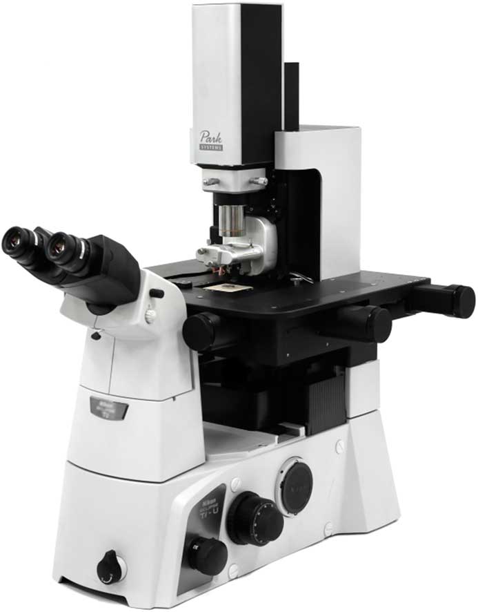
The Park NX12 is an inverted optical microscope based SPM platform for SICM, SECM, and SECCM, in addition to atomic force microscopy for research on a broad range of materials from organic to inorganic, transparent to opaque, and soft to hard. Park NX12 is suited for advanced research on materials such as membranes, organic devices and electronics, and biological and pathological samples in both air and liquid, plus a solution and platform for pipette-based SPM techniques.
Park Systems
Olympus BX53 Microscope with High-Luminosity LED
With an LED illuminator equivalent to a 100-watt halogen lamp, the Olympus BX53 microscope delivers outstanding brightness and true-to-life images. The BX53’s ergonomic design and ease of use make it an ideal system for clinical laboratories, while the LED illuminator’s brightness makes the system an excellent solution for up to 26 observation-head teaching systems. The BX53’s integrated Light Intensity Manager and ergonomic design make it comfortable and convenient to use in transmitted light applications.

Olympus Corporation
Helios G4 Plasma FIB System
The Helios G4 plasma FIB system is Thermo Fisher’s latest-generation DualBeam microscope. It can perform a wide variety of failure analysis applications, from high-speed delayering to SEM cross-section imaging of devices and TEM sample preparation. Semiconductor delayering is an increasingly important application in fault localization at sub-14 nm technology nodes. The plasma FIB and proprietary Dx chemistry is used to expose metallization layers, allowing electrical fault isolation and analysis to be performed with Thermo Fisher nanoprobing tools.

Thermo Fisher Scientific Inc.
SRRF-Stream – Andor’s Super-Resolution Microscopy Camera
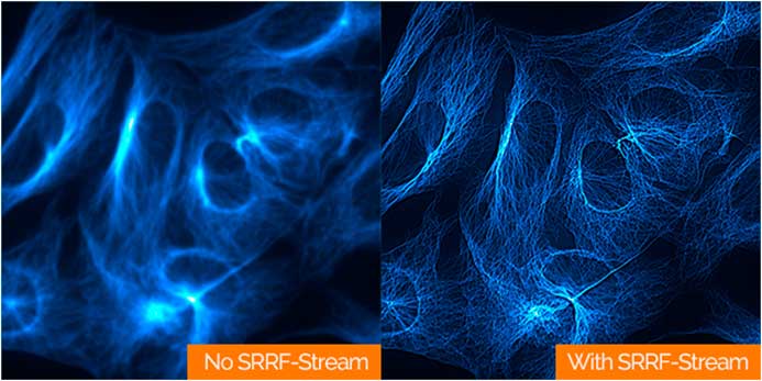
Andor Technology announced the launch of a new super-resolution microscopy technology, available on single-photon sensitive iXon EMCCD cameras. SRRF-Stream unlocks real-time, super-resolution fluorescence microscopy on most modern microscopes, using conventional fluorophores at low illumination intensities, thus making it highly compatible with live cell imaging. A resolution improvement from 2- to 6-fold (50–150 nm final resolution) can be expected for most datasets.
Andor Technology, an Oxford Instruments company
ImageXpress® Nano Automated Imaging System
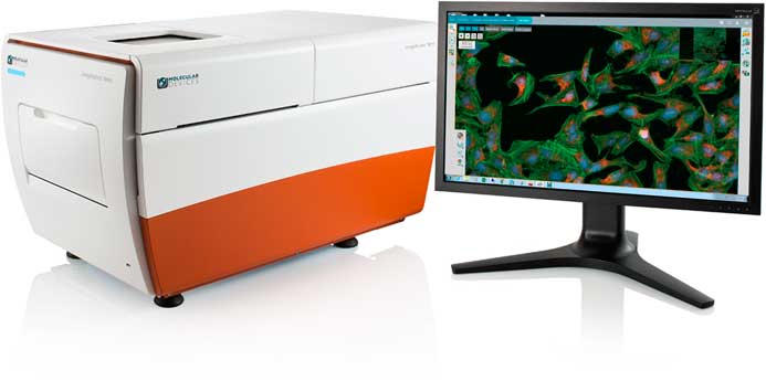
The ImageXpress® Nano Automated Imaging System enables researchers to get better data faster and improve collaborations with peers—anywhere, anytime. This cost-effective automated microscopy system allows researchers to scale up their experiments to multi-slide or microplate-level evaluations. The new integrated, browser-based imaging and analysis software is also easy to learn and can be remotely run using predefined protocols for specific types of experiments while having the flexibility to build custom protocols.
Molecular Devices, LLC
Phenom-World Announces Phenom Pro and ProX SEMs
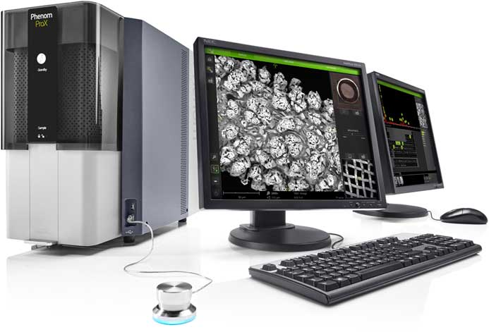
Phenom-World introduced its fifth-generation Phenom Pro and ProX SEMs. The systems’ enhanced imaging performance provide 20 percent resolution improvement thanks to new electronics and an improved lens, larger choice of detectors, and new software. This significantly widens their application range, while still maintaining the ease-of-use that Phenom-World’s SEMs are known for. Their performance offers a serious alternative to floor model SEMs in applications that include materials science, industrial manufacturing, electronics, earth science, life sciences, education, and more.
Phenom-World
ZEISS Celldiscoverer 7 for Live Cell Imaging
ZEISS Celldiscoverer 7 combines the user-friendly automation features of a boxed microscope with the image quality and flexibility of an inverted research microscope. The new optical design of ZEISS Celldiscoverer 7, together with advanced automatic calibration routines, helps researchers to achieve reproducible high-quality data even from complex long-term, time-lapse imaging experiments. A new hardware-based autofocus not only finds and keeps the focus automatically, but also detects the thickness and optical properties of the sample carrier.
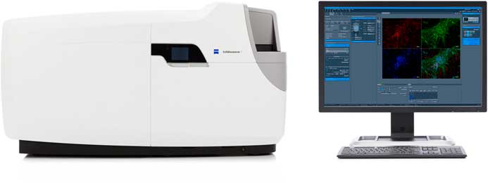
Carl Zeiss AG
XEI Scientific Introduces the Evactron® E50 Plasma Cleaner for Fast Hydrocarbon Removal
The Evactron E50 Hollow Cathode Remote Plasma Source from XEI Scientific uses up to 50 watts to generate flowing afterglow cleaning with air, removing carbon compounds from vacuum chambers operating with turbomolecular pumps. The unique new system has instant ignition from any vacuum level. Compact and powerful, it uses a 50-watt external hollow cathode electrode to produce plasma for fast, reliable, high-performance cleaning.
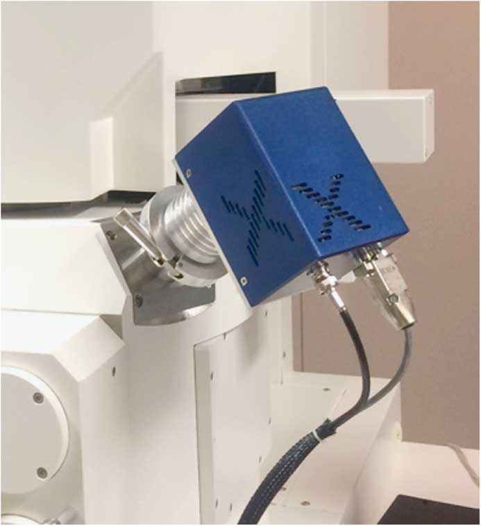
XEI Scientific, Inc.
Thermo Fisher Themis S TEM
The Themis S system is Thermo Fisher’s latest addition to the industry-standard Themis TEM platform. Targeted at the needs of semiconductor failure analysis labs working at the sub-20 nm technology node, the Themis S system is designed for high-volume semiconductor imaging and analysis and includes an integrated vibration isolation enclosure and full remote operation capability. The probe-corrected 80–200 kV column, automated alignments, XFEG source, and DualX X-ray spectrometer provide robust, sub-Ångström imaging and fast, accurate elemental and strain analysis.
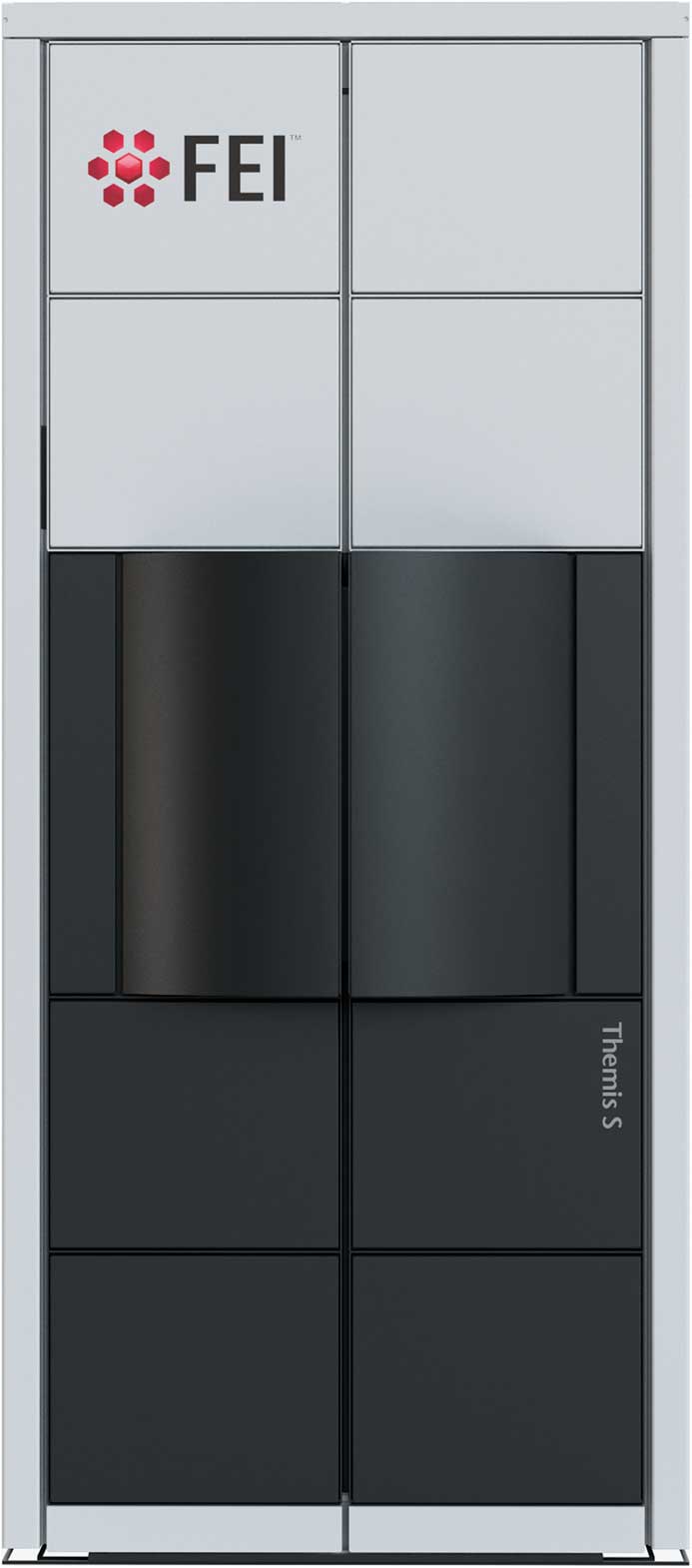
Thermo Fisher Scientific Inc.
Unique Adjustable Device Replaces Monochromators and Bandpass Filters
Spectrolight’s Flexible Wavelength Selector is ideal for any kind of spectral imaging. With a FWS, a microscopist can take wide-field images at different wavelengths and with different spectral bandwidths without resorting to filter wheels and such. It can also be used with broadband light sources such as the Spectrolight Mighty Light to produce a tunable beam of narrowband light with real-time user control of center wavelength and bandwidth.
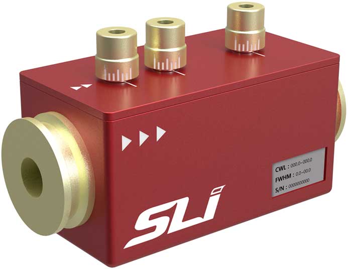
Spectrolight, Inc
Oxford Instruments Enables Real-Time Chemical Imaging
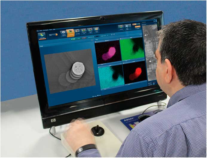
Oxford introduced a real-time navigation and imaging system for the SEM that helps users interactively explore their sample based on video-rate X-ray maps, live-colored by element to see what elements are present, rather than just the black and white electron image alone. This unique capability is enabled by the combination of two new products, a new fast silicon drift detector, Ultim® Max, and the real-time EDS analysis software, AZtecLive.
Oxford Instruments NanoAnalysis
Olympus DSX510 Digital Microscope
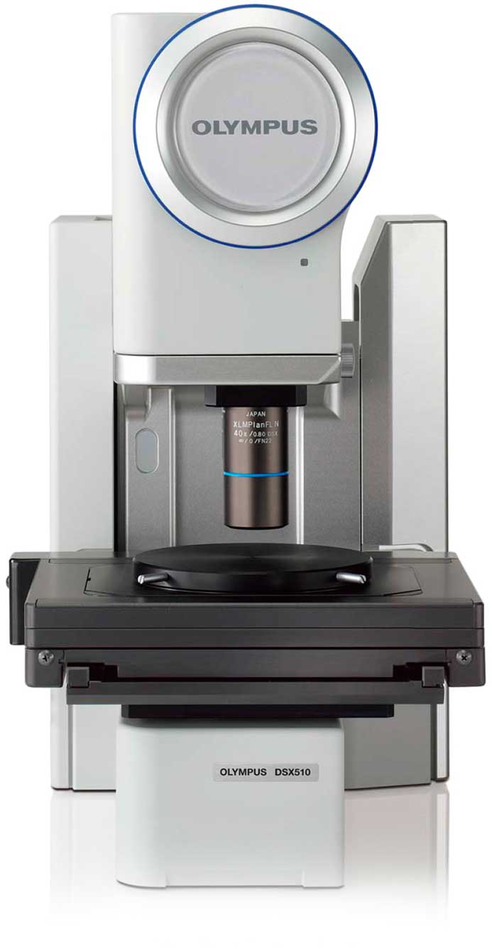
The Olympus DSX510 digital microscope system allows even first-time users to immediately produce superior images and highly reliable results. The DSX510 delivers efficient observation, simple image capture, extremely accurate measurement, and easy sharing of results. With lenses that have higher NA and lower aberration than current digital microscopes, plus improved evenness of light intensity, the DSX510 series of digital microscopes offer high resolution equal to the very best light microscopes.
Olympus Corporation
TMC Introduces SEM-Base® VI Active Piezoelectric Vibration Cancelation System
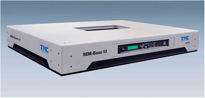
TMC introduced SEM-Base® VI, the next generation of its STACIS® active piezoelectric vibration cancelation product line. SEM-Base VI provides, on average, 6 dB improved vibration isolation performance over previous models, depending on the frequency and direction of the input. Additionally, TMC’s next-generation controller, the DC-2020, features a new dual-core processor and provides tool owners and researchers with a very simple and easy-to-use graphical interface for fast system assessment and operational surety.
TMC is a unit of AMETEK Ultra Precision Technologies
Quartz Tuning Forks for AFM/STM
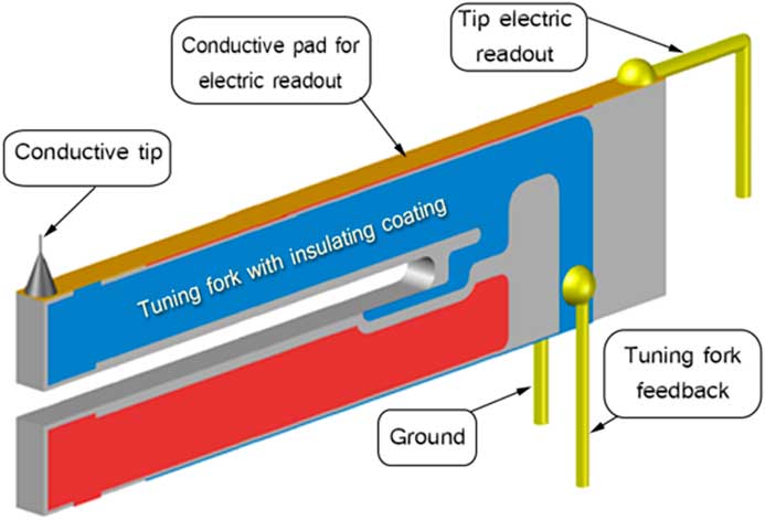
NaugaNeedles now offers tuning-fork-based probes for SPM applications. A third electrode has been added to the top of one of the tuning fork prongs. It is connected to the tip for tunneling current/conductivity readout without cross talk with the fork electrodes. Because the additional mass to the fork is minimal, the quality factor and the resonance frequency do not change significantly. The tip sharpness can be as small as 15 nm.
NaugaNeedles
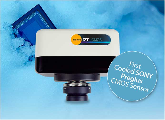
SPOT RT sCMOS Uses Sony’s Breakthrough Pregius™ CMOS Sensor from SPOT Imaging
The SPOT RT sCMOS camera uses Sony’s breakthrough Pregius™ CMOS sensor. Now you can experience unprecedented speed and sensitivity in a scientific CMOS camera with MP live and captured image resolution. Deep cooling allows dim images to be seen without becoming obscured by dark current. A global shutter ensures undistorted images of moving specimens. The RT sCMOS is optimal for fluorescence microscopy, FISH, GFP imaging, immunofluorescence, and 3D deconvolution applications.
SPOT Imaging
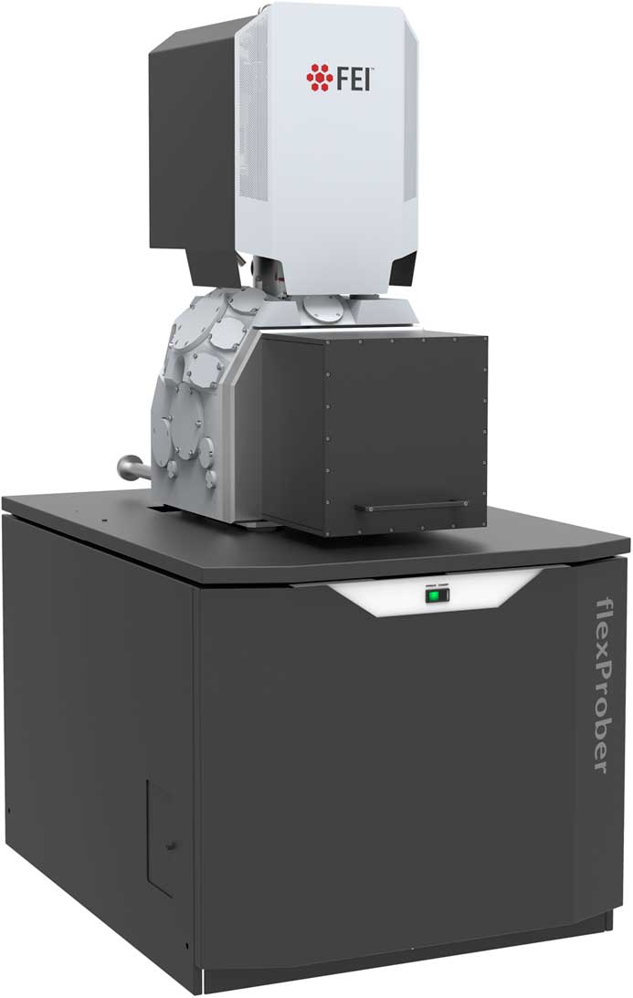
Flexprober System is Used for Fast Electrical Fault Isolation
The new flexProber system is designed to help engineers quickly locate and identify electrical faults, using an SEM to position fine mechanical probes on exposed circuit elements. Accurately locating the fault can improve productivity and cost-effectiveness in subsequent analysis by ensuring that the fault is included when a thin section is extracted for high-resolution imaging in a TEM. The flexProber system includes a SEM column designed for probing applications.
Thermo Fisher Scientific Inc.


