Introduction
Microscopists are looking at smaller features every day, and the potential to see artifacts that degrade or obscure these small features has never been greater. Because many current microscopy techniques are conducted in vacuum environments, residual gaseous components in the vacuum chamber may have substantial effects on the data collected. This brief review will describe some approaches to keep the quality of data collected in electron and ion-beam microscopes at the highest possible levels. In short, we will attempt to “keep it clean.”
Carbon Contamination
The problem of hydrocarbon contamination inside the electron microscope is well documented and has been an issue from the earliest days of electron microcscopy. This artifact is the result of the electron (or ion) beam striking unwanted contaminant molecules and promoting the growth of carbonaceous materials on the surface of the sample. Because fewer low-energy, secondary electrons reach the detector from the sample surface, the contamination region often appears as a darkened area in the secondary electron image. Typically, in a beam-scanning instrument, this contamination layer is in the shape of the rastered pattern on the sample—a rectangle. The effect is more pronounced at high magnifications and lower accelerating voltages—just the conditions under which smaller surface features are often analyzed. Further, if the contaminated area is measured in an atomic force microscope (AFM), the build-up of this material has been shown to be quite pronounced [Reference Amman1]. Of course, the first method of reducing contamination is to use lint-free or powder-free gloves when handling specimens and any microscope part inside the vacuum. Cleaning the specimen before placing it in the scanning electron microscope (SEM) helps, but there is always a small amount of hydrocarbon in the system coming from the scope itself (machining processes in chamber manufacture, lubricants in the stage, O-rings, etc.) that are difficult to eliminate. Hence, there is the requirement for chamber cleaning.
Methods for Removing Contamination
Conventional plasma cleaners. The original work by Zaluzec [2] resulted in a patent and commercial products that use plasma to clean samples and parts destined for use in electron microscopes. This has allowed scientists to remove substantial contamination artifacts from their images. Follow-on work, such as that of Isabell and Fischione [Reference Isabell and Fischione3], continued to demonstrate the value of plasma cleaning to remove problematic hydrocarbons and even to clean contaminated samples if they have already suffered beam-induced carbonaceous polymerization on their surfaces. Most of these units use argon or argon/oxygen mixtures. In these systems, the energetic ions interact with the surfaces to eliminate the unwanted artifacts.
Plasma cleaners are now common in the electron microscope (EM) suite with users in many disciplines cleaning both samples and sample holders prior to microscopy. These ex-situ plasma cleaners are available from several manufacturers at various levels of sophistication and price. However, these instruments do not address the internal surfaces of the microscope, which may harbor miniscule amounts of mobile hydrocarbon contaminants that cannot be completely removed. Disassembly and manual cleaning is not very practical, and, needless to say, highly labor intensive (read: expensive).
Other methods. In reducing contaminants inside the microscope, early work centered on the use of cryo surfaces or cold traps to capture the hydrocarbons and sequester them in areas where they would do no immediate harm. Still used in many tools, this method does provide some relief from the visible build up of contamination (see Figure 1), but the cold surfaces eventually become saturated, and the adsorbed contaminants must be removed, usually by warming them up and removing the materials with other cleaning methods. Clearly, it would be best to remove the unwanted materials completely.

Figure 1: Carbon contamination on a sample in the SEM. (a) The before image shows the build-up after scanning for 10 minutes at 5 kV and 10 pA with no cryo trap. (b) Contamination is less but still noticeable when the cryo trap was used during the same 10-minute exposure. (c) After downstream plasma cleaning technique alone. This result was obtained after cleaning both the chamber and the sample after the same exposure and contamination as (a).
Some success was shown using prolonged purging with dry nitrogen. However, this was a slow and inefficient process that required overnight, or even several-day, periods of flowing gases.
Downstream Plasma Cleaning
In 1999, XEI Scientific patented and introduced a radio frequency (RF) plasma product that used the technique of secondary or downstream plasma cleaning to address the problem of cleaning internal surfaces of vacuum chambers in electron microscopes. The current XEI Scientific product is called the Evactron® De-Contaminator, and a schematic of its downstream plasma process is shown in Figure 2.

Figure 2: Schematic representation of the downstream plasma cleaning process.
How it works. This type of system produces the active plasma in a remote chamber (called a Plasma Radical Source or PRS) and transfers the active species to the cleaning chamber via gas flow, relying primarily on the chemical activity of the reactive radicals produced by the plasma for the cleaning action (Figure 2). Experiments with different gases to create the plasma have shown room air to be an excellent source of oxygen to create reactive radicals and efficiently crack hydrocarbon molecules. It has the benefits of being available, free, and safe. Also, via the choice of other non-corrosive gases for producing radicals, different chemical etch processes may be selected and benign regimes for sensitive components may be obtained as well as optimized chemistries for the fast removal of unwanted contaminants.
While the energetic ions are contained in the external PRS, reactive gas radicals are allowed to drift through the vacuum chamber and come into contact with the sample and internal surfaces. Photons in the plasma are in the Vacuum UV (VUV) wavelengths, and VUV energy is very effective in breaking most organic bonds, that is, C-H, C-C, C=C, C-O, and C-N. Thus, high molecular weight contaminants are broken into smaller components. A second cleaning action is carried out by the various oxygen species created in the plasma (O2+, O2−, O3, O, O+, O−, ionized ozone, meta-stably-excited oxygen, and free electrons), which combine with organic contaminants to form H2O, CO, CO2, and low molecular weight hydrocarbons. Exhibiting relatively high vapor pressure, these compounds are easily pumped out of the microscope by the vacuum system.
Cleaning cycle. Cleaning is done at higher pressures than those that typically exist when the microscope is in operation. However, the process is quite fast and often can be accomplished immediately after a vent or sample exchange cycle. Also, once a system is initially cleaned, maintenance can usually be accomplished with a weekly cleaning of 10 minutes or less. This obviously depends on the type and cleanliness of samples being inserted into the scope. Figure 3 shows an example of improvement in the image of a gold on carbon SEM resolution sample. The image on the left was taken before cleaning with the Evactron process, and the image on the right is the result after plasma cleaning.

Figure 3: Removal of contamination from a “gold on carbon” resolution test sample. The image on the left was taken on a “well used” standard that had be exposed for a prolonged period in a modern and supposedly clean FE SEM. It was removed and cleaned for several minutes in a chamber equipped with a downstream plasma source and reinserted into the microscope for examination (seen in the photo on the right). No damage occurs to the carbon substrate, and the resolution on the sample is clearly improved.
Use in metrology. Workers at NIST are strong believers in removing all contamination from both samples and chambers. The work of the NIST nanoscale metrology group needs highly accurate scanning electron and helium ion microscopy down to sub-1 nm resolution [Reference Vladar, Archie and Ming4], and this demands contamination-free operation. Repeatable results require a high degree of cleanliness so that over the few minutes of measurement, the sample does not change noticeably. This also includes the need for consistent secondary electron emission (yield). The group is one of the leading proponents of plasma-based SEM cleaning. Prior to using XEI's Evactron, NIST had tried nearly every known cleaning method, including a liquid nitrogen trap, clean nitrogen gas bleeding, cryo, special pump oil, oil free methods, and so on. No method worked to the standards required at NIST with respect to its SEM research. The Evactron downstream plasma cleaning method allows the NIST contamination specifications to be met. By implementation and regular use of these methods, it is possible to get rid of electron beam induced contamination. (Author's note: NIST does not endorse any specific products or brands that it may use.)
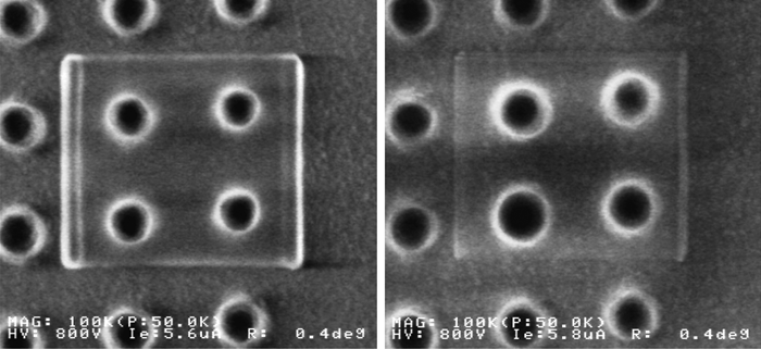
CD measurements. Comparison imaging to examine the effects of contamination on critical dimension (CD) measurements has shown that these image artifacts can affect dimensional measurements [Reference Vladar5]. In CD work, modification of dimensions by the SEM imaging process causes a loss of precision in the measurement. Using a very clean Hitachi 6280 (Figure 4 left), the test pattern began to show filling-in of the holes after a 20-minute scan. After Evactron in situ cleaning of the chamber and the specimen, a repeat of the measurement showed no filling of the holes and a much-reduced scan mark (Figure 4 right).

Figure 4: A CD test pattern began to show filling-in of the holes after a 20-minute scan (left). After Evactron in situ cleaning of the chamber and the specimen, a repeat of the measurement (right) showed no filling of the holes and a much-reduced scan mark.
Other applications
It is also well established that cleaner vacuum systems can assist in removing spurious analytical artifacts. Often, carbon analysis can contain contributions that do not originate from the sample but can be due to contamination. Work by Strein and Allred [Reference Strein and Allred6] showed that the antechamber of an X-ray photoelectron spectroscopy system was introducing a thin layer of carbon onto samples, making carbon analysis unreliable. Downstream plasma treatment in the antechamber removed the contamination.
Horiuchi et al. [Reference Horiuchi7] have also shown that analytical transmission electron microscopy (TEM) results on polymer brush samples could be accomplished with a system cleaned using downstream plasma. Electron energy loss spectrometry (EELS) in imaging mode could be used for high-resolution carbon mapping.
Having pristine surfaces is an absolute requirement for nanomanipulation and nanofabrication. Recent work by Mancevski has shown that downstream plasma cleaning was essential for successful vapor phase cutting of carbon nanotubes using a nano-manipulator system [Reference Mancevski and Rack8]. Also, electrical measurements, made by positioning minute probes on circuits, using nanopositioning systems located inside SEMs and FIBs, require that the probes be free of contamination in order to make good contacts. In situ cleaning of these devices is a requirement for accurate measurements.
Plasma radical sources are rapidly evolving to solve new problems and are now available in a number of configurations for most makes and models of electron and ion microscopes. Standard PRS units commonly allow for air, pure oxygen, and oxygen/argon mixtures. These units have KF 40 flanges adapted for most SEMs and Dual Beam FIB/SEMs. Further, because Evactron units are portable, these systems may be moved around the laboratory, cleaning a number of different electron microscopes. An ultra-high vacuum (UHV) Conflat flange version is available for surface analysis tools or other HV chambers and provides for the use of hydrogen gas. Versions of the PRS specific for TEMs, where a patented hollow cathode is inserted through the sample insertion port, are becoming available on most major TEM models. This unique design for the TEM delivers the cleaning capability directly to the hard-to-reach area where beam and sample interact in the TEM. Two of the most recent configurations are summarized in Figure 5. These show a TEM Wand and a PRS mounted in a benchtop unit.

Figure 5: Examples of two different types of plasma radical source (PRS) configurations: the TEM Wand (left) and the SoftClean benchtop system (right). Images courtesy of XEI Scientific, Inc.
The downstream plasma technique has proven extremely useful and is now well accepted. There are over 1,100 installations of the XEI tool on nearly all makes and models of SEM and Dual Beam FIB/SEMs. In fact, today most new high-resolution tools come equipped with some form of downstream plasma cleaning upon delivery from the factory. Further, service personnel often carry a portable version of the Evactron system when they make service and preventative visits in the field in order to maximize SEM performance.
Conclusions
Using reactive gas plasma systems has proven to be one of the most effective methods to remove contamination artifacts that hamper imaging and analysis in electron microscopes. The latest systems combine downstream cleaning in microscopes with benchtop sample cleaning, bringing together the attributes of clean samples and clean microscope environments in a single product. Now it is easier than ever for electron microscopists to “keep it clean.”


