No CrossRef data available.
Published online by Cambridge University Press: 09 September 2022

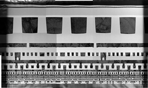
Warping is a limiting factor when preparing transmission electron microscopy (TEM) samples using focused ion beam (FIB)/scanning electron microscope (SEM) systems. The conventional FIB sputtering process leaves at least one side of the lamella too thin to provide structural support to offset inherent stresses. As a result, warping can occur impacting imagining and reducing the potential size of lamellae. For example, capturing more than a few back-end metal layers in a 3 μm wide cross-section lamella can be difficult. Frequently, TEM analysts must perform multiple stage adjustments to analyze such a sample. In this paper, two methods are presented that enable FIB/SEM operators to prepare TEM samples where the thinned region of interest is surrounded by thick structures. As a result, these methods reduce warping and enable the fabrication of TEM lamellae not possible by conventional means. For example, these methods have been used to produce a 10 μm wide by 8 μm tall cross-section TEM sample that captured front-end transistors and 14 back-end metal layers. Furthermore, warping was so limited that only one alignment was needed by the TEM analyst to complete the imaging of the sample. The methods, called the horizontal bracing and window methods, make use of the deposition of low-Z amorphous films that are electron transparent in the TEM.